
Main menu

  • Home
  • Content
    • Current
    • Ahead of print
    • Past Issues
    • JNM Supplement
    • SNMMI Annual Meeting Abstracts
    • Continuing Education
    • JNM Podcasts
  • Subscriptions
    • Subscribers
    • Institutional and Non-member
    • Rates
    • Journal Claims
    • Corporate & Special Sales
  • Authors
    • Submit to JNM
    • Information for Authors
    • Assignment of Copyright
    • AQARA requirements
  • Info
    • Reviewers
    • Permissions
    • Advertisers
  • About
    • About Us
    • Editorial Board
    • Contact Information
  • More
    • Alerts
    • Feedback
    • Help
    • SNMMI Journals
  • SNMMI
    • JNM
    • JNMT
    • SNMMI Journals
    • SNMMI

User menu

  • Subscribe
  • My alerts
  • Log in
  • Log out
  • My Cart

Search

  • Advanced search
Journal of Nuclear Medicine
  • SNMMI
    • JNM
    • JNMT
    • SNMMI Journals
    • SNMMI
  • Subscribe
  • My alerts
  • Log in
  • Log out
  • My Cart
Journal of Nuclear Medicine

Advanced Search

  • Home
  • Content
    • Current
    • Ahead of print
    • Past Issues
    • JNM Supplement
    • SNMMI Annual Meeting Abstracts
    • Continuing Education
    • JNM Podcasts
  • Subscriptions
    • Subscribers
    • Institutional and Non-member
    • Rates
    • Journal Claims
    • Corporate & Special Sales
  • Authors
    • Submit to JNM
    • Information for Authors
    • Assignment of Copyright
    • AQARA requirements
  • Info
    • Reviewers
    • Permissions
    • Advertisers
  • About
    • About Us
    • Editorial Board
    • Contact Information
  • More
    • Alerts
    • Feedback
    • Help
    • SNMMI Journals
  • View or Listen to JNM Podcast
  • Visit JNM on Facebook
  • Join JNM on LinkedIn
  • Follow JNM on Twitter
  • Subscribe to our RSS feeds
Research ArticleCLINICAL INVESTIGATIONS

Predicting Seizure-Free Status for Temporal Lobe Epilepsy Patients Undergoing Surgery: Prognostic Value of Quantifying Maximal Metabolic Asymmetry Extending over a Specified Proportion of the Temporal Lobe

Tina W. Lin, Michelle A. Kung de Aburto, Magnus Dahlbom, Lynn L. Huang, Michael M. Marvi, Michael Tang, Johannes Czernin, Michael E. Phelps and Daniel H.S. Silverman
Journal of Nuclear Medicine May 2007, 48 (5) 776-782; DOI: https://doi.org/10.2967/jnumed.106.034249
Tina W. Lin
  • Find this author on Google Scholar
  • Find this author on PubMed
  • Search for this author on this site
Michelle A. Kung de Aburto
  • Find this author on Google Scholar
  • Find this author on PubMed
  • Search for this author on this site
Magnus Dahlbom
  • Find this author on Google Scholar
  • Find this author on PubMed
  • Search for this author on this site
Lynn L. Huang
  • Find this author on Google Scholar
  • Find this author on PubMed
  • Search for this author on this site
Michael M. Marvi
  • Find this author on Google Scholar
  • Find this author on PubMed
  • Search for this author on this site
Michael Tang
  • Find this author on Google Scholar
  • Find this author on PubMed
  • Search for this author on this site
Johannes Czernin
  • Find this author on Google Scholar
  • Find this author on PubMed
  • Search for this author on this site
Michael E. Phelps
  • Find this author on Google Scholar
  • Find this author on PubMed
  • Search for this author on this site
Daniel H.S. Silverman
  • Find this author on Google Scholar
  • Find this author on PubMed
  • Search for this author on this site
  • Article
  • Figures & Data
  • Info & Metrics
  • PDF
Loading

Article Figures & Data

Figures

  • Tables
  • FIGURE 1. 
    • Download figure
    • Open in new tab
    • Download powerpoint
    FIGURE 1. 

    Axial tomographic brain slices were displayed in original linear (or inverse linear) gray scale (A), according to a statistically parameterized scale (B), and by an asymmetry parameterized scale (C), as described in the text. Numerically labeled color bars were displayed adjacent to the parameterized scales (D). Scan interpreter could specify the proportion of cortex (x%) for which maximal asymmetry was to be quantified as well as certain display features that would not directly affect quantification, using interactive slider controls (E).

  • FIGURE 2. 
    • Download figure
    • Open in new tab
    • Download powerpoint
    FIGURE 2. 

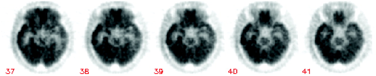
    Scan interpreter selects plane(s) reflecting maximal temporal asymmetry.

  • FIGURE 3. 
    • Download figure
    • Open in new tab
    • Download powerpoint
    FIGURE 3. 

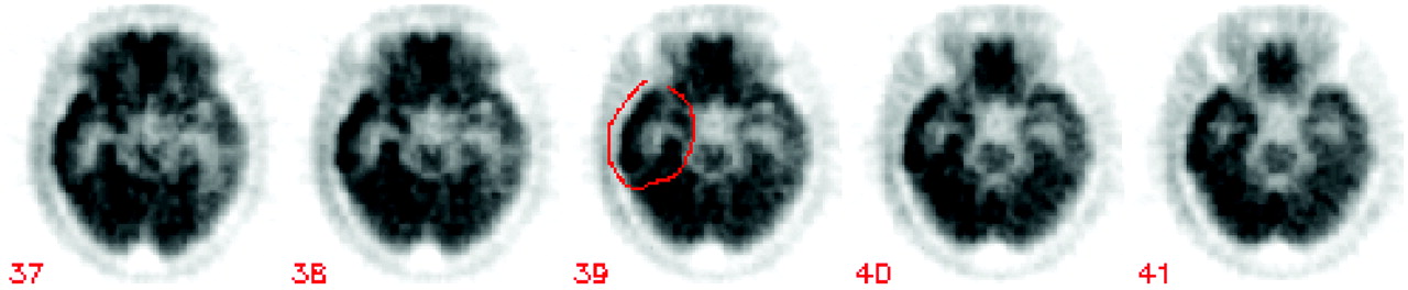
    Interpreter draws loose region around temporal cortex in one of maximally asymmetric planes, on side of brain visually possessing greater activity, to initiate calculation of T-AIx (temporal lobe asymmetry index).

  • FIGURE 4. 
    • Download figure
    • Open in new tab
    • Download powerpoint
    FIGURE 4. 

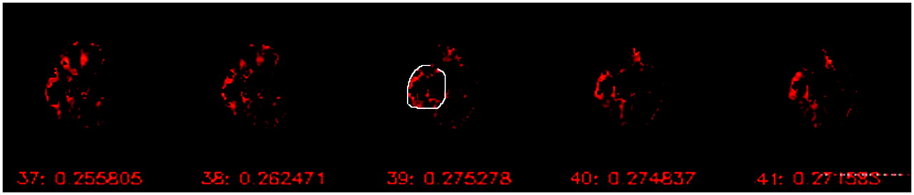
    T-AIx (temporal lobe asymmetry index) values are automatically generated for 5 adjacent planes and displayed immediately below image planes (in this case, equaling 0.275, as seen below central image, plane 39).
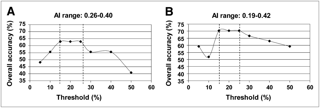
  • FIGURE 5. 
    • Download figure
    • Open in new tab
    • Download powerpoint
    FIGURE 5. 

    Extent specification yielding highest overall accuracy was consistently determined to be 20% by systematic tests in both first (A) and second (B) PET patient series.

  • FIGURE 6. 
    • Download figure
    • Open in new tab
    • Download powerpoint
    FIGURE 6. 

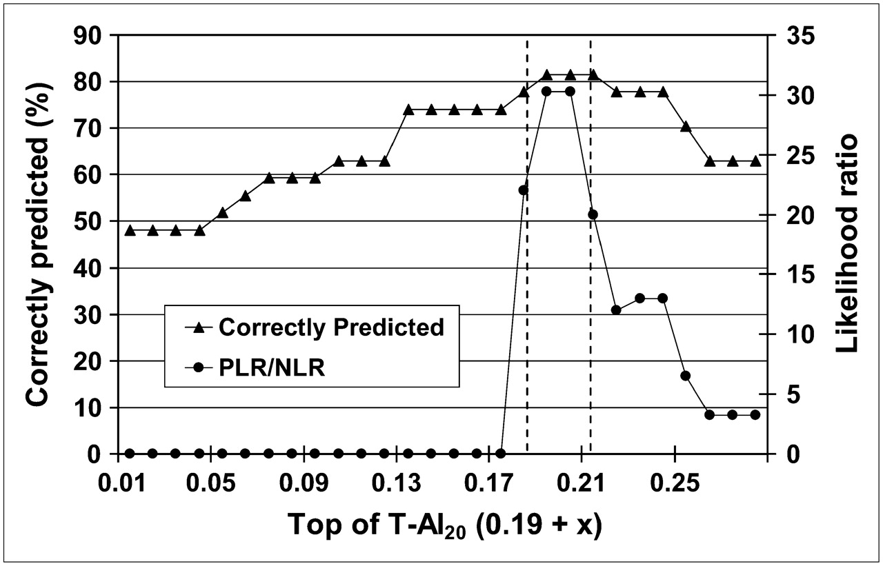
    Optimal determined extent specification (20%) was applied systematically to identify optimal T-AI range for achieving seizure-free outcome in second PET patient series. By both overall accuracy and likelihood ratio measures, this optimal range extended from 0.19 to 0.40 (0.19 + 0.21). PLR = positive likelihood ratio; NLR = negative likelihood ratio; numbers on right-sided y-axis correspond to 10 × PLR/NLR.
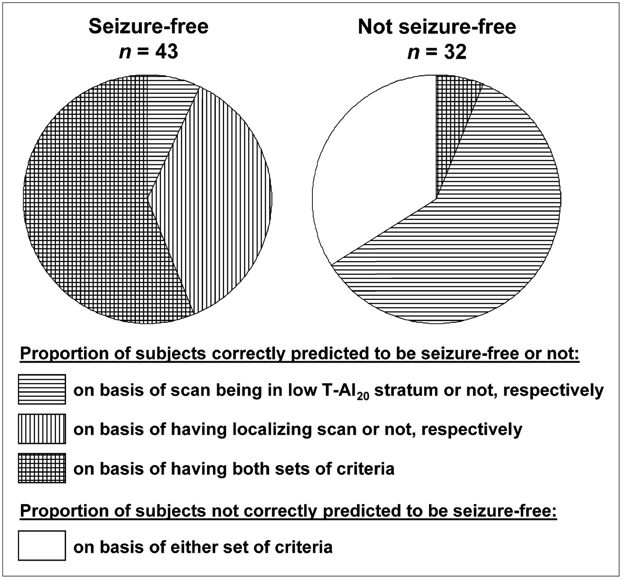
  • FIGURE 7. 
    • Download figure
    • Open in new tab
    • Download powerpoint
    FIGURE 7. 

    Distribution of prognoses based on visual and quantitative interpretations of PET scans, among patients who achieved seizure-free status (left, n = 43) and patients who continued to have seizures (right, n = 32) after surgical therapy. A high T-AI20 value (>0.40) was identified before surgery in most patients who continued to seize postsurgically, despite having unilaterally localizing patterns of hypometabolism on visual assessment of presurgical PET scans (horizontally lined segment in right pie chart), but in only a minority of patients who became seizure free (vertically lined area in left pie chart)—an indication of the added prognostic value of the T-AI20 index over visual assessment alone, in predicting seizure-free status.

Tables

  • Figures
    • View popup
    TABLE 1

    Patient Characteristics (n = 75)

    VariableFirst PET patient series* (n = 47)Second PET patient series* (n = 28)
    Sex
     Male25 (53)13 (46)
     Female22 (47)15 (54)
    Mean age ± SD at time of PET23.9 ± 15.922.6 ± 15.0
     <2 y4 (9)2 (7)
     2–12 y10 (21)6 (21)
     >12 y33 (70)20 (72)
    Clinical follow-up time (y)3.83.8
    • ↵* Values in parentheses are percentages.

PreviousNext
Back to top

In this issue

Journal of Nuclear Medicine: 48 (5)
Journal of Nuclear Medicine
Vol. 48, Issue 5
May 2007
  • Table of Contents
  • Table of Contents (PDF)
  • About the Cover
  • Index by author
Print
Download PDF
Article Alerts
Sign In to Email Alerts with your Email Address
Email Article

Thank you for your interest in spreading the word on Journal of Nuclear Medicine.

NOTE: We only request your email address so that the person you are recommending the page to knows that you wanted them to see it, and that it is not junk mail. We do not capture any email address.

Enter multiple addresses on separate lines or separate them with commas.
Predicting Seizure-Free Status for Temporal Lobe Epilepsy Patients Undergoing Surgery: Prognostic Value of Quantifying Maximal Metabolic Asymmetry Extending over a Specified Proportion of the Temporal Lobe
(Your Name) has sent you a message from Journal of Nuclear Medicine
(Your Name) thought you would like to see the Journal of Nuclear Medicine web site.
Citation Tools
Predicting Seizure-Free Status for Temporal Lobe Epilepsy Patients Undergoing Surgery: Prognostic Value of Quantifying Maximal Metabolic Asymmetry Extending over a Specified Proportion of the Temporal Lobe
Tina W. Lin, Michelle A. Kung de Aburto, Magnus Dahlbom, Lynn L. Huang, Michael M. Marvi, Michael Tang, Johannes Czernin, Michael E. Phelps, Daniel H.S. Silverman
Journal of Nuclear Medicine May 2007, 48 (5) 776-782; DOI: 10.2967/jnumed.106.034249

Citation Manager Formats

  • BibTeX
  • Bookends
  • EasyBib
  • EndNote (tagged)
  • EndNote 8 (xml)
  • Medlars
  • Mendeley
  • Papers
  • RefWorks Tagged
  • Ref Manager
  • RIS
  • Zotero
Share
Predicting Seizure-Free Status for Temporal Lobe Epilepsy Patients Undergoing Surgery: Prognostic Value of Quantifying Maximal Metabolic Asymmetry Extending over a Specified Proportion of the Temporal Lobe
Tina W. Lin, Michelle A. Kung de Aburto, Magnus Dahlbom, Lynn L. Huang, Michael M. Marvi, Michael Tang, Johannes Czernin, Michael E. Phelps, Daniel H.S. Silverman
Journal of Nuclear Medicine May 2007, 48 (5) 776-782; DOI: 10.2967/jnumed.106.034249
Twitter logo Facebook logo LinkedIn logo Mendeley logo
  • Tweet Widget
  • Facebook Like
  • Google Plus One
Bookmark this article

Jump to section

  • Article
    • Abstract
    • MATERIALS AND METHODS
    • RESULTS
    • DISCUSSION
    • CONCLUSION
    • Acknowledgments
    • References
  • Figures & Data
  • Info & Metrics
  • PDF

Related Articles

  • This Month in JNM
  • Google Scholar

Cited By...

  • PET of Serotonin 1A Receptors and Cerebral Glucose Metabolism for Temporal Lobectomy
  • Voxel-Based Analysis of Asymmetry Index Maps Increases the Specificity of 18F-MPPF PET Abnormalities for Localizing the Epileptogenic Zone in Temporal Lobe Epilepsies
  • Google Scholar

More in this TOC Section

  • Feasibility of Ultra-Low-Activity 18F-FDG PET/CT Imaging Using a Long–Axial-Field-of-View PET/CT System
  • Cardiac Presynaptic Sympathetic Nervous Function Evaluated by Cardiac PET in Patients with Chronotropic Incompetence Without Heart Failure
  • Validation and Evaluation of a Vendor-Provided Head Motion Correction Algorithm on the uMI Panorama PET/CT System
Show more Clinical Investigations

Similar Articles

SNMMI

© 2025 SNMMI

Powered by HighWire