


Article Figures & Data
Figures
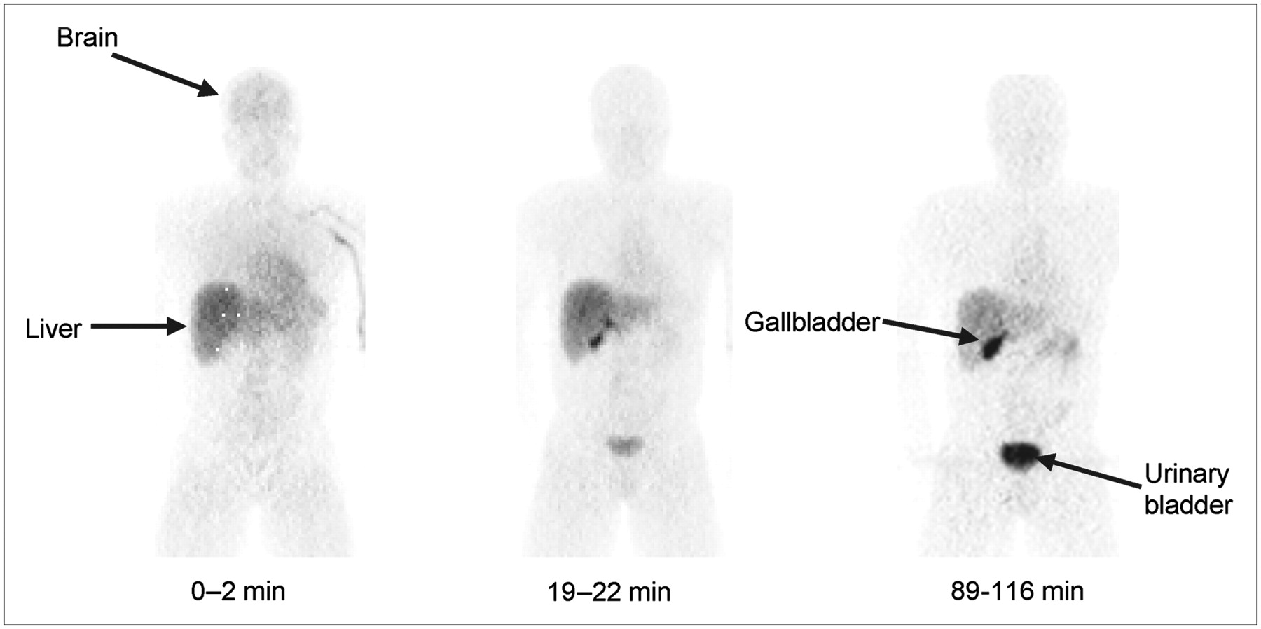
- FIGURE 1.
Compressed anteroposterior whole-body 2D planar images of subject 4, a typical healthy individual. Images were obtained at 0–2, 19–22, and 89–116 min after intravenous injection of 703 MBq 11C-PBR28.
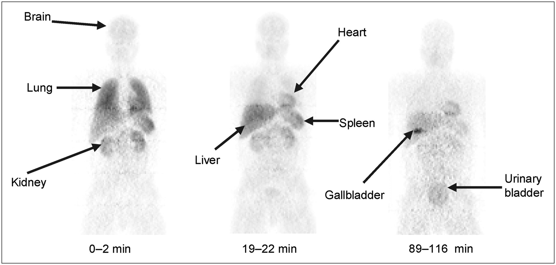
- FIGURE 2.
Compressed anteroposterior whole-body 2D planar images for unusual healthy subject 7 with minimal or no 11C-PBR28 binding. Images were obtained approximately 0–2, 19–22, and 89–116 min after intravenous injection of 544 MBq.
- FIGURE 3.
Decay-corrected time–activity curves for visually identifiable organs from the average of subjects 1–6 (A and B) and the unusual subject 7 (C and D). Kidneys (C) and spleen (D) could not be seen in subject 7. Graph becomes too cluttered if SD error bars are included. Distribution of data is exemplified by the % coefficient of variation (SD/mean) at 60 min for A and B, with lungs at 40%, liver at 28%, gallbladder at 29%, and brain at 35%.
- FIGURE 4.
Decay-corrected time–activity curves of lung (A) and brain (B) in subjects 1–6 (•) and unusual subject 7 (□). Symbols represent mean + SD for subjects 1–6.
- FIGURE 5.
Compressed anteroposterior whole-body 2D planar images in same monkey at baseline (A) and after receptor blockade (B). For the latter, nonradioactive PK 11195 (10.7 mg/kg intravenously) was administered 3 min before the radioligand. Injected activity of 11C-PBR28 was 348 MBq (A) and 332 MBq (B).
- FIGURE 6.
Decay-corrected time–activity curves for visually identifiable organs in monkeys from the average of 3 baseline (A and B) and 1 receptor-blocked study (C and D).


Tables
Residence time (h) Organ Subjects 1–6 Subject 7 Ratio* Brain 0.020 ± 0.007 0.013 0.7 Heart wall 0.011 ± 0.004 0.017 1.5 Kidneys 0.058 ± 0.023 — — Liver 0.069 ± 0.015 0.122 1.8 Gallbladder 0.004 ± 0.003 0.015 4.0 Urinary bladder 0.018 ± 0.011 0.021 1.2 Lungs 0.083 ± 0.031 0.033 0.4 Spleen 0.016 ± 0.004 — — Remainder in body 0.213 ± 0.074 0.270 1.3 ↵* Ratio of subject 7 to mean of subjects 1–6.
Values are mean ± SD of 6 subjects. Subject 7 lacked PBR binding and was analyzed separately.
Dose Organ μSv/MBq mrem/mCi Adrenals 4.1 ± 0.2 15.0 ± 0.9 Brain 4.8 ± 1.5 17.9 ± 5.5 Breasts 1.9 ± 0.2 7.1 ± 0.7 Gallbladder wall 12.2 ± 6.8 45.3 ± 25 Lower large intestine wall 1.9 ± 0.3 7.0 ± 1.2 Small intestine 2.3 ± 0.3 8.4 ± 1.2 Stomach 2.7 ± 0.2 10.0 ± 0.7 Upper large intestine wall 2.4 ± 0.3 8.7 ± 1.2 Heart wall 11.2 ± 3.0 41.6 ± 11 Kidneys 52.6 ± 20 194.7 ± 75 Liver 13.1 ± 2.6 48.4 ± 9.5 Lungs 22.0 ± 7.6 81.6 ± 28 Muscle 1.9 ± 0.2 7.1 ± 0.9 Ovaries 2.0 ± 0.2 7.4 ± 1.3 Pancreas 3.9 ± 0.1 14.6 ± 0.5 Red marrow 2.1 ± 0.1 7.6 ± 0.5 Osteogenic cells 2.5 ± 0.5 9.2 ± 1.8 Skin 1.4 ± 0.3 5.2 ± 1.0 Spleen 25.9 ± 5.6 95.9 ± 21 Testes 1.5 ± 0.3 5.4 ± 1.3 Thymus 2.3 ± 0.2 8.5 ± 0.9 Thyroid 1.7 ± 0.4 6.1 ± 1.3 Urinary bladder wall* 13.3 ± 6.7 49.2 ± 25 Uterus 2.3 ± 0.2 8.5 ± 0.7 Total body 2.9 ± 0.1 10.7 ± 0.4 Effective dose equivalent 11.0 ± 2.4 40.7 ± 8.9 Effective dose 6.6 ± 1.7 24.3 ± 6.3 ↵* Dynamic urinary bladder model was not used.
Values are expressed as mean ± SD.
Residence time (h) Organ Baseline Preblocked Brain 0.086 ± 0.006 0.024 Heart wall 0.021 ± 0.003 0.008 Kidneys 0.048 ± 0.008 0.042 Urinary bladder 0.012 ± 0.003 0.018 Lungs 0.277 ± 0.01 0.067 Liver* 0.020 0.051 Spleen 0.006 ± 0.004 — Remainder in body 0.022 ± 0.12 0.282 ↵* Liver was visible in only 1 study.
Residence times in baseline conditions were averaged over 3 studies using 2 monkeys. One preblocked study was performed by injecting 10.7 mg/kg of nonradioactive PK 11195 three minutes before the radioligand.
Supplemental Data
Files in this Data Supplement:


Jump to section
Related Articles
Cited By...
- Early brain neuroinflammatory and metabolic changes identified by dual tracer microPET imaging in mice with acute liver injury
- Study protocol for a phase II, double-blind, randomised controlled trial of cannabidiol (CBD) compared with placebo for reduction of brain neuroinflammation in adults with chronic low back pain
- Quantification of ONO-2952 Occupancy of 18-kDaTranslocator Protein in Conscious Monkey Brains using Positron Emission Tomography
- 11C-ER176, a Radioligand for 18-kDa Translocator Protein, Has Adequate Sensitivity to Robustly Image All Three Affinity Genotypes in Human Brain
- Glial activation colocalizes with structural abnormalities in amyotrophic lateral sclerosis
- In Vivo Detection of Age- and Disease-Related Increases in Neuroinflammation by 18F-GE180 TSPO MicroPET Imaging in Wild-Type and Alzheimer's Transgenic Mice
- Influence of TSPO Genotype on 11C-PBR28 Standardized Uptake Values
- Propofol Decreases In Vivo Binding of 11C-PBR28 to Translocator Protein (18 kDa) in the Human Brain
- Brain and Whole-Body Imaging of Nociceptin/Orphanin FQ Peptide Receptor in Humans Using the PET Ligand 11C-NOP-1A
- Radiation Dosimetry and Biodistribution of the TSPO Ligand 11C-DPA-713 in Humans
- 18F-FEAC and 18F-FEDAC: PET of the Monkey Brain and Imaging of Translocator Protein (18 kDa) in the Infarcted Rat Brain
- Fetal Dose Estimates for 18F-Fluoro-L-Thymidine Using a Pregnant Monkey Model
- PET-Derived Biodistribution and Dosimetry of the Benzodiazepine Receptor-Binding Radioligand 11C-(R)-PK11195 in Children and Adults
- Human Brain Imaging and Radiation Dosimetry of 11C-N-Desmethyl-Loperamide, a PET Radiotracer to Measure the Function of P-Glycoprotein