


Abstract
We evaluated two 18F-labeled PET ligands, N-benzyl-N-ethyl-2-[7,8-dihydro-7-(2-18F-fluoroethyl)-8-oxo-2-phenyl-9H-purin-9-yl]acetamide (18F-FEAC) and N-benzyl-N-methyl-2-[7,8-dihydro-7-(2-18F-fluoroethyl)-8-oxo-2-phenyl-9H-purin-9-yl]acetamide (18F-FEDAC), by investigating their kinetics in the monkey brain and by performing in vitro and in vivo imaging of translocator protein (18 kDa) (TSPO) in the infarcted rat brain. Methods: Dissection was used to determine the distribution of 18F-FEAC and 18F-FEDAC in mice, whereas PET was used for a monkey. With each 18F-ligand, in vitro autoradiography and small-animal PET were performed on infarcted rat brains. Results: 18F-FEAC and 18F-FEDAC had a high uptake of radioactivity in the heart, lung, and other TSPO-rich organs of mice. In vitro autoradiography showed that the binding of each 18F-ligand significantly increased on the ipsilateral side of rat brains, compared with the contralateral side. In a small-animal PET study, PET summation images showed the contrast of radioactivity between ipsilateral and contralateral sides. Pretreatment with TSPO ligands N-benzyl-N-ethyl-2-(7-methyl-8-oxo-2-phenyl-7,8-dihydro-9H-purin-9-yl)acetamide (AC-5216) or (R)-N-methyl-N-(1-methylpropyl)-1-(2-chlorophenyl)isoquinoline-3-carboxamide (PK11195) diminished the difference in uptake between the 2 sides. The PET study showed that each 18F-ligand had uptake and distribution patterns in the monkey brain similar to those of 11C-AC-5216. After injection into the monkey during PET, the uptake of each 18F-ligand in the brain decreased over time whereas 11C-AC-5216 did not. In the brain homogenate of mice, the percentage of the fraction corresponding to intact 18F-FEAC and 18F-FEDAC was 68% and 75% at 30 min after injection. In monkey plasma, each 18F-ligand was scarcely metabolized until the end of the PET scan. Conclusion: 18F-FEAC and 18F-FEDAC produced in vitro and in vivo signals allowing visualization of the increase in TSPO expression in the infarcted rat brain. The kinetics of both 18F-ligands in the monkey brain and tolerance for in vivo metabolism suggested their usefulness for imaging studies of TSPO in primates.
The translocator protein (18 kDa) (TSPO) (1), formerly known as peripheral-type benzodiazepine receptor, is widely distributed in peripheral tissues and the brain (2,3). In the brain, TSPO is located mainly in glial cells, and TSPO expression was increased in microglial cells activated by brain injury or neuroinflammation (4–7). Neuropathologic evidence has demonstrated that an increase in TSPO and concurrent microglial and astroglial activation accompanies the neurodegenerative process in Alzheimer disease (7–9). These findings motivated the development of imaging probes labeled by positron-emitting radionuclides and have enabled visualization of the distribution and change in TSPO induced by microglial activation in normal and abnormal brains of humans and animals (10–18).
11C-labeled (R)-N-methyl-N-(1-methylpropyl)-1-(2-chlorophenyl)isoquinoline-3-carboxamide (PK11195) was the first PET ligand used for clinical imaging of TSPO (19). However, 11C-PK11195 has several limitations, such as relatively low brain uptake, high nonspecific binding, high plasma protein binding, and very high lipophilicity (20). To precisely characterize TSPO with an improved PET ligand, several new ligands labeled with 11C or 18F have been reported (10,11). Of these ligands, 11C-labeled N,N-diethyl-2-[2-(4-methoxyphenyl)-5,7-dimethylpyrazolo[1,5-α]pyrimidin-3-yl]acetamide (DPA-713) and 18F-labeled N,N-diethyl-2-(2-(4-(2-fluoroethoxy)phenyl)-5,7-dimethylpyrazolo[1,5-α]pyrimidin-3-yl)acetamide (DPA-714) are promising PET ligands with potent binding affinity and suitable kinetics in rodent and primate brains for TSPO imaging (14,15,17,18). We have developed N-(2-11C,5-dimethoxybenzyl)-N-(5-fluoro-2-phenoxyphenyl) acetamide (11C-DAA1106), N-(5-fluoro-2-phenoxyphenyl)-N-(2-18F-fluoroethyl-5-methoxybenzyl)acetamide (18F-FEDAA1106), and N-benzyl-N-ethyl-2-(7-11C-methyl-8-oxo-2-phenyl-7,8-dihydro-9H-purin-9-yl)acetamide (11C-AC-5216) for the clinical investigation of TSPO in our institute (20–23). However, these PET ligands had little dissociation in the primate brain, which makes the assessment of their brain kinetics difficult (24–26). Thus, PET studies with ligands such as 11C-AC-5216 create complications in the quantitative analysis of the measurement of TSPO expression and function in health and disease.
We recently synthesized two 18F-labeled oxopurine analogs: N-benzyl-N-ethyl-2-[7,8-dihydro-7-(2-18F-fluoroethyl)-8-oxo-2-phenyl-9H-purin-9-yl]acetamide (18F-FEAC) and N-benzyl-N-methyl-2-[7,8-dihydro-7-(2-18F-fluoroethyl)-8-oxo-2-phenyl-9H-purin-9-yl]acetamide (18F-FEDAC) (27). 18F-FEAC (inhibition constant [Ki], 0.2 nM) and 18F-FEDAC (Ki, 1.3 nM) had potent in vitro binding affinity for TSPO with high selectivity (Ki, >8,700 nM for central benzodiazepine receptor). A previous small-animal PET study for a neuroinflammatory rat model demonstrated that both 18F-ligands had high uptake of radioactivity in a kainic acid–infused striatum, a brain region where TSPO expression was augmented (27).
In this study, we performed in vitro autoradiography and small-animal PET to visualize elevated TSPO expression in infarcted rat brains and evaluate the potential of 8F-FEAC and 18F-FEDAC for the imaging of TSPO. We investigated the brain kinetics of both 18F-ligands and compared their uptake and kinetics with those of 11C-AC-5216 in the same monkey. In addition, we determined biodistribution in whole mice and performed metabolite analysis in mouse brain and plasma and in monkey plasma using 18F-FEAC and 18F-FEDAC.
MATERIALS AND METHODS
Radioligands
18F-FEAC.
For irradiation, 95% enriched 18O-H2O was used. 18F was produced by the 18O(p,n)18F nuclear reaction using a CYPRIS HM-18 cyclotron (Sumitomo Heavy Industries). The radioactive intermediate 18F-FCH2CH2Br (22) was reacted with a precursor (1 mg) and NaOH (7 μL, 0.5N aqueous solution) in dimethylformamide (300 μL) at 90°C for 15 min. After purification by high-performance liquid chromatography (HPLC), 920–1,460 MBq (n = 23) of 18F-FEAC were obtained as an injectable solution at a beam current of 15 μA and proton bombardment of 15 min. The radiochemical purity and specific activity of 18F-FEAC were at least 98% and 290–420 GBq/μmol at the end of synthesis.
18F-FEDAC.
The radiosynthesis procedure for 18F-FEDAC was similar to that for 18F-FEAC except that a different precursor was used. At the end of synthesis, 840–1,520 MBq (n = 25) of 18F-FEDAC were obtained with a radiochemical purity of at least 97% and specific activity of 330–450 GBq/μmol.
Animal Experiments
All animal experiments were performed according to the recommendations of the Committee for the Care and Use of Laboratory Animals, National Institute of Radiologic Sciences. Animals were maintained and handled in accordance with the recommendations of the National Institutes of Health and the institutional guidelines of the National Institute of Radiologic Sciences.
Biodistribution Study on Mice
A saline solution of 18F-FEAC (1.7 MBq/200 μL, 350 GBq/μmol) or 18F-FEDAC (2.1 MBq/200 μL, 330 GBq/μmol) was injected into ddY mice (7 wk old, male) through the tail vein. At 1, 5, 15, 30, 60, and 90 min after injection, 3 mice per time point were sacrificed by cervical dislocation. Whole brain, heart, lung, liver, kidney, spleen, bone, muscle, small intestine, testicle, and blood samples were quickly removed and weighed carefully. The radioactivity in these tissues was measured using a 1480 Wizard γ-counter (Perkin-Elmer) and expressed as a percentage of the injected dose (%ID) per gram of wet tissue. All radioactivity measurements were corrected for decay.
In Vitro Autoradiography Study on Infarcted Rat Brains
Transient focal ischemia in rats was produced by intraluminal occlusion of the middle cerebral artery for 30 min using a 3-vessel occlusion/reperfusion model (5,18). After ischemic surgery, the filament was removed for reperfusion. During this process, no behavioral change was observed in these animals. The body temperature of these rats was monitored and maintained. The rats were used 7 d after the surgery.
The rats were sacrificed by decapitation under diethyl ether anesthesia, and the brains were quickly removed and frozen on powdered dry ice. Coronal brain sections (20 μm) were cut with a cryostat (HM560; Carl Zeiss) at −15°C. Brain sections were immersed in a buffer of 50 mM Tris(hydroxymethyl)aminomethane hydrochloride (Trizma; Sigma-Aldrich) for 20 min at 25°C. 18F-FEAC (320 GBq/μmol) or 18F-FEDAC (350 MBq/μmol) with the same radioactivity (3.7 MBq) was added to the incubation solutions. To determine the specific binding of each 18F-ligand to TSPO, we added AC-5216 or PK11195 (10 μM) to the incubation solutions in advance. Brain sections were incubated at 25°C for 30 min. After incubation, the sections were washed twice for 2 min each time with cold buffer, dipped in cold distilled water, and dried with cold air. These sections were then placed in contact with imaging plates (BAS-MS2025; Fujifilm). Autoradiograms were obtained and quantified using a Bio-Imaging Analyzer System (BAS5000; Fujifilm). Radioactivity concentrations in the brain regions were measured and expressed as photostimulated luminescence per area (mm2). Regions of interest (ROIs) were placed on the infarcted areas (10.3 ± 2.3 mm2/slice) of ipsilateral sides and the same areas of the contralateral sides. Finally, these sections were stained with cresyl violet.
Small-Animal PET Study on Infarcted Rat Brains
PET Scans.
Anatomic template images of rat brains were generated by a high-resolution MRI system. Briefly, a rat was anesthetized with sodium pentobarbital (50 mg/kg, intraperitoneally) and scanned with a 40-cm-bore, 7-T horizontal magnet (NIRS/KOBELCO; Bruker BioSpin) equipped with 12-cm-diameter gradients (Bruker BioSpin). A 7.2-cm-diameter coil was used for radiofrequency transmission, and signals were received by a 4-channel surface coil. Coronal T2-weighted MR images were obtained by a fast spin-echo sequence with the following imaging parameters: repetition time, 8,000 min; effective echo time, 15 min; field of view, 3.5 × 3.5 cm; and slice thickness, 0.6 mm.
PET scans were performed using an Inveon small-animal PET scanner (Siemens Medical Solutions), which provides 159 transaxial slices 0.796 mm (center-to-center) apart, a 10-cm transaxial field of view, and a 12.7-cm axial field of view. Before the scans, the rats were anesthetized with 5% (v/v) isoflurane, and maintained thereafter by 1%–2% (v/v) isoflurane. After transmission scans for attenuation using a 57Co point source, emission scans were performed for 60 min after the intravenous injection of each 18F-ligand (17 ± 3 MBq, 310–400 GBq/μmol). PET images were obtained by summing the uptake between 0 and 60 min. In pretreatment studies, unlabeled TSPO ligands (AC-5216, 1 mg/kg/2 mL, or PK11195, 3 mg/kg/2 mL; dissolved in distilled water containing 10% ethanol and 5% polysorbate 80) were injected 30 s before injection of 18F-ligand (17 ± 2 MBq, 310–390 GBq/μmol). Because pretreatment with AC-5216 or PK11195 affected the initial uptake of 18F-ligand, PET images in this study were obtained by summing the uptake between 10 and 60 min after injection of 18F-ligand.
All list-mode acquisition data were sorted into 3-dimensional sinograms, which were then Fourier-rebinned into 2-dimensional sinograms (frames × min: 4 × 1, 8 × 2, 8 × 5). Dynamic images were reconstructed with filtered backprojection using a Ramp filter and a Nyquist cutoff of 0.5 cycle/pixel. ROIs were placed on the infarcted areas (9.2 ± 1.5 mm2/slice) of ipsilateral sides and the same areas of contralateral sides using ASIPro VM (Analysis Tools and System Setup/Diagnostics Tool; Siemens Medical Solutions) with reference to the MRI template. Each PET image for each 18F-ligand was overlaid on the MR image in the rat brain, and a time–activity curve for each brain region was determined.
Brain uptake of radioactivity was decay-corrected to the injection time and expressed as the standardized uptake value, normalized for injected radioactivity and body weight: (radioactivity per cubic centimeter tissue/injected radioactivity) × gram body weight.
PET Data Modeling.
Binding potential relative to nondisplaceable binding (BPnd) was estimated for the kinetics of the ipsilateral and contralateral ROIs using the PMOD software package (version 3.1; PMOD Technologies). The simplified reference-tissue model was used, and the contralateral mirrored ROI was used as the reference region to determine BPnd. All statistical examinations in this PET study were performed using GraphPad Prism, vision 5 (GraphPad Software Inc.).
PET Study on Monkey
PET was performed using a high-resolution SHR-7700 PET camera (Hamamatsu Photonics) designed for laboratory animals. This camera provides 31 transaxial slices 3.6 mm (center-to-center) apart and a 33.1-cm field of view. An MR image of the brain was obtained using a Gyroscan S15/ACS II (1.5 T; Philips) with a 3-dimensional T1-weighted axial MR image sequence. A male rhesus monkey (Macaca mulatta) weighing about 8 kg was anesthetized with ketamine (10 mg/kg/h, intramuscularly) every hour throughout the session. Transmission scans for attenuation correction were subsequently performed for 20 min using a 74-MBq 68Ge–68Ga source. A dynamic transmission scan in the 3-dimensional acquisition mode was performed for 90 min (1 min × 4 scans, 2 min × 8 scans, 5 min × 8 scans, 10 min × 3 scans). Emission scan images were reconstructed with a Colsher filter of 4 mm, and circular ROIs with a 5-mm diameter were placed over the occipital cortex and frontal cortex using image analysis software (20). A solution of 11C-AC-5216 (100 MBq, 140 GBq/μmol), 18F-FEAC (110 MBq, 380 GBq/μmol), or 18F-FEDAC (115 MBq, 350 GBq/μmol) was injected into the monkey, and time-sequential tomographic scanning was performed for 90 min on a transverse section of the brain. ROIs were placed on the occipital cortex and frontal cortex using the image analysis software PMOD with reference to the MR image of the monkey brain. Each PET image for each 18F-ligand was overlaid on the MR image in the monkey brain, and a time–activity curve for each brain region was determined. Brain uptake of radioactivity was decay-corrected to the injection time and expressed as %ID per volume (mL).
Metabolite Assay
Mouse Plasma and Brain.
After intravenous injection of 18F-ligand (5 MBq/200 μL, 330–380 GBq/μmol) into ddy mice (n = 4), these mice were sacrificed by cervical dislocation at 1, 5, 15, 30 and 60 min. Blood (0.5–1.0 mL) and whole-brain samples were removed quickly. The blood sample was centrifuged at 15,000 rpm for 1 min at 4°C to separate plasma, 250 μL of which were collected in a test tube containing CH3CN (500 μL) and unlabeled authentic sample (10 μL of a 1 mg/5.0 mL concentration of CH3CN). After the tube was stirred in a vortex mixer for 15 s and centrifuged at 15,000 rpm for 2 min for deproteinization, the supernatant was collected. The extraction efficiency of radioactivity into the CH3CN supernatant ranged from 84% to 96% of total radioactivity in the plasma. On the other hand, the cerebellum and forebrain were dissected from the mouse brain and homogenized together in ice-cooled 1:1 CH3CN/H2O (1.0 mL) solution. The homogenate was centrifuged at 15,000 rpm for 2 min at 4°C, and the supernatant was collected. The recovery of radioactivity into the supernatant was more than 80% based on total radioactivity in the brain homogenate.
An aliquot of the supernatant (100–300 μL) prepared from the plasma or brain homogenate was injected into the analytic HPLC system (Capcell Pak C18 [Shiseido Co., Ltd.], 4.6-mm internal diameter × 250 mm, 254 nm; 1:1 CH3CN/H2O; 1.5 mL/min). The percentage ratio of intact 18F-ligand to total radioactivity (corrected for decay) on the HPLC chromatogram was calculated as (peak area for 18F-ligand/total peak area) × 100.
Monkey Plasma.
After intravenous injection of 18F-ligand (110,130 MBq) into the monkey, arterial blood samples (1 mL) were collected at 2, 5, 15, 30, 60, and 90 min. All samples were centrifuged at 15,000 rpm for 1 min at 4°C to separate plasma, 250 μL of which were collected in a test tube containing CH3CN (0.5 mL). The plasma was treated as described above.
RESULTS
Biodistribution Study on Mice
The radioactivity distribution of 18F-FEAC and 18F-FEDAC was measured over 90 min in mice. As shown in Table 1, both 18F-ligands had a similar distribution pattern in the examined regions. High uptake (>5 %ID/g) was found in the lung, heart, and kidney, which are known to be TSPO-enriched organs. Subsequently, radioactivity in the lung and heart declined rapidly until 5 min and then decreased slowly until the end of the experiment. Uptake in the liver and kidney peaked at 5 min and then decreased slowly. In the blood, radioactivity decreased immediately after injection and remained low. High initial uptake (>1 %ID/g) was determined in the mouse brain, the target tissue in this study. No significant increase in radioactivity was detected in bone.
Distribution in Mice
In Vitro Autoradiography Study on Infarcted Rat Brains
After evaluation in normal mice, we visualized elevated levels of TSPO expression in the infarcted rat brain after ischemia using each 18F-ligand. At day 7 after ischemic surgery, infarction was observed on the ipsilateral side but not on the contralateral side. By cresyl violet staining, neuronal loss and gliosis were confirmed in the infarcted brain sections. Figure 1 shows the in vitro autoradiographic results. For each 18F-ligand, binding was higher on the ipsilateral side than on the contralateral side: the ratios of radioactivity between the 2 sides were 1.80 ± 0.2 (n = 4) for 18F-FEAC and 1.73 ± 0.3 (n = 4) for 18F-FEDAC. The difference in radioactivity between contralateral and ipsilateral sides was abolished by coincubation with an excess of AC-5216 or PK11195. These treatments reduced the radioactivity on the 2 sides. As shown in Figure 2G, the decrease in binding by AC-5216 was more significant on the ipsilateral side (75% decrease for 18F-FEAC and 73% for 18F-FEDAC) than on the contralateral side (56% for 18F-FEAC and 40% for 18F-FEDAC). PK11195 also inhibited the radioactivity level of each 18F-ligand on the ipsilateral and contralateral sides.
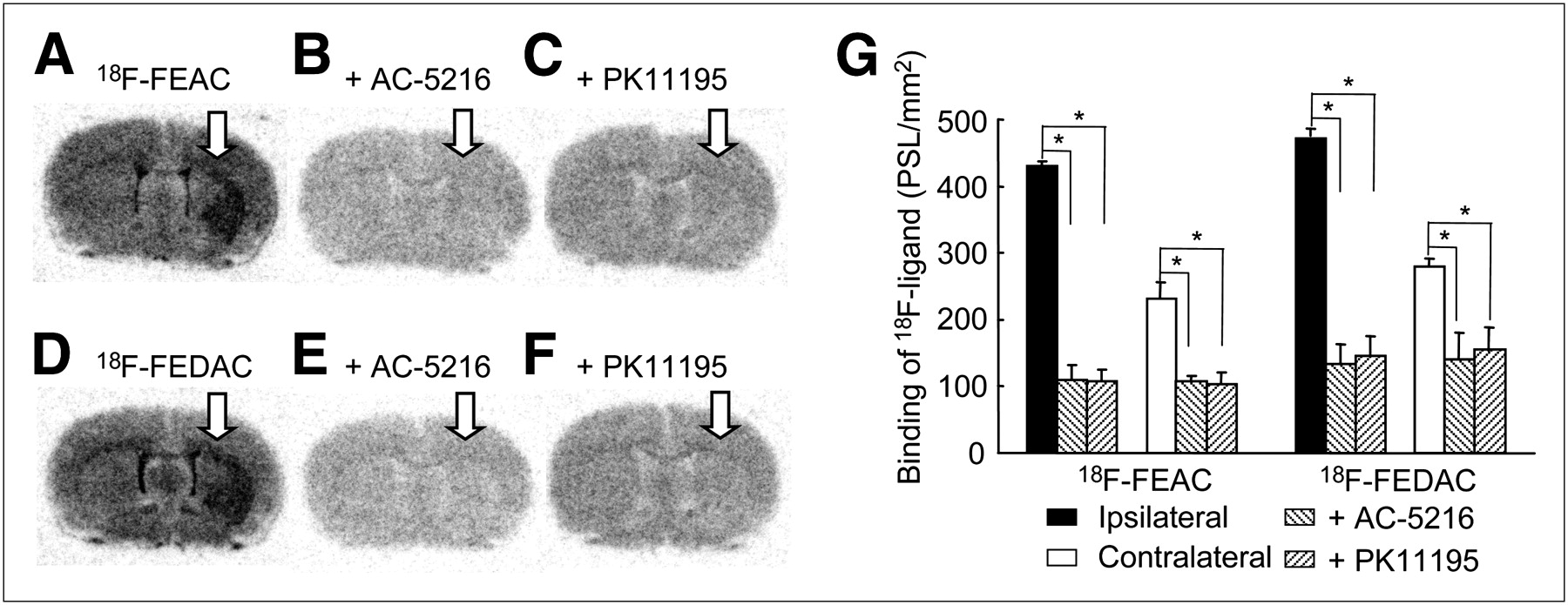
(A–F) Coronal autoradiographic results in infarcted rat brains, with arrows indicating infarcted areas: 18F-FEAC (A), 18F-FEAC with AC-5216 (B), 18F-FEAC with PK11195 (C), 18F-FEDAC (D), 18F-FEDAC with AC-5216 (E), and 18F-FEDAC with PK11195 (F). (G) Radioactivity concentration (photostimulated luminescence/mm2; mean ± SD, n = 4) was determined in these regions. *P < 0.001, compared with contralateral side in each group by Student paired t test.

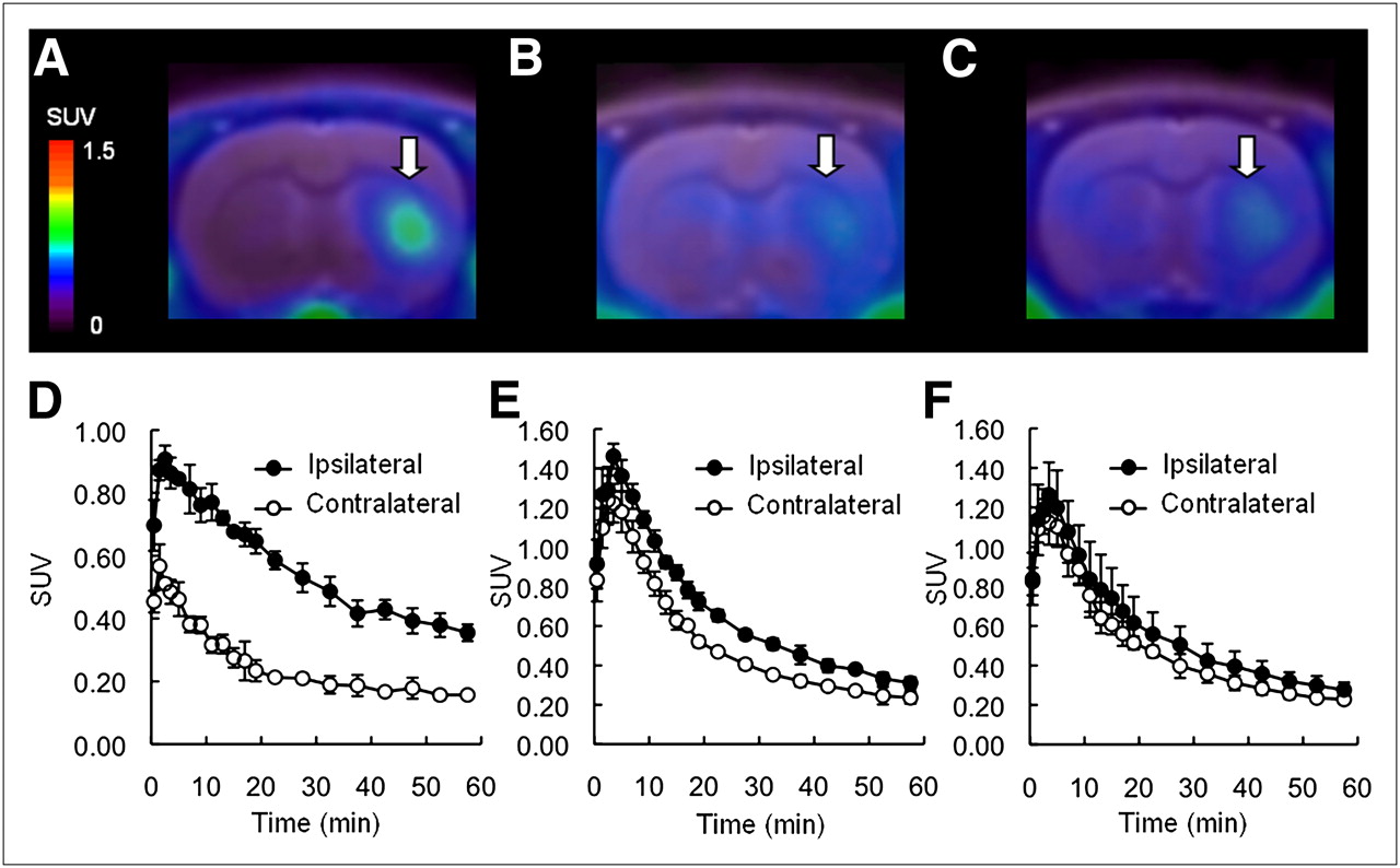
Representative PET images and time–activity curves of 18F-FEAC in infarcted rat brains. PET images were generated by averaging whole scan and were overlaid on MR images. Arrows indicate infarcted areas. Shown are control rats (n = 4) (A and D), rats pretreated with AC-5216 (n = 4) (B and E), and rats pretreated with PK11195 (n = 4) (C and F).
Small-Animal PET Study on Infarcted Rat Brains
Figures 2 and 3 show representative PET images and time–activity curves of 18F-FEAC and 18F-FEDAC, respectively, in the infarcted rat brain. PET images (Figs. 2A and 3A) displayed a contrast of radioactivity between ipsilateral and contralateral sides. Each 18F-ligand had higher uptake on ipsilateral sides than on contralateral sides. Their time–activity curves (Figs. 2D and 3D) were similar in rat brains. Each 18F-ligand entered the brain rapidly, and the radioactivity level in the brain peaked within 1–3 min after injection. Maximum uptake on the ipsilateral and contralateral sides was about 0.87 and 0.49 standardized uptake value, respectively, for 18F-FEAC and 0.91 and 0.57 standardized uptake value, respectively, for 18F-FEDAC. Compared with the contralateral side, where radioactivity decreased rapidly after its low initial uptake, radioactivity on the ipsilateral side decreased slowly. At the end of the PET scan, the uptake decreased to 72% (18F-FEAC) and 66% (18F-FEDAC) of the corresponding maximum uptake on the ipsilateral side, respectively. The uptake ratios of radioactivity between ipsilateral and contralateral sides reached 3.03 for 18F-FEAC and 2.76 for 18F-FEDAC at 20 min after injection (Fig. 4). Time–activity curves were analyzed using the simplified reference-tissue model to estimate the BPnd of each 18F-ligand. The BPnd values were 1.70 ± 0.19 (n = 4) for 18F-FEAC and 1.37 ± 0.06 (n = 4) for 18F-FEDAC. This difference in BPnd between 18F-ligands was statistically significant (P < 0.05, unpaired t test with Welch correction).

Representative PET images and time–activity curves of 18F-FEDAC in infarcted rat brains. PET images were generated by averaging whole scan and were overlaid on MR images. Arrows indicate infarcted areas. Shown are control rats (n = 4) (A and D), rats pretreated with AC-5216 (n = 4) (B and E), and rats pretreated with PK11195 (n = 4) (C and F).

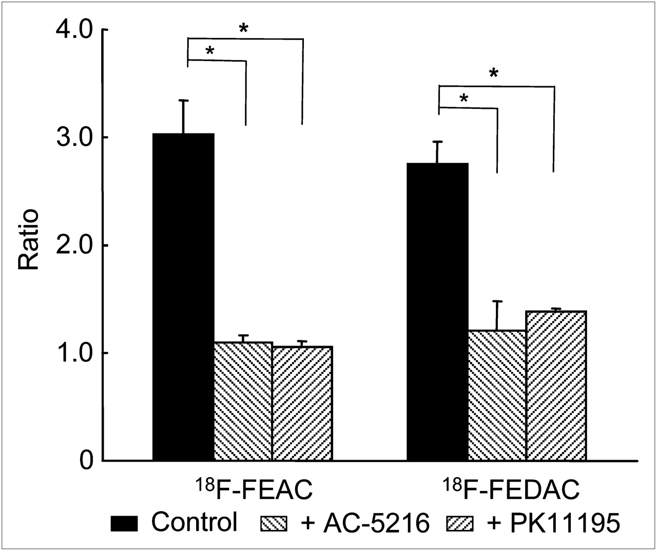
Uptake ratios (mean ± SD, n = 4) between ipsilateral and contralateral sides of infarcted rat brains. Ratio was calculated from uptake data of 18F-FEAC or 18F-FEDAC at 20 min after injection. *P < 0.001, compared with control group (18F-FEAC or 18F-FEDAC only) in each group (pretreatment with AC-5216 or PK11195) by Student paired t test.
The effects of unlabeled TSPO ligands (AC-5216, 1 mg/kg; PK11195, 3 mg/kg) on the uptake and kinetics of each 18F-ligand were determined. As shown in their PET images (18F-FEAC: Figs. 2B and 2C; 18F-FEDAC: Figs. 3B and 3C) and time–activity curves (18F-FEAC: Figs. 2E and 2F; 18F-FEDAC: Figs. 3E and 3F), AC-5216 or PK11195 diminished the contrast of radioactivity between ipsilateral and contralateral sides. By pretreatment with these TSPO ligands, the uptake ratio of each 18F-ligand between the 2 sides was reduced to 1.0–1.4 (Fig. 4).
PET Study on Monkey
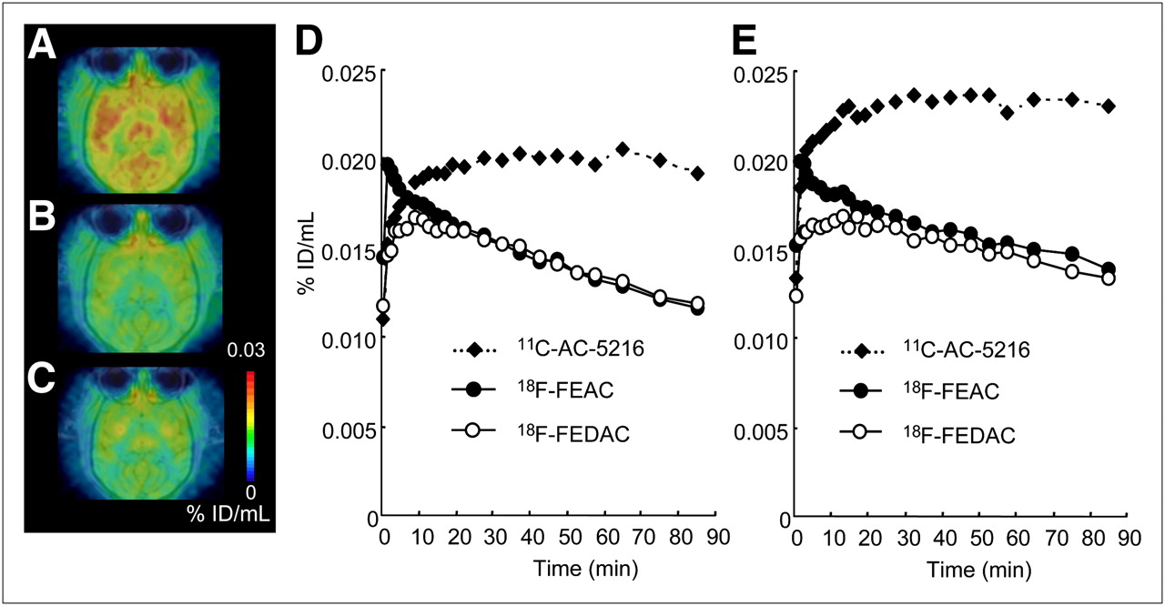
The uptake and kinetics of 18F-FEAC and 18F-FEDAC in the monkey brain were examined using PET. For comparison, PET with 11C-AC-5216 was preliminarily performed for the same monkey. Figure 5 shows the summation images and time–activity curves of the 3 PET ligands in the monkey brain. Like 11C-AC-5216, 18F-FEAC and 18F-FEDAC displayed a near-homogeneous pattern in the monkey brain. As seen from time–activity curves in the occipital cortex, the maximum uptake of 18F-FEDAC (0.02% dose/mL) immediately after injection was similar to that of 11C-AC-5216 at about 20 min and was higher than that of 18F-FEAC (0.016% dose/mL) at 5 min. After reaching the maximum, the uptake of 11C-AC-5216 maintained that level until the end of the PET scan. In contrast to 11C-AC-5216, each 18F-ligand showed decreased uptake over time in the brain. At 90 min after injection, the radioactivity of 18F-FEAC and 18F-FEDAC decreased to 70% and 61%, respectively, of the maximum. The uptake of each 18F-ligand decreased with time in the occipital cortex and in the frontal cortex, striatum, thalamus, and cerebellum of the monkey brain.

(A–C) PET images showing distribution of 11C-AC-5216 (A), 18F-FEAC (B), and 18F-FEDAC (C) in brain of same rhesus monkey. These images were generated by averaging whole scan and were overlaid on MR images. (D and E) Time–activity curves for 11C-AC-5216, 18F-FEAC, and 18F-FEDAC in occipital (D) and frontal cortex (E) of monkey brain.
As seen on the PET images, no significant accumulation of radioactivity was observed in the monkey skull during the scan with each 18F-ligand.
Metabolite Analysis
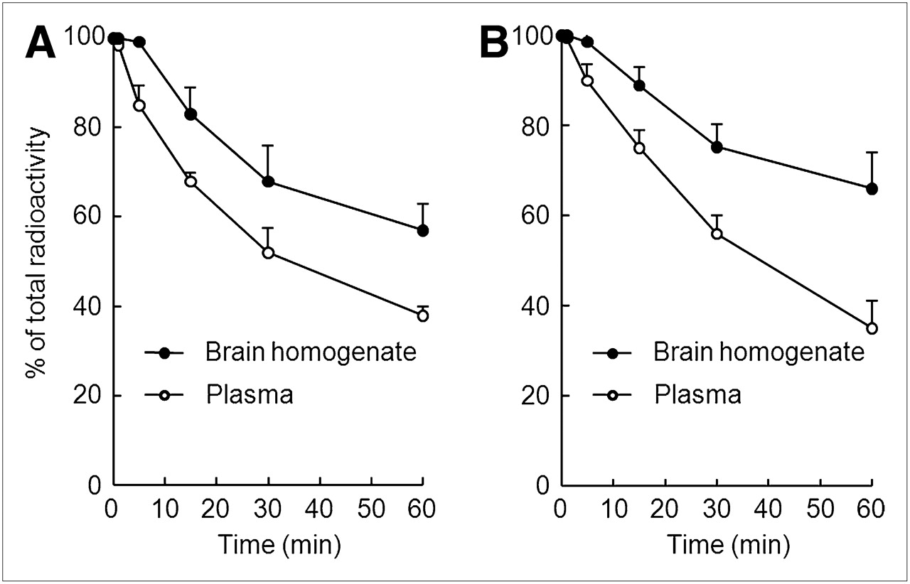
Figure 6 shows the percentages of unmetabolized 18F-ligand in the plasma and brain homogenate of mice analyzed by radio-HPLC. After injection into the mice, the fraction corresponding to intact 18F-ligand in the plasma decreased over time. In the brain homogenate, the percentages of intact 18F-FEAC and 18F-FEDAC were 68% and 75% at 30 min after injection, respectively. A radioactive metabolite with high polarity was observed on the HPLC chart of each 18F-ligand.

Percentages (mean ± SD, n = 4) of unmetabolized 18F-ligand in mouse plasma and brain homogenate at several time points after injection of 18F-ligand: 18F-FEAC (A) and 18F-FEDAC (B).
Table 2 shows the results of metabolite analysis in monkey plasma. The fraction corresponding to the unmetabolized 18F-FEAC and18F-FEDAC in the plasma remained at 96% of the total radioactivity at 30 min after injection. The percentages of intact forms were 87% and 80%, respectively, for 18F-FEAC and 18F-FEDAC at the end (90 min) of the PET scans. Only a small amount of the same radiolabeled metabolite of each 18F-ligand was detected.
Percentages of Intact 18F-Ligand in Monkey Plasma
DISCUSSION
Although several dozen PET ligands have been developed for in vivo TSPO imaging (10,11), radioligands such as 11C-AC-5216, 11C-DAA1106, and 18F-FEDAA1106 had little dissociation in the primate brain (23–26). The slow kinetics of these PET ligands is a significant disadvantage. The present study demonstrated that, compared with 11C-AC-5216, 18F-FEAC and 18F-FEDAC had improved kinetics in the monkey brain. Both 18F-ligands produced signals to allow in vitro and in vivo visualization of elevated TSPO expression in the infarcted rat brain.
Uptake of 18F-FEAC and 18F-FEDAC in normal mice agreed with the distribution of TSPO in rodents as reported previously (Table 1) (21,23). The highest uptake of radioactivity was in the lung and heart and may have been due to the high mitochondrial content of TSPO in these organs. Both 18F-ligands had a relatively high level (>1 %ID/g) of radioactivity in the brain, revealing that they could pass the blood–brain barrier and enter the brain rapidly—a prerequisite for a favorable PET ligand in brain imaging. Uptake in the brain may be related to the suitable lipophilicity of these ligands (calculated distribution coefficient ClogD: 3.6 for 18F-FEAC and 3.2 for 18F-FEDAC) (27).
Because of the relatively low density of TSPO in the normal rodent brain, we used an ischemic model to validate the usefulness of 18F-FEAC and 18F-FEDAC for in vitro and in vivo TSPO imaging. This is a well-known rat model in which TSPO expression is elevated by neuroinflammation with microglial activation in the infarcted areas of the brain (5,18). In vitro autoradiography showed that the ipsilateral side gave rise to a TSPO-rich area that could be readily and similarly visualized using 18F-FEAC or 18F-FEDAC (Fig. 1). Treatment with TSPO-specific ligands, AC-5216 or PK11195, blocked their binding on the ipsilateral and contralateral sides, suggesting that both 18F-ligands had in vitro TSPO-specific binding in both inflammatory and noninflammatory areas of the infarcted brains.
In a small-animal PET study on infarcted brains, PET summation images showed a significant contrast of radioactivity between contralateral and ipsilateral sides (Figs. 2A and 3A). The uptake ratio of each 18F-ligand between the 2 sides peaked at 20 min after injection. This ratio was significantly reduced by pretreatment with AC-5216 or PK11195 (Fig. 4). These results indicated that the in vivo binding of both 18F-ligands was specific to TSPO and that increased binding was caused by overexpression of TSPO in the infarcted brains. The increase in the initial uptake of 18F-ligand by pretreatment with AC-5216 (Figs. 2E and 3E) or PK11195 (Figs. 2F and 3F) could be explained by precluding radioactivity from peripheral organs, because high radioactivity relative to TSPO binding was present in organs such as the lung and heart. Likely caused by the flow of radioactivity from the peripheral organs to the brain, the uptake ratio of 18F-FEDAC between ipsilateral and contralateral sides was not reduced to 1 by pretreatment with AC-5216 or PK11195 (Fig. 4). On the other hand, the BPnd value of 18F-FEAC was slightly higher than that of 18F-FEDAC, which might be due to the higher binding affinity of FEAC (Ki, 0.49 nM) than of FEDAC (1.34 nM) for TSPO (27).
To evaluate the potential of both 18F-ligands in the primate brain, we compared the distribution pattern and kinetics of 18F-FEAC and 18F-FEDAC with those of 11C-AC-5216 in the same monkey brain (Fig. 5). PET images showed that the distribution patterns of both 18F-ligands were consistent with that of 11C-AC-5216 in the monkey brain and were also similar to those of 11C-AC-5216 in the human brain, reported previously (26). This result, in addition to the structural similarity of the 3 radioligands, confirmed that the uptake of both 18F-ligands reflected the distribution of TSPO in the monkey brain. The maximum uptake of 18F-FEAC and 18F-FEDAC in the occipital cortex, frontal cortex, and other regions of the monkey brain were 3–4 times higher than that of 11C-PK11195 in the same monkey, as calculated from our previous studies (20). The radioactivity of 18F-FEDAC in the brain peaked immediately after injection and then decreased. 18F-FEAC also displayed a slow decrease in the brain after the maximum level had been reached at 5 min. In contrast to both 18F-ligands, 11C-AC-5216, like 11C-DAA1106 and 18F-FEDAA1106, did not dissociate in any brain regions (23). The kinetics of both 18F-ligands in the monkey brain may be preferable to those of 11C-AC-5216 for PET assessment.
The relatively rapid kinetics may be due to the decreased binding affinity of both 18F-ligands with TSPO. FEDAC and FEAC displayed slightly lower affinities with TSPO (Ki, 1.34 and 0.49 nM, respectively) than did AC-5216 (0.20 nM), DAA1106 (0.16 nM), and FEDAA1106 (0.08 nM). Reduction of the binding affinity may result in the dissociation of both 18F-ligands from TSPO in the brain. FEAC (cLogD, 3.6) and FEDAC (3.2) became less lipophilic than did DAA1106 (3.7) and FEDAA1106 (3.8)—a characteristic that could reduce nonspecific binding in the brain. Thus, suitable lipophilicity may also partly contribute to the clearance of both 18F-ligands in the brain.
As seen from the PET summation images (Figs. 2, 3, and 5), no significant accumulation of 18F-F− was observed in the skulls of rat and monkey. Distribution in mice by the dissection method also demonstrated no significant radioactivity in bones (Table 1). On the other hand, although the radiolabeled metabolite of each 18F-ligand was found in the brain homogenate of mice, intact 18F-ligand was represented as the main component (Fig. 6). The percentage of intact 18F-FEAC or 18F-FEDAC was 83% or 89% in the homogenate at 15 min after injection, but the maximum uptake ratio of each 18F-ligand between ipsilateral and contralateral sides was achieved within 15–20 min. These results indicated that the specific binding of 18F-FEAC or 18F-FEDAC with TSPO in the infarcted rat brain was mainly due to these radioligands themselves. Further, in vivo metabolite analysis confirmed the relative stability of each 18F-ligand in the monkey plasma (Table 2). Thus, PET for TSPO in the primate brain was unlikely to be affected by their radiolabeled metabolites.
The present PET results suggested that 18F-FEAC and 18F-FEDAC are useful for in vivo imaging and quantification of TSPO in the brain. Based on their kinetics in the brain, a PET study using both 18F-ligands may be suitable for quantitative analysis of the expression and change in TSPO, in comparison with 11C-AC-5216. Although these 18F-ligands with relatively rapid dissociation may show limited specificity for TSPO, and their brain kinetics may be easily influenced by blood flow, it is worth comparing their kinetics and BPnd with those of 11C-AC-5216 in the human brain. Whether the elevated TSPO expression in the neuroinflammatory brains could be detected efficiently using both 18F-ligands needs to be elucidated. Recently, some TSPO PET ligands, such as the pyrazolopyrimidine analogs 11C-DPA-713 and 18F-DPA-714, have already shown suitable kinetics in the primate brain (14,15,17,18). It would be valuable to compare the oxopurine analogs 18F-FEAC and 18F-FEDAC with pyrazolopyrimidine and other structure radioligands using the same experimental animal model prepared under the same conditions.
CONCLUSION
18F-FEAC and 18F-FEDAC provided sufficient signals to allow visualization of TSPO in infarcted rat brains in vitro and in vivo. The kinetics in the monkey brain and tolerance for in vivo metabolism suggest their usefulness for imaging studies of TSPO in normal and abnormal brains. Thus, the prospects for human clinical investigations using both 18F-ligands are promising. We expect that in vivo TSPO imaging in humans can be used to quantitatively estimate the neuroinflammation and outcomes of disease-modifying treatments in neurodegenerative disorders such as Alzheimer disease.
Acknowledgments
We thank Takashi Okauchi and Hidekatsu Wakisaka (National Institute of Radiological Sciences) for their help with PET scans. We also thank the staff of the National Institute of Radiological Sciences for their help with radiosynthesis and animal experiments. This study was supported in part by a Grant-in-Aid for the Molecular Imaging Program from the Ministry of Education, Culture, Sports, Science and Technology, Government of Japan.
- © 2010 by Society of Nuclear Medicine
REFERENCES
- Received for publication October 31, 2009.
- Accepted for publication May 10, 2010.


Jump to section
Related Articles
Cited By...
- 18F-FEDAC as a Targeting Agent for Activated Macrophages in DBA/1 Mice with Collagen-Induced Arthritis: Comparison with 18F-FDG
- Imaging Microglial Activation with TSPO PET: Lighting Up Neurologic Diseases?
- In Vivo Measurement of the Affinity and Density of Metabotropic Glutamate Receptor Subtype 1 in Rat Brain Using 18F-FITM in Small-Animal PET