
Main menu

  • Home
  • Content
    • Current
    • Ahead of print
    • Past Issues
    • JNM Supplement
    • SNMMI Annual Meeting Abstracts
    • Continuing Education
    • JNM Podcasts
  • Subscriptions
    • Subscribers
    • Institutional and Non-member
    • Rates
    • Journal Claims
    • Corporate & Special Sales
  • Authors
    • Submit to JNM
    • Information for Authors
    • Assignment of Copyright
    • AQARA requirements
  • Info
    • Reviewers
    • Permissions
    • Advertisers
  • About
    • About Us
    • Editorial Board
    • Contact Information
  • More
    • Alerts
    • Feedback
    • Help
    • SNMMI Journals
  • SNMMI
    • JNM
    • JNMT
    • SNMMI Journals
    • SNMMI

User menu

  • Subscribe
  • My alerts
  • Log in
  • My Cart

Search

  • Advanced search
Journal of Nuclear Medicine
  • SNMMI
    • JNM
    • JNMT
    • SNMMI Journals
    • SNMMI
  • Subscribe
  • My alerts
  • Log in
  • My Cart
Journal of Nuclear Medicine

Advanced Search

  • Home
  • Content
    • Current
    • Ahead of print
    • Past Issues
    • JNM Supplement
    • SNMMI Annual Meeting Abstracts
    • Continuing Education
    • JNM Podcasts
  • Subscriptions
    • Subscribers
    • Institutional and Non-member
    • Rates
    • Journal Claims
    • Corporate & Special Sales
  • Authors
    • Submit to JNM
    • Information for Authors
    • Assignment of Copyright
    • AQARA requirements
  • Info
    • Reviewers
    • Permissions
    • Advertisers
  • About
    • About Us
    • Editorial Board
    • Contact Information
  • More
    • Alerts
    • Feedback
    • Help
    • SNMMI Journals
  • View or Listen to JNM Podcast
  • Visit JNM on Facebook
  • Join JNM on LinkedIn
  • Follow JNM on Twitter
  • Subscribe to our RSS feeds
OtherFOCUS ON MOLECULAR IMAGING

Comparison of Imaging Techniques for Tracking Cardiac Stem Cell Therapy

Sarah J. Zhang and Joseph C. Wu
Journal of Nuclear Medicine December 2007, 48 (12) 1916-1919; DOI: https://doi.org/10.2967/jnumed.107.043299
Sarah J. Zhang
  • Find this author on Google Scholar
  • Find this author on PubMed
  • Search for this author on this site
Joseph C. Wu
  • Find this author on Google Scholar
  • Find this author on PubMed
  • Search for this author on this site
  • Article
  • Figures & Data
  • Info & Metrics
  • PDF
Loading

Article Figures & Data

Figures

  • FIGURE 1. 
    • Download figure
    • Open in new tab
    • Download powerpoint
    FIGURE 1. 

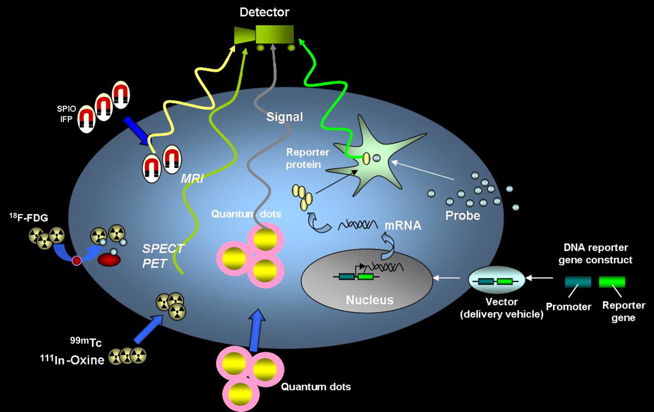
    Schematic for noninvasive imaging of stem cell fate in myocardium. The 4 different techniques include magnetic particle labeling, radionuclide labeling, QD labeling, and reporter gene labeling. SPIO = superparamagnetic iron oxide; IFP = iron fluorescent particles.

  • FIGURE 2. 
    • Download figure
    • Open in new tab
    • Download powerpoint
    FIGURE 2. 
    • Download figure
    • Open in new tab
    • Download powerpoint
    FIGURE 2. 
    • Download figure
    • Open in new tab
    • Download powerpoint
    FIGURE 2. 
    • Download figure
    • Open in new tab
    • Download powerpoint
    FIGURE 2. 

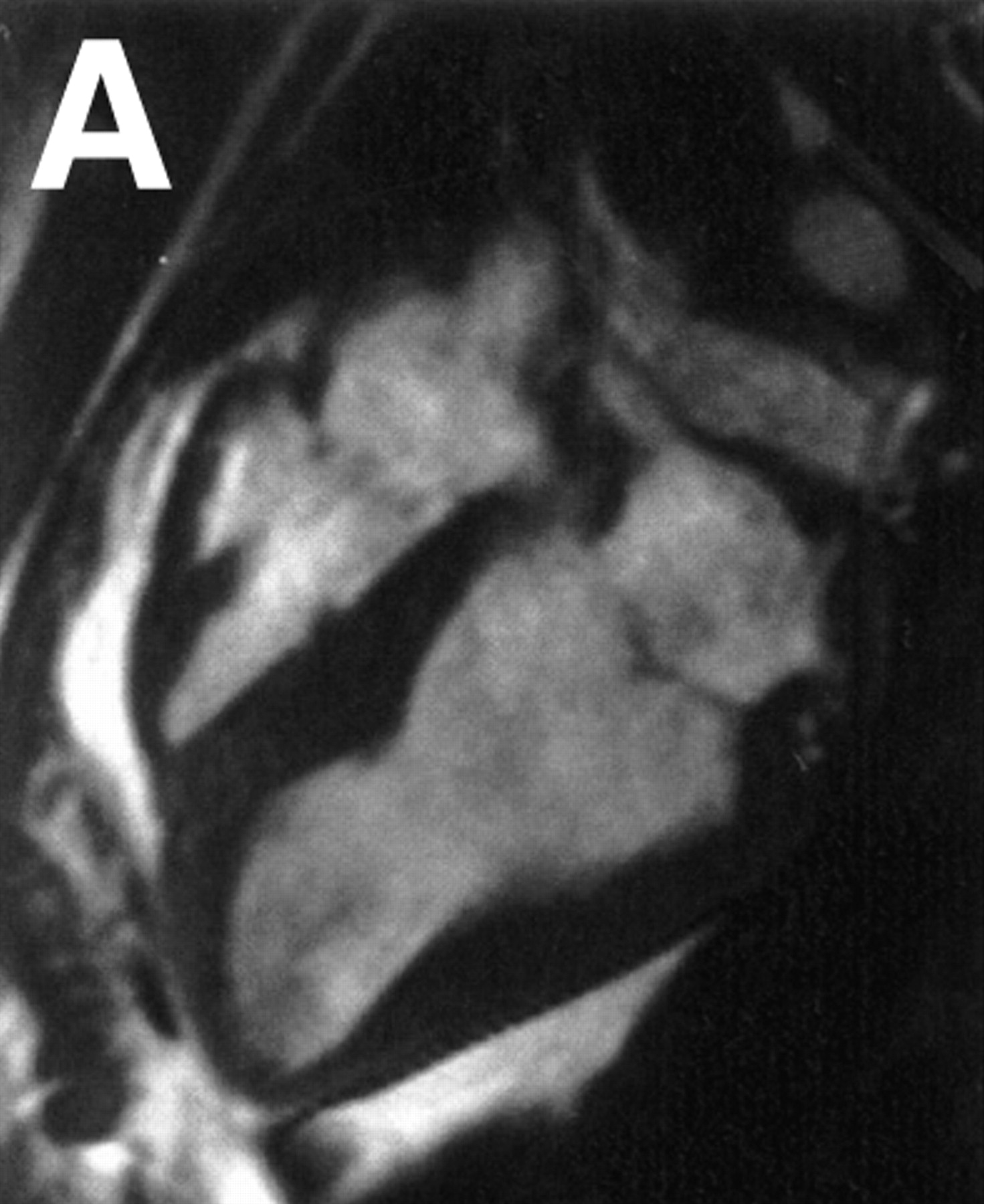
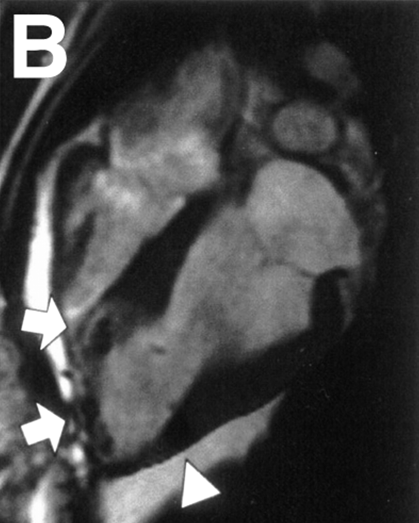
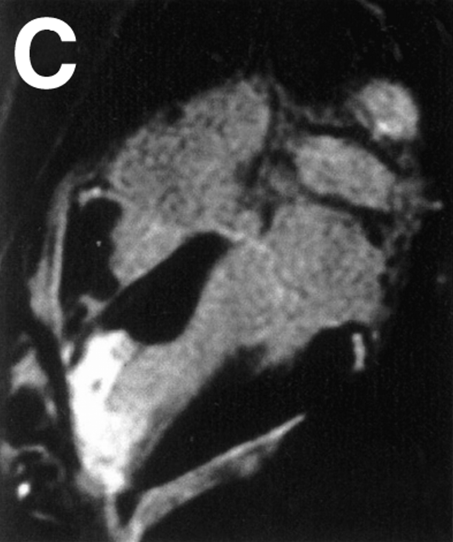
    MRI of transplanted cardiac stem cells in pigs. (A and B) Long-axis view of left ventricle before (A) and after (B) transcatheter injection of 4 × 10⁶ iron fluorescent particle (IFP)–labeled mesenchymal stem cells (MSCs) into infarct at apex (arrows) and into adjoining normal myocardium (arrowhead). (C and D) Long-axis view with delayed, hyperenhanced inversion recovery highlights areas of nonviable infarcted myocardium before (C) and after (D) injection of IFP-labeled MSCs. MSCs appear dark against hyperenhanced infarct. (Reprinted with permission from (10).)

  • FIGURE 3. 
    • Download figure
    • Open in new tab
    • Download powerpoint
    FIGURE 3. 
    • Download figure
    • Open in new tab
    • Download powerpoint
    FIGURE 3. 
    • Download figure
    • Open in new tab
    • Download powerpoint
    FIGURE 3. 
    • Download figure
    • Open in new tab
    • Download powerpoint
    FIGURE 3. 

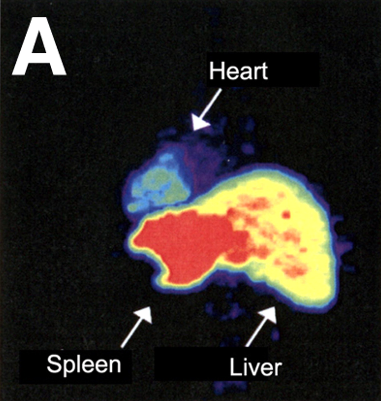
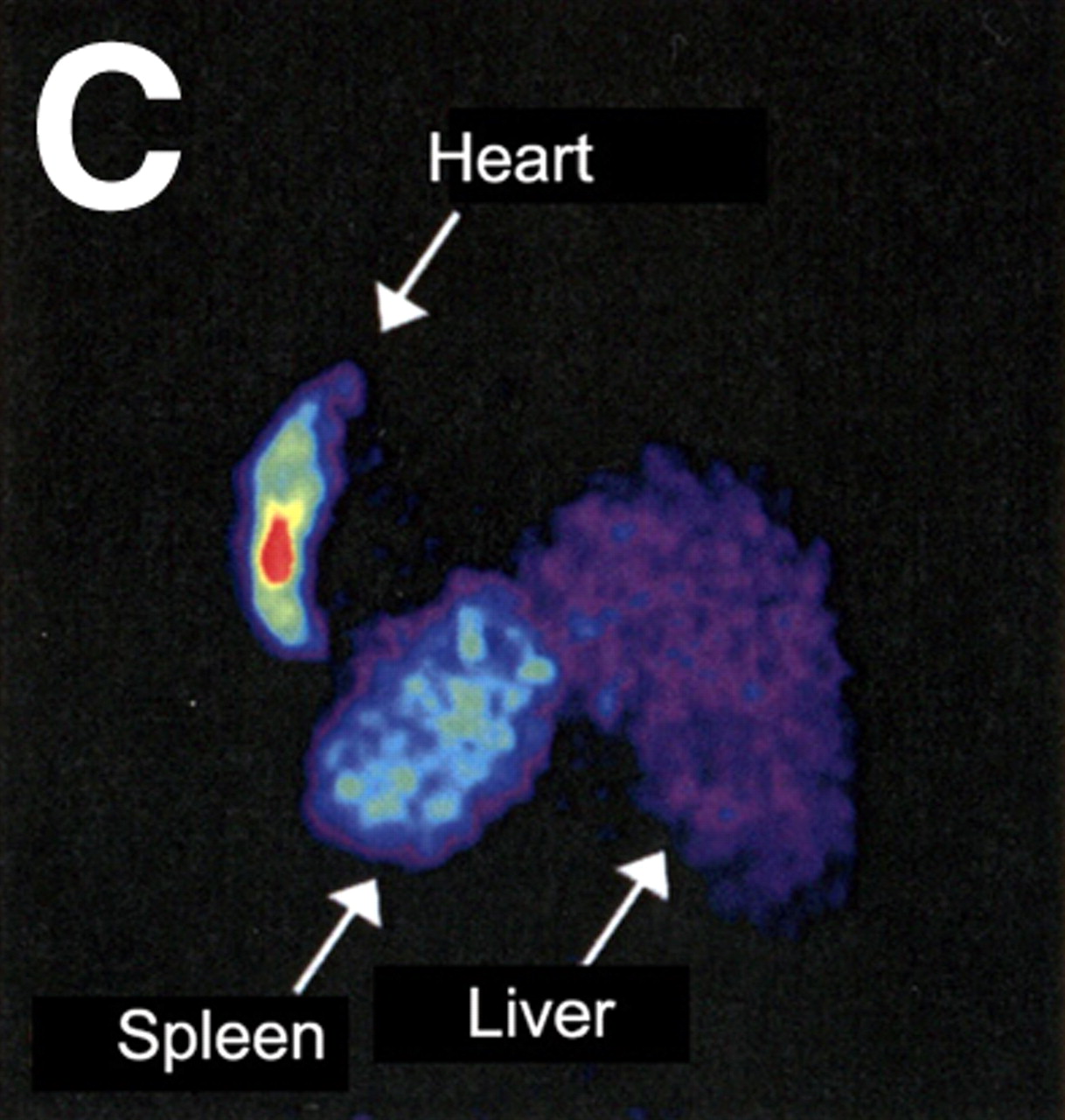
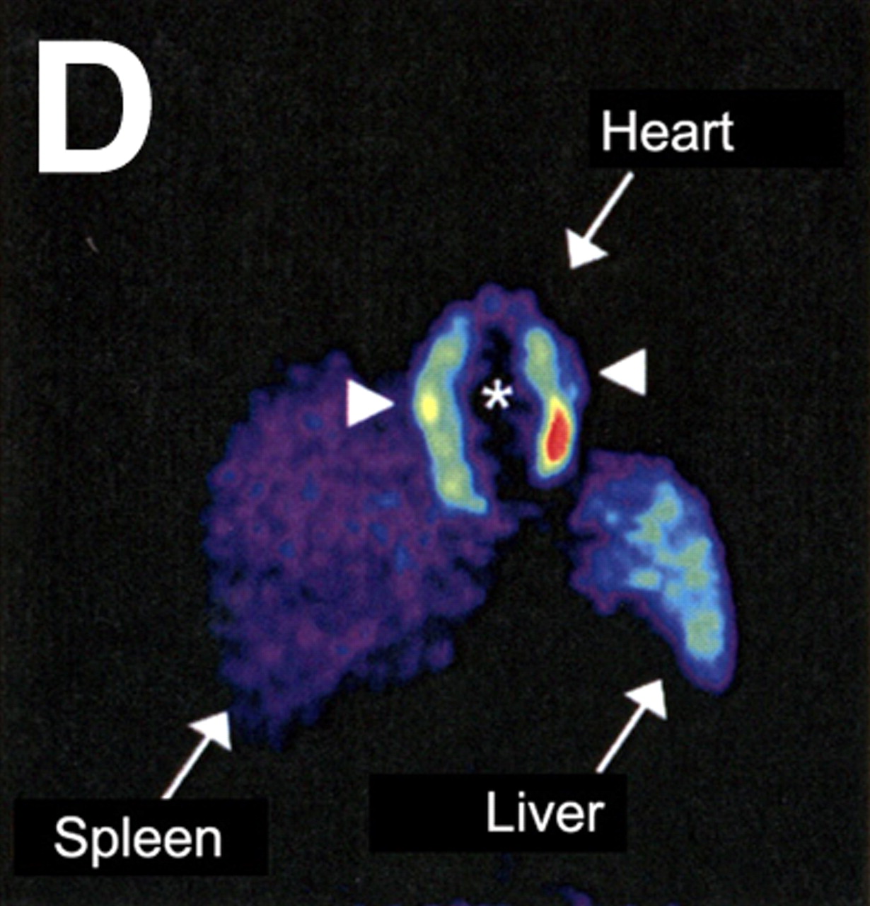
    Evaluation of myocardial homing and biodistribution of 18F-FDG–labeled bone marrow cells after intracoronary delivery in patient after myocardial infarction. (A and B) Representative left posterior oblique (A) and left anterior oblique (B) views at 65 min after injection of 18F-FDG–labeled unselected bone marrow cells into the left circumflex coronary artery. (C and D) Representative left posterior oblique (C) and left anterior oblique (D) views at 70 min after injection of 18F-FDG–labeled CD34-positive subpopulation of bone marrow cells into left anterior descending coronary artery. (Reprinted with permission from (17).)

  • FIGURE 4. 
    • Download figure
    • Open in new tab
    • Download powerpoint
    FIGURE 4. 

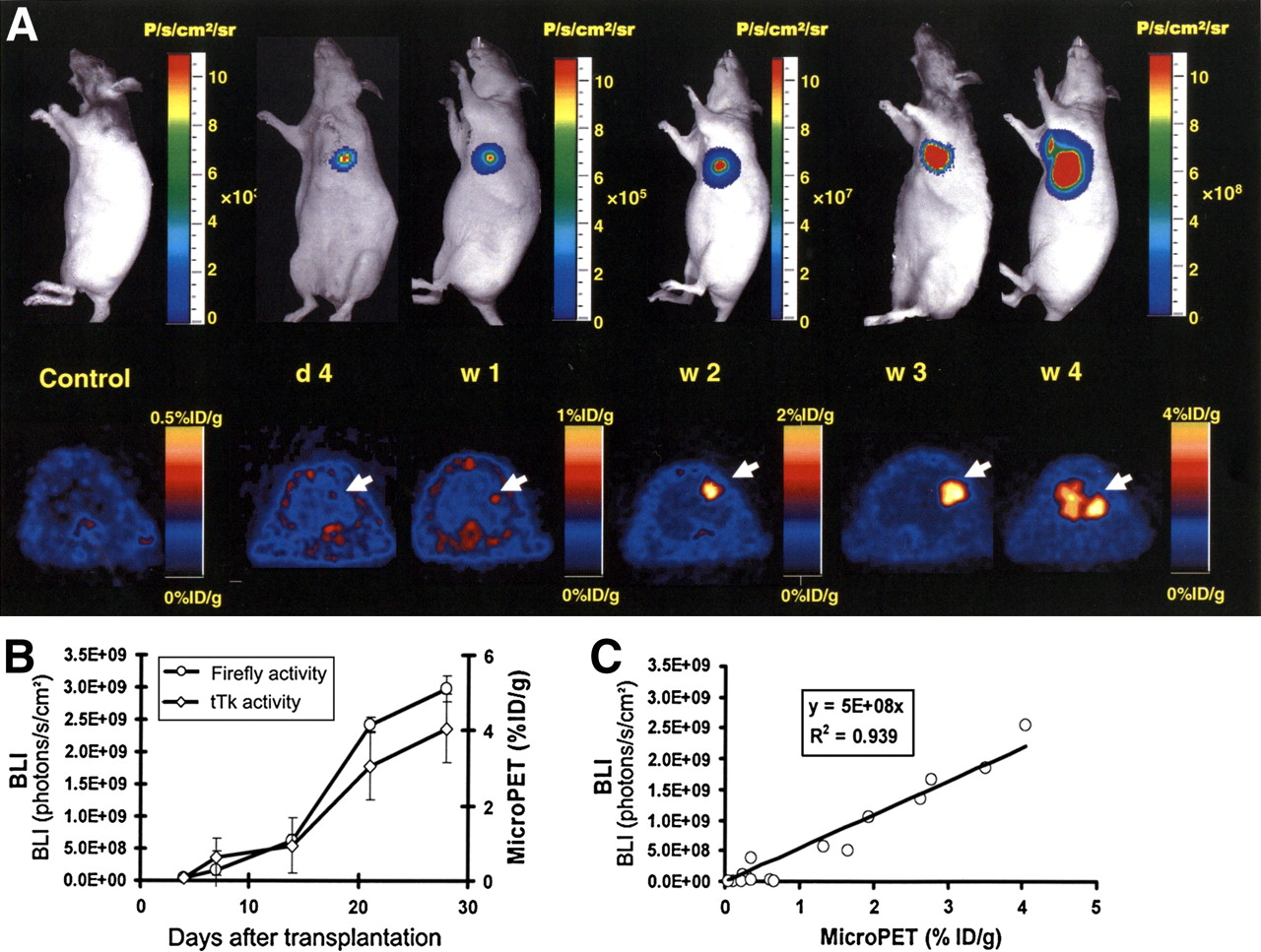
    Tracking of transplanted embryonic stem cells in rats by multimodality molecular imaging. (A) Bioluminescence imaging (top) and small-animal PET (bottom) of embryonic stem cell fate in living animals. Representative images demonstrate imaging signal activities in these mouse embryonic stem cells that stably express firefly luciferase (bioluminescence imaging), monomeric red fluorescence protein (fluorescence), and herpes simplex virus truncated thymidine kinase (PET) triple-fusion reporter gene. (B) Significant increase of firefly luciferase and truncated thymidine kinase activities were seen from week 2 to week 4, indicative of in vivo teratoma formation. (C) In vivo correlation between bioluminescence and PET imaging signals. BLI = bioluminescence imaging; d = day; P = photons; sr = steridian; tTk = truncated thymidine kinase; w = week. (Reprinted with permission from (24).)

PreviousNext
Back to top

In this issue

Journal of Nuclear Medicine: 48 (12)
Journal of Nuclear Medicine
Vol. 48, Issue 12
December 2007
  • Table of Contents
  • Table of Contents (PDF)
  • About the Cover
  • Index by author
Print
Download PDF
Article Alerts
Sign In to Email Alerts with your Email Address
Email Article

Thank you for your interest in spreading the word on Journal of Nuclear Medicine.

NOTE: We only request your email address so that the person you are recommending the page to knows that you wanted them to see it, and that it is not junk mail. We do not capture any email address.

Enter multiple addresses on separate lines or separate them with commas.
Comparison of Imaging Techniques for Tracking Cardiac Stem Cell Therapy
(Your Name) has sent you a message from Journal of Nuclear Medicine
(Your Name) thought you would like to see the Journal of Nuclear Medicine web site.
Citation Tools
Comparison of Imaging Techniques for Tracking Cardiac Stem Cell Therapy
Sarah J. Zhang, Joseph C. Wu
Journal of Nuclear Medicine Dec 2007, 48 (12) 1916-1919; DOI: 10.2967/jnumed.107.043299

Citation Manager Formats

  • BibTeX
  • Bookends
  • EasyBib
  • EndNote (tagged)
  • EndNote 8 (xml)
  • Medlars
  • Mendeley
  • Papers
  • RefWorks Tagged
  • Ref Manager
  • RIS
  • Zotero
Share
Comparison of Imaging Techniques for Tracking Cardiac Stem Cell Therapy
Sarah J. Zhang, Joseph C. Wu
Journal of Nuclear Medicine Dec 2007, 48 (12) 1916-1919; DOI: 10.2967/jnumed.107.043299
Twitter logo Facebook logo LinkedIn logo Mendeley logo
  • Tweet Widget
  • Facebook Like
  • Google Plus One
Bookmark this article

Jump to section

  • Article
    • PRINCIPLES OF STEM CELL IMAGING
    • FUTURE PERSPECTIVES
    • Footnotes
    • References
  • Figures & Data
  • Info & Metrics
  • PDF

Related Articles

  • This Month in JNM
  • PubMed
  • Google Scholar

Cited By...

  • Differential responses of transplanted stem cells to the diseased environment unveiled by a single molecular NIR II cell tracker
  • Hybrid PET/MR Imaging of the Heart: Potential, Initial Experiences, and Future Prospects
  • Transplantation and Tracking of Human-Induced Pluripotent Stem Cells in a Pig Model of Myocardial Infarction: Assessment of Cell Survival, Engraftment, and Distribution by Hybrid Single Photon Emission Computed Tomography/Computed Tomography of Sodium Iodide Symporter Transgene Expression
  • Imaging Gene Expression in Live Cells and Tissues
  • Cardiovascular Molecular Imaging: Focus on Clinical Translation
  • Current Perspectives on Imaging Cardiac Stem Cell Therapy
  • Timing of Bone Marrow Cell Delivery Has Minimal Effects on Cell Viability and Cardiac Recovery After Myocardial Infarction
  • Multimodality Cardiovascular Molecular Imaging, Part II
  • Google Scholar

More in this TOC Section

  • From Genome to Phenome: Opportunities and Challenges of Molecular Imaging
  • Imaging the Activity of Efflux Transporters at the Blood–Brain Barrier in Neurologic Diseases: Radiotracer Selection Criteria
  • Molecular Imaging of Pulmonary Fibrosis
Show more Focus on Molecular Imaging

Similar Articles

SNMMI

© 2025 SNMMI

Powered by HighWire