
Main menu

  • Home
  • Content
    • Current
    • Ahead of print
    • Past Issues
    • JNM Supplement
    • SNMMI Annual Meeting Abstracts
    • Continuing Education
    • JNM Podcasts
  • Subscriptions
    • Subscribers
    • Institutional and Non-member
    • Rates
    • Journal Claims
    • Corporate & Special Sales
  • Authors
    • Submit to JNM
    • Information for Authors
    • Assignment of Copyright
    • AQARA requirements
  • Info
    • Reviewers
    • Permissions
    • Advertisers
  • About
    • About Us
    • Editorial Board
    • Contact Information
  • More
    • Alerts
    • Feedback
    • Help
    • SNMMI Journals
  • SNMMI
    • JNM
    • JNMT
    • SNMMI Journals
    • SNMMI

User menu

  • Subscribe
  • My alerts
  • Log in
  • My Cart

Search

  • Advanced search
Journal of Nuclear Medicine
  • SNMMI
    • JNM
    • JNMT
    • SNMMI Journals
    • SNMMI
  • Subscribe
  • My alerts
  • Log in
  • My Cart
Journal of Nuclear Medicine

Advanced Search

  • Home
  • Content
    • Current
    • Ahead of print
    • Past Issues
    • JNM Supplement
    • SNMMI Annual Meeting Abstracts
    • Continuing Education
    • JNM Podcasts
  • Subscriptions
    • Subscribers
    • Institutional and Non-member
    • Rates
    • Journal Claims
    • Corporate & Special Sales
  • Authors
    • Submit to JNM
    • Information for Authors
    • Assignment of Copyright
    • AQARA requirements
  • Info
    • Reviewers
    • Permissions
    • Advertisers
  • About
    • About Us
    • Editorial Board
    • Contact Information
  • More
    • Alerts
    • Feedback
    • Help
    • SNMMI Journals
  • View or Listen to JNM Podcast
  • Visit JNM on Facebook
  • Join JNM on LinkedIn
  • Follow JNM on Twitter
  • Subscribe to our RSS feeds
OtherBASIC SCIENCE INVESTIGATIONS

Dynamic Tracking During Intracoronary Injection of 18F-FDG-Labeled Progenitor Cell Therapy for Acute Myocardial Infarction

Brendan Doyle, Brad J. Kemp, Panithaya Chareonthaitawee, Cynthia Reed, Jeffrey Schmeckpeper, Paul Sorajja, Stephen Russell, Philip Araoz, Stephen J. Riederer and Noel M. Caplice
Journal of Nuclear Medicine October 2007, 48 (10) 1708-1714; DOI: https://doi.org/10.2967/jnumed.107.042838
Brendan Doyle
  • Find this author on Google Scholar
  • Find this author on PubMed
  • Search for this author on this site
Brad J. Kemp
  • Find this author on Google Scholar
  • Find this author on PubMed
  • Search for this author on this site
Panithaya Chareonthaitawee
  • Find this author on Google Scholar
  • Find this author on PubMed
  • Search for this author on this site
Cynthia Reed
  • Find this author on Google Scholar
  • Find this author on PubMed
  • Search for this author on this site
Jeffrey Schmeckpeper
  • Find this author on Google Scholar
  • Find this author on PubMed
  • Search for this author on this site
Paul Sorajja
  • Find this author on Google Scholar
  • Find this author on PubMed
  • Search for this author on this site
Stephen Russell
  • Find this author on Google Scholar
  • Find this author on PubMed
  • Search for this author on this site
Philip Araoz
  • Find this author on Google Scholar
  • Find this author on PubMed
  • Search for this author on this site
Stephen J. Riederer
  • Find this author on Google Scholar
  • Find this author on PubMed
  • Search for this author on this site
Noel M. Caplice
  • Find this author on Google Scholar
  • Find this author on PubMed
  • Search for this author on this site
  • Article
  • Figures & Data
  • Supplemental
  • Info & Metrics
  • PDF
Loading

Article Figures & Data

Figures

  • Tables
  • Additional Files
  • FIGURE 1. 
    • Download figure
    • Open in new tab
    • Download powerpoint
    FIGURE 1. 

    Labeling with 18F-FDG and in vivo detection of human and porcine progenitor cells. All P values are nonsignificant except *P < 0.05. (A) Comparison of labeling efficiency at incubation temperatures of 20°C, 28°C, and 37°C. (B) Comparison of labeling efficiency after incubation for periods of 10, 20, 30, 45, and 60 min at 37°C. (C) Elution of radiolabel by CPC at 1 and 2 h. (D) Detection of intracoronary injected 18F-FDG progenitor cells in inferolateral territory of normal heart in vivo.

  • FIGURE 2. 
    • Download figure
    • Open in new tab
    • Download powerpoint
    FIGURE 2. 

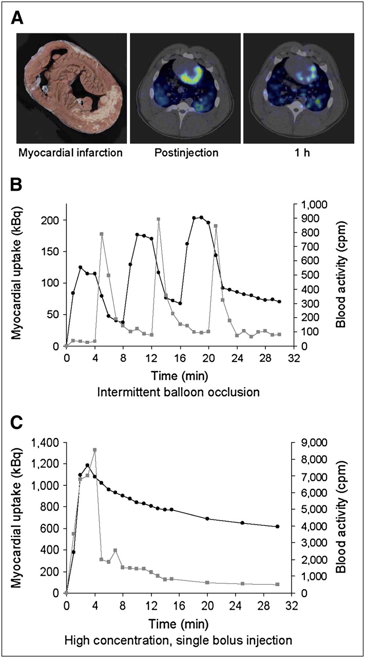
    Dynamic tracking of 18F-FDG-labeled CPC during intracoronary injection. (A) From left to right: gross, unstained specimen shows extensive inferolateral MI (image is oriented to match subsequent PET/CT images); single frame of dynamic 3D PET/CT from same animal taken immediately after intracoronary injection shows myocardial activity localized to infarct territory and border zone; single frame from same animal taken 1 h after injection shows significant decrease in regional activity when compared with postinjection image. (B) Time–activity curves for myocardium (black line) and peripheral blood (gray line) during intracoronary injection using 3-cycle balloon-occlusion technique. Balloon was inflated for 4 min during coronary injection and subsequently deflated for 4 min during each cycle. Blood samples were drawn at 1-min intervals. (C) Time–activity curves for myocardium (black line) and peripheral blood (gray line) during high-concentration, single-bolus intracoronary injection.
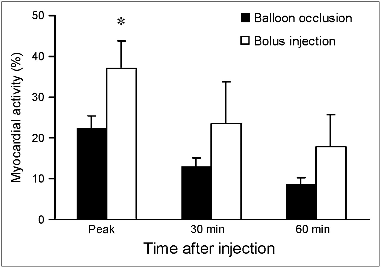
  • FIGURE 3. 
    • Download figure
    • Open in new tab
    • Download powerpoint
    FIGURE 3. 

    Comparison of myocardial activity (measured as percentage of total injected activity) after balloon-occlusion delivery or high-concentration, single-bolus therapy at various time points. *P < 0.05 (ANOVA) for comparison of injection techniques at any given time point.

Tables

  • Figures
  • Additional Files
    • View popup
    TABLE 1

    Characteristics of Experimental Animals at Baseline

    CharacteristicGroup 1: balloon occlusionGroup 2: single bolusP
    Pig body mass (kg)42.1 ± 0.241.7 ± 0.5NS
    Ejection fraction (%)46.6 ± 4.244.9 ± 5.1NS
    Left ventricular mass (g)89.1 ± 6.186.5 ± 5.3NS
    Infarct size (g)14.5 ± 2.917.8 ± 1.9NS
    Infarct size (% of LV mass)16.3 ± 3.420.6 ± 2.7NS
    End-diastolic volume (mL)64.2 ± 2.967.3 ± 5.2NS
    End-systolic volume (mL)35.2 ± 5.833.8 ± 4.9NS
    Cardiac output (L/min)2.9 ± 0.12.7 ± 0.5NS
    Stroke volume (mL)28.7 ± 2.325.5 ± 2.2NS
    • P value denotes comparison of mean baseline values in balloon-occlusion delivery group with mean baseline values in single-bolus delivery group using ANOVA.

    • NS = not significant.

    • View popup
    TABLE 2

    Labeling of CPC with 18F-FDG Among Experimental Animals

    Animal no.Delivery strategyBaseline labeling (%)Cell viability (%)Specific activity (kBq/107 NCs)Cell bound: 1 h (%)Cell bound: 2 h (%)
    1Balloon occlusion9298.6336.78171
    2Balloon occlusion9199.185.18580
    3Balloon occlusion9699.329.68174
    4Single bolus9398.933.37268
    5Single bolus9499.677.78571
    6Single bolus9299.1270.18374
    • NCs = nucleated cells.

Additional Files

  • Figures
  • Tables
  • Supplemental Data

    Files in this Data Supplement:

    • Supplemental MRI Data
    • Supplemental Figure 1
    • Supplemental Figure Legend
PreviousNext
Back to top

In this issue

Journal of Nuclear Medicine: 48 (10)
Journal of Nuclear Medicine
Vol. 48, Issue 10
October 2007
  • Table of Contents
  • Table of Contents (PDF)
  • About the Cover
  • Index by author
Print
Download PDF
Article Alerts
Sign In to Email Alerts with your Email Address
Email Article

Thank you for your interest in spreading the word on Journal of Nuclear Medicine.

NOTE: We only request your email address so that the person you are recommending the page to knows that you wanted them to see it, and that it is not junk mail. We do not capture any email address.

Enter multiple addresses on separate lines or separate them with commas.
Dynamic Tracking During Intracoronary Injection of 18F-FDG-Labeled Progenitor Cell Therapy for Acute Myocardial Infarction
(Your Name) has sent you a message from Journal of Nuclear Medicine
(Your Name) thought you would like to see the Journal of Nuclear Medicine web site.
Citation Tools
Dynamic Tracking During Intracoronary Injection of 18F-FDG-Labeled Progenitor Cell Therapy for Acute Myocardial Infarction
Brendan Doyle, Brad J. Kemp, Panithaya Chareonthaitawee, Cynthia Reed, Jeffrey Schmeckpeper, Paul Sorajja, Stephen Russell, Philip Araoz, Stephen J. Riederer, Noel M. Caplice
Journal of Nuclear Medicine Oct 2007, 48 (10) 1708-1714; DOI: 10.2967/jnumed.107.042838

Citation Manager Formats

  • BibTeX
  • Bookends
  • EasyBib
  • EndNote (tagged)
  • EndNote 8 (xml)
  • Medlars
  • Mendeley
  • Papers
  • RefWorks Tagged
  • Ref Manager
  • RIS
  • Zotero
Share
Dynamic Tracking During Intracoronary Injection of 18F-FDG-Labeled Progenitor Cell Therapy for Acute Myocardial Infarction
Brendan Doyle, Brad J. Kemp, Panithaya Chareonthaitawee, Cynthia Reed, Jeffrey Schmeckpeper, Paul Sorajja, Stephen Russell, Philip Araoz, Stephen J. Riederer, Noel M. Caplice
Journal of Nuclear Medicine Oct 2007, 48 (10) 1708-1714; DOI: 10.2967/jnumed.107.042838
Twitter logo Facebook logo LinkedIn logo Mendeley logo
  • Tweet Widget
  • Facebook Like
  • Google Plus One
Bookmark this article

Jump to section

  • Article
    • Abstract
    • MATERIALS AND METHODS
    • RESULTS
    • DISCUSSION
    • CONCLUSION
    • Acknowledgments
    • Footnotes
    • References
  • Figures & Data
  • Supplemental
  • Info & Metrics
  • PDF

Related Articles

  • This Month in JNM
  • PubMed
  • Google Scholar

Cited By...

  • An off-the-shelf artificial cardiac patch improves cardiac repair after myocardial infarction in rats and pigs
  • CellGPS: Whole-body tracking of single cells by positron emission tomography
  • 18F-FDG Labeling of Mesenchymal Stem Cells and Multipotent Adult Progenitor Cells for PET Imaging: Effects on Ultrastructure and Differentiation Capacity
  • Microfluidic Single-Cell Analysis Shows That Porcine Induced Pluripotent Stem Cell-Derived Endothelial Cells Improve Myocardial Function by Paracrine Activation
  • Cell Tracking and the Development of Cell-Based Therapies: A View From the Cardiovascular Cell Therapy Research Network
  • Current Perspectives on Imaging Cardiac Stem Cell Therapy
  • Assessment and Optimization of Cell Engraftment After Transplantation Into the Heart
  • Reporter Gene PET for Monitoring Survival of Transplanted Endothelial Progenitor Cells in the Rat Heart After Pretreatment with VEGF and Atorvastatin
  • Tracking Cell Fate With Noninvasive Imaging
  • In Vivo Imaging of Stem Cells and Beta Cells Using Direct Cell Labeling and Reporter Gene Methods
  • The Year in Molecular Imaging
  • Collagen-Based Matrices Improve the Delivery of Transplanted Circulating Progenitor Cells: Development and Demonstration by Ex Vivo Radionuclide Cell Labeling and In Vivo Tracking With Positron-Emission Tomography
  • Google Scholar

More in this TOC Section

  • Tumor Uptake of Anti-CD20 Fabs Depends on Tumor Perfusion
  • How Sensitive Is the Upper Gastrointestinal Tract to 90Y Radioembolization? A Histologic and Dosimetric Analysis in a Porcine Model
  • 11C-Methionine PET of Myocardial Inflammation in a Rat Model of Experimental Autoimmune Myocarditis
Show more Basic Science Investigations

Similar Articles

SNMMI

© 2025 SNMMI

Powered by HighWire