


Article Figures & Data
Figures
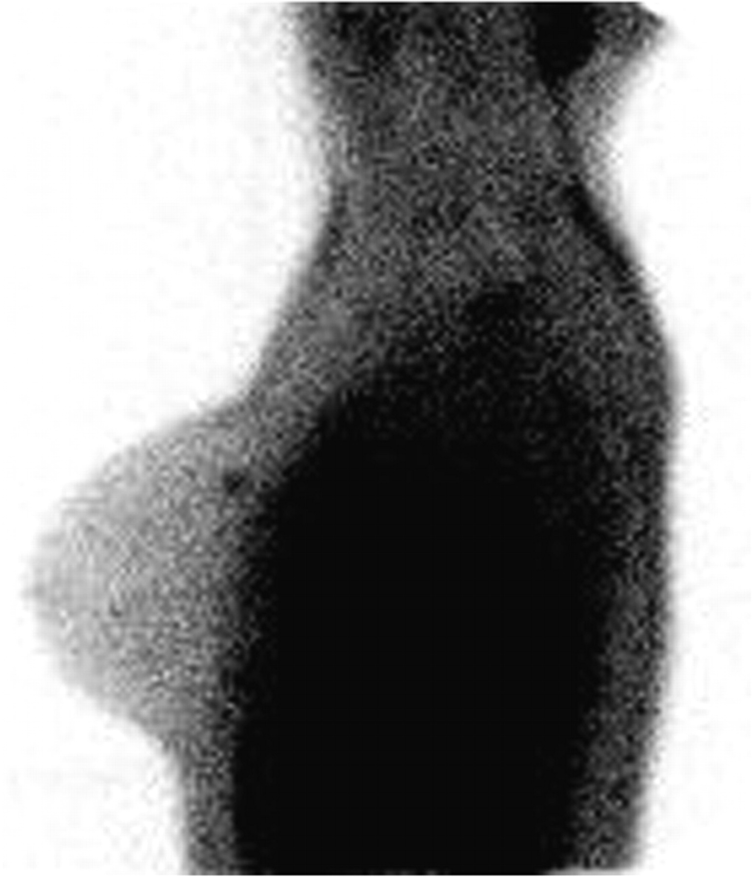
- FIGURE 1.
Patient 1: Infiltrative ductal carcinoma (8 mm). Lateral projection.
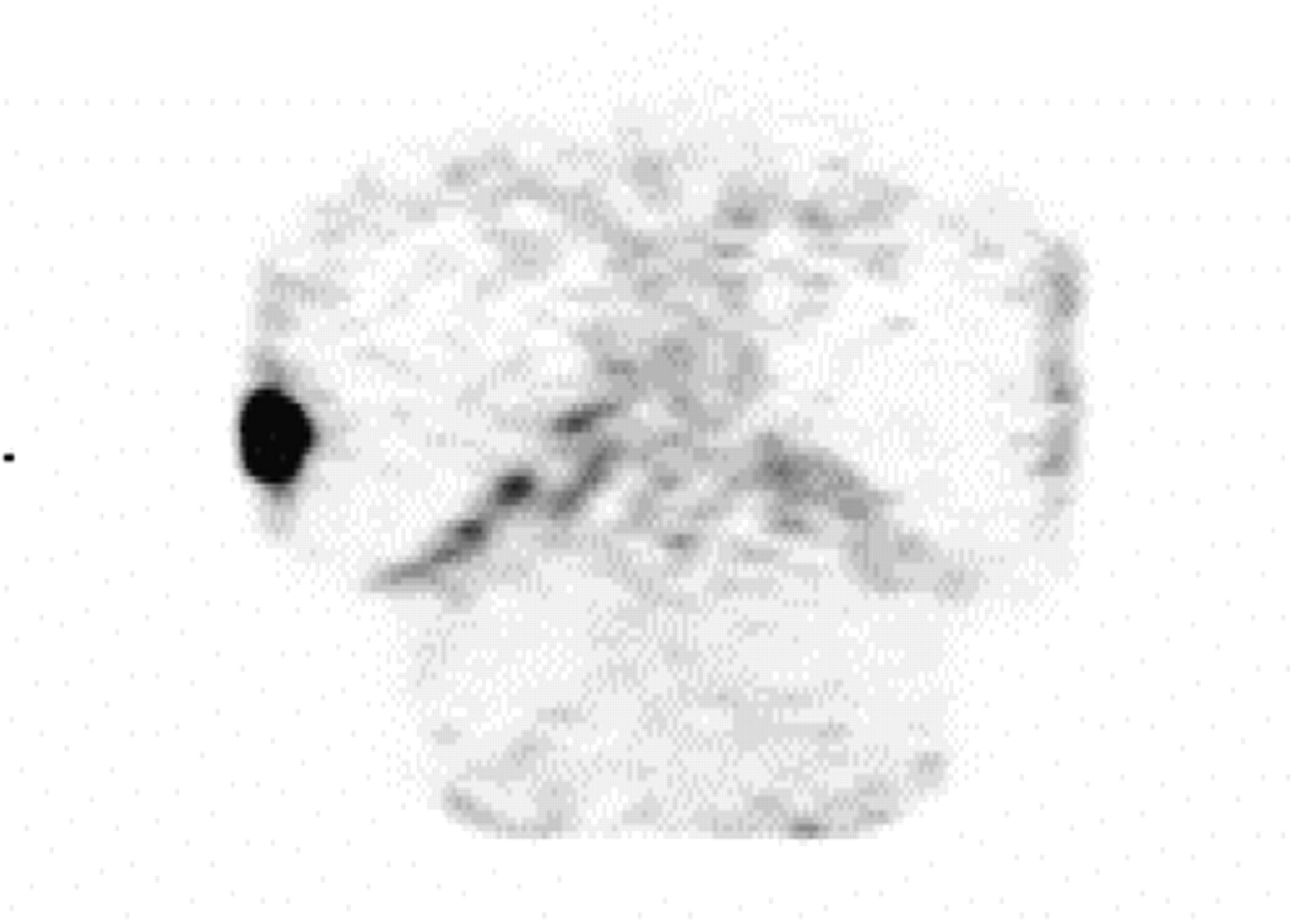
- FIGURE 2.
Patient 3: Infiltrative ductal carcinoma (10 mm), with surrounding ductal carcinoma in situ. Frontal projection.
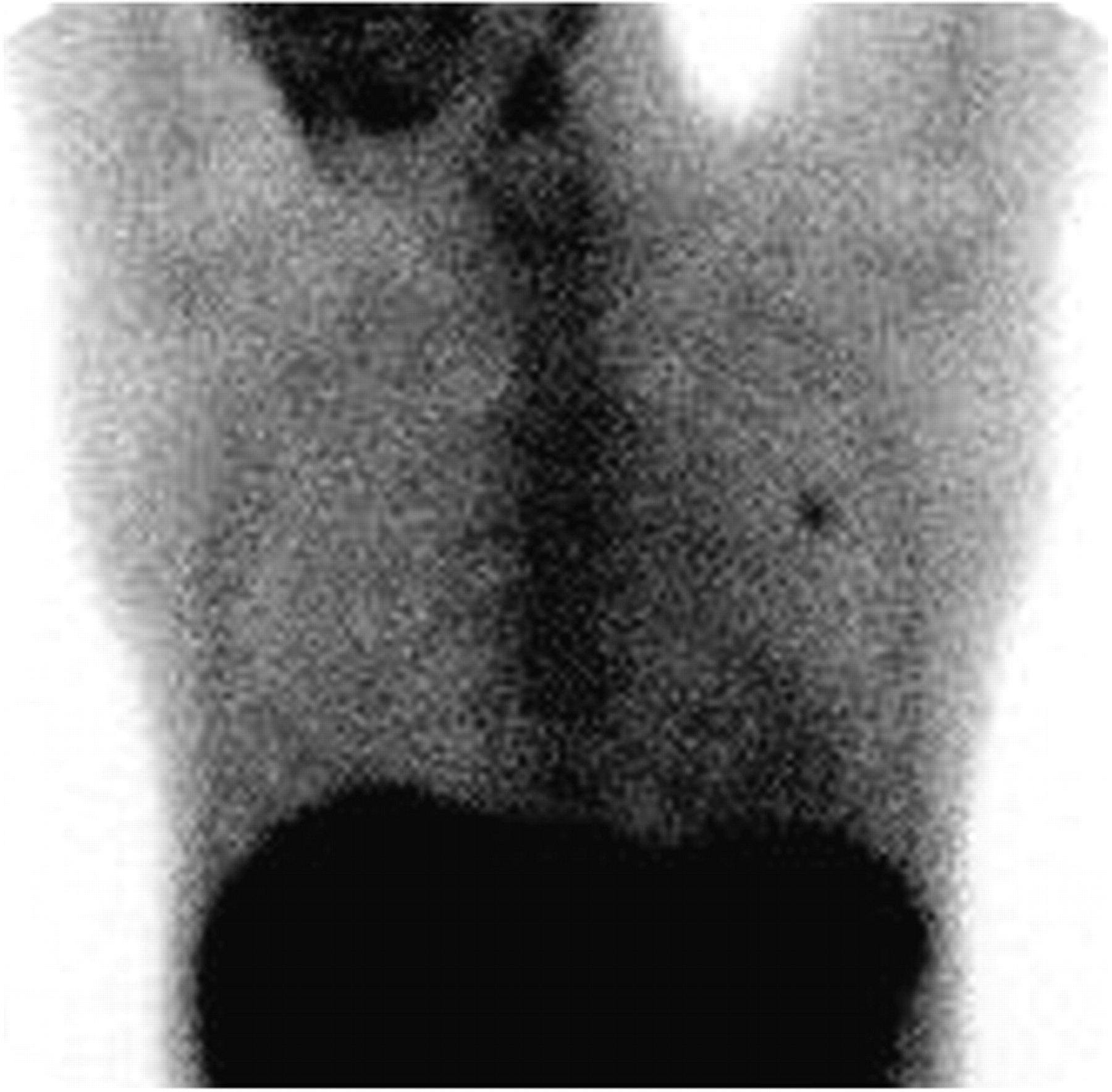
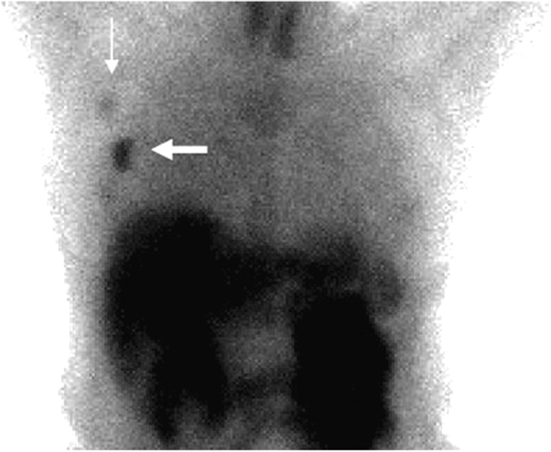
- FIGURE 3.
Patient 14: Large infiltrative ductal carcinoma (35 mm) (large arrow) with uptake in axillary lymph nodes (small arrow). Frontal projection.
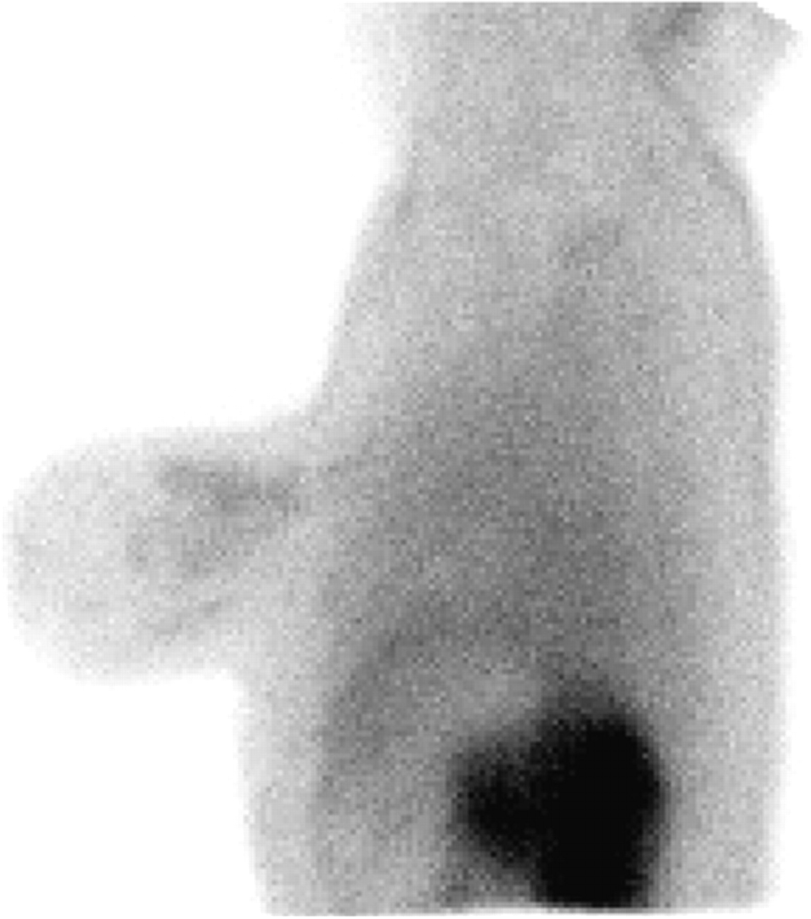
- FIGURE 4.
Patient 10: Diffuse, heterogeneous uptake in fibrocystic disease, with no focal pathology. Lateral projection.
- FIGURE 5.
Patient 19: Intense uptake in infected cyst. SPECT, coronal projection.


Tables
Serum biochemistry Hematology Urine analysis Creatinine Hematocrit Bilirubin Urea nitrogen Hemoglobin Protein Uric acid RBC Ketone Bilirubin (total, direct, indirect) WBC Occult blood WBC differential count Specific gravity Protein (total) pH Albumin Coagulation parameters Microscopy ASAT ALAT Protrombin time Alkaline phosphatases Activated partial tromboplastin time γ-GT Chloride Calcium Phosphorus Bicarbonate LD CPK Amylase Glucose Sodium Potassium RBC = red blood cell count; WBC = white blood cell count; ASAT = aspartate aminotransferase; ALAT = alanine aminotransferase; γ-GT = γ-glutamyltransferase; LD = lactate dehydrogenase; CPK = creatine phosphokinase.
Physical examination included general appearance, cardiovascular, lung, and abdominal examination, motor function, cranial nerves (II–XII), and reflexes (biceps, triceps, patellar, ankle).
Lesion size (mm) Visual uptake grading Patient no. Pathology US* XMM† SMM‡ 1 IDC 8 4 5 3 2 IDC 20 5 5 3 3 IDC (+ DCIS) 10 (+ 12) 5 5 3 4 L: DCIS 10 4 1 3, R: IDC 25 5 5 3 DCIS§ 2.5 5 ILC × 2 20, 23 5, 3 5, 5 3, 3 ILC§ 11 1 1 2 6 IDC (+ DCIS) 14 (+ 10) 5 5 3 7 IDC 6 5 5 1 8 IDC 7 5 5 3 9 IDC × 2 (widely DCIS) 12, 14 5, 4 5, 5 3, 3 10 IDC (+ DCIS) 40 5 5 3 11 ILC 20 5 5 3 12 IDC 28 5 5 3 13 IDC (+ DCIS) 40 5 5 3 14 IDC 35 5 5 3 15 IDC (+ DCIS) 40 5 5 3 16 IDC 40 5 5 3 ILC 40 3 4 3 ↵* US: 5-scale grading, where grade 3 = indeterminate and grade 5 = malignant.
↵† XMM: findings are graded according to BIRADS, where grade 1 = negative, grade 2 = benign finding, grade 3 = probably benign finding, grade 4 = suspicious abnormality (biopsy should be considered), and grade 5 = highly suggestive of malignancy.
↵‡ SMM with NC100692 administration (study drug): 3-grade scale, where grade 1 = no obvious uptake, grade 2 = uncertain/heterogeneous uptake, and grade 3 = focal lesion uptake.
↵§ Lesions were found during histopathologic examination but were not observed by any imaging modality.
BIRADS = Breast Imaging Reporting and Data System; IDC = infiltrative ductal carcinoma; ILC = infiltrative lobular carcinoma; DCIS = ductal carcinoma in situ.
Visual uptake grading Patient no. Diagnosis US* XMM† SMM‡ 10 Fibrocystic changes, other (fat) No lesion No lesion 2 13 Fibrocystic changes 2 2 1 17 4 Fibrocystic changes 2 2 2 (FNAC)§, 3 others 18 Fibroadenoma 2 2 1 19 Infectious cyst 2 2 3 20 Fibroadenoma 3 2 1 ↵* US: 5-scale grading, where grade 2 = benign finding and grade 3 = indeterminate.
↵† XMM: findings are graded according to BIRADS, where grade 1 = negative, grade 2 = benign finding, grade 3 = probably benign finding, grade 4 = suspicious abnormality (biopsy should be considered), and grade 5 = highly suggestive of malignancy.
↵‡ SMM with NC100692 administration (study drug): 3-grade scale, where grade 1 = no obvious uptake, grade 2 = uncertain/heterogeneous uptake, and grade 3 = focal lesion uptake.
↵§ FNAC = lesion had fine-needle aspiration cytology before scintigraphy.
BIRADS = Breast Imaging Reporting and Data System.


Jump to section
Related Articles
Cited By...
- A Bridge Not Too Far: Linking Disciplines Through Molecular Imaging Probes
- Comparing the Diagnostic Potential of 68Ga-Alfatide II and 18F-FDG in Differentiating Between Non-Small Cell Lung Cancer and Tuberculosis
- Integrin {alpha}v{beta}3 Imaging of Radioactive Iodine-Refractory Thyroid Cancer Using 99mTc-3PRGD2
- 99mTc-3PRGD2 for Integrin Receptor Imaging of Lung Cancer: A Multicenter Study
- Radiopeptide Imaging and Therapy in Europe
- Novel insights on imaging sex hormone-dependent tumourigenesis in vivo
- A Bridge Not Too Far: Linking Disciplines Through Molecular Imaging Probes
- Serial Noninvasive Targeted Imaging of Peripheral Angiogenesis: Validation and Application of a Semiautomated Quantitative Approach
- Molecular Imaging and the Failing Heart: Through the Looking Glass
- The Biodistribution and Radiation Dosimetry of the Arg-Gly-Asp Peptide 18F-AH111585 in Healthy Volunteers
- Patterns of {alpha}v{beta}3 Expression in Primary and Metastatic Human Breast Cancer as Shown by 18F-Galacto-RGD PET