


Article Figures & Data
Figures
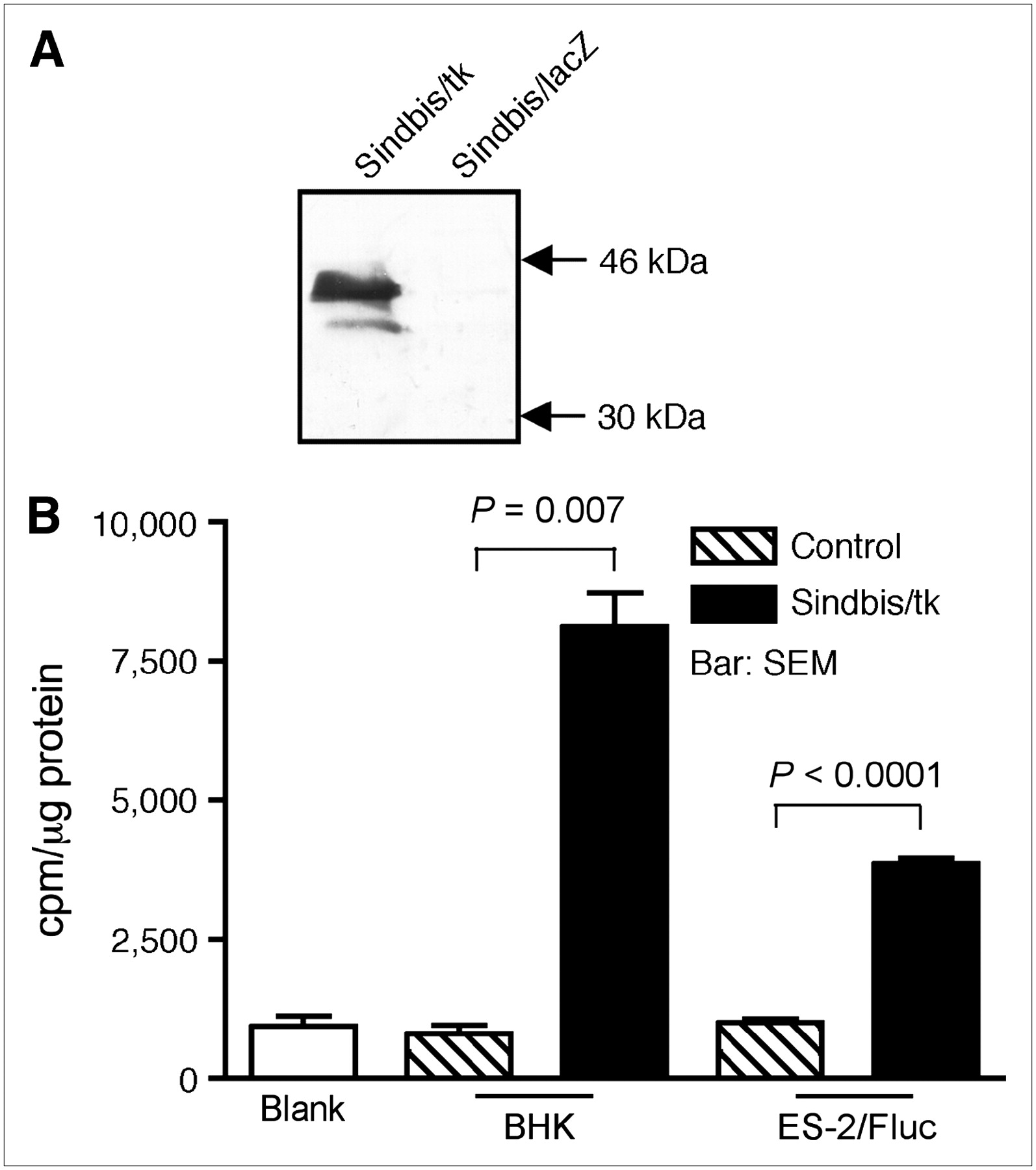
- FIGURE 1.
Sindbis/tk vector efficiently transduces HSVtk activity to infected cells. (A) Western blot analysis of HSVtk expression. Cell lysates from BHK cells infected with Sindbis/tk, which carries HSV-1 thymidine kinase gene, or with Sindbis/lacZ, which carries bacterial β-galactosidase gene, were separated on 12% SDS-PAGE before probing with a polyclonal antibody specific for HSVtk. Specific ∼46-kDa band corresponding to HSVtk was observed in Sindbis/tk-infected cells but not in Sindbis/lacZ-infected cells. (B) Thymidine kinase activities in cell lysates from BHK (n = 2) or ES-2 (n = 3) cells infected with Sindbis/tk. Blank assays contained no cell lysate in reaction mixtures. BHK control (n = 2) and ES-2 control (n = 3) assays used lysates from cells that were not infected with Sindbis/tk.
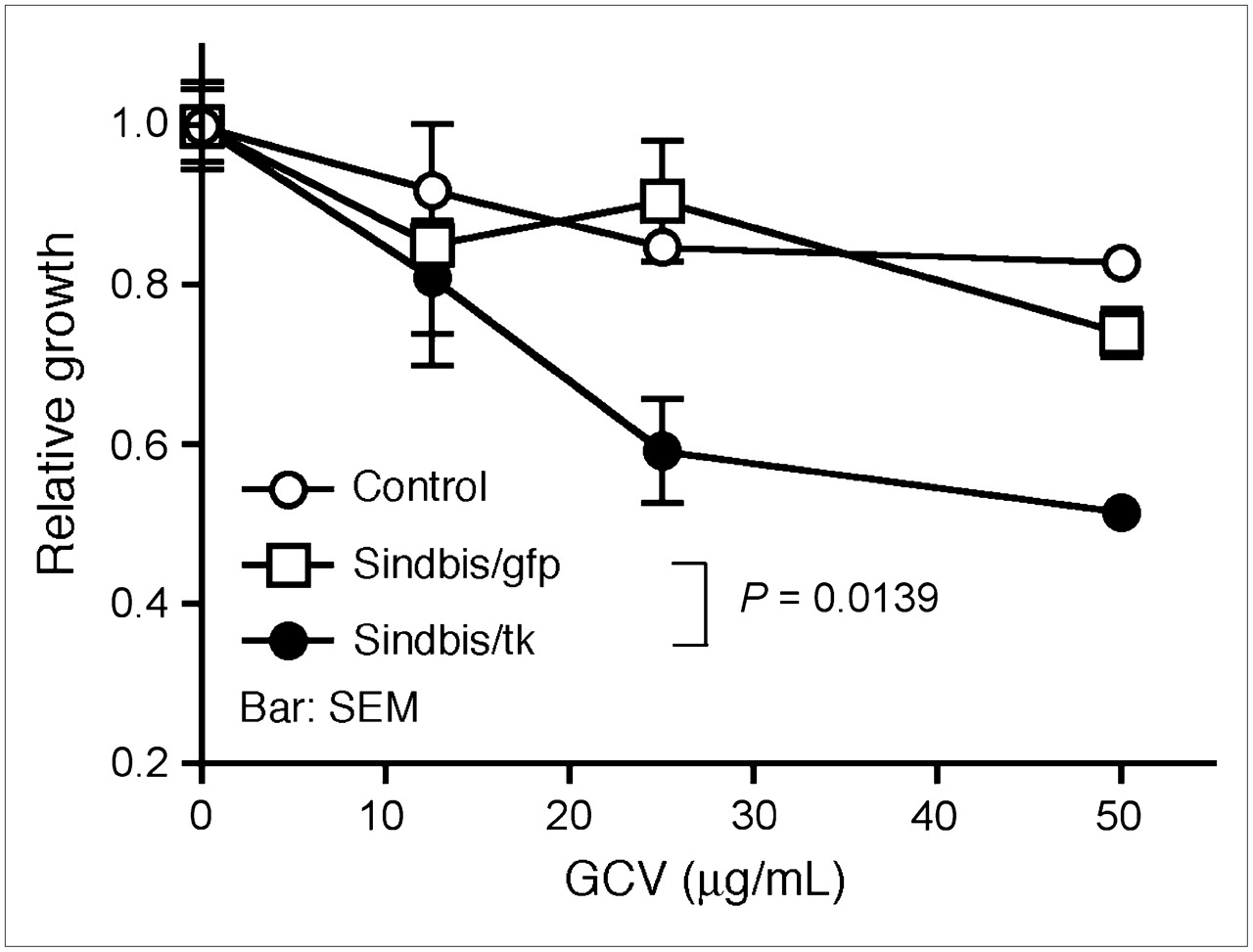
- FIGURE 2.
GCV, a prodrug activated by HSVtk, specifically enhances the cytotoxicity of Sindbis/tk vector against tumor cells. GCV enhances killing of Sindbis/tk-infected ES-2/Fluc cells in vitro, as determined by MTS respiratory assay. ES-2/Fluc cells, which stably express firefly luciferase gene, were left uninfected (control) or infected with Sindbis/tk vector Sindbis/gfp carrying a green fluorescent protein gene. After infection, cells were cultured for 2 d in medium containing different concentrations of GCV (0–50 μg/mL). Relative cell growth was determined using MTS assay that measures respiration of survived cells. Error bars represent SEM of triplicate data.
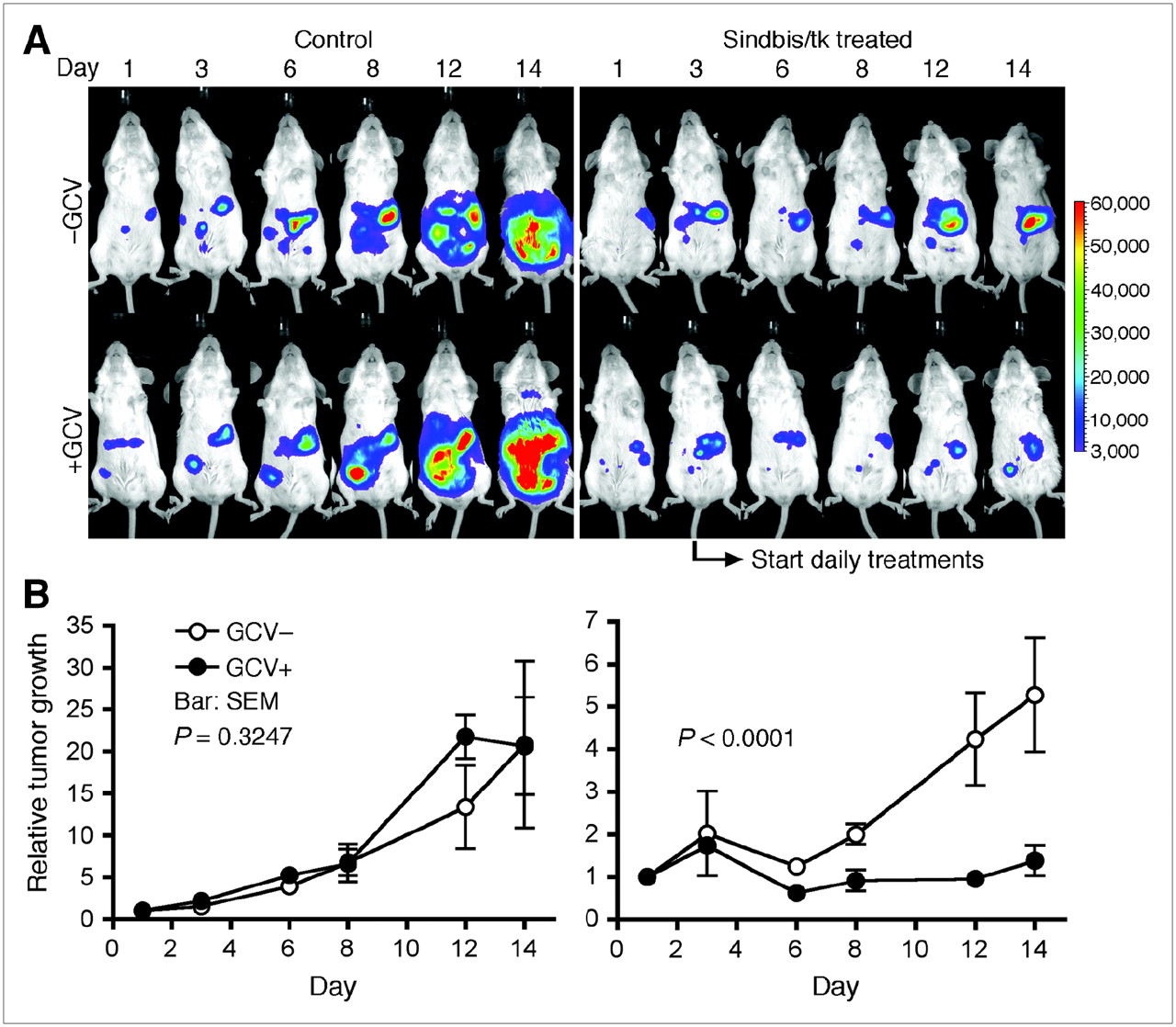
- FIGURE 3.
GCV enhances killing of Sindbis/tk-infected ES-2/Fluc cells in vivo, as determined by the IVIS system capable of noninvasive detection of bioluminescent signal generated by ES-2/Fluc tumors. (A) SCID mice were inoculated with ES-2/Fluc on day 0. We started daily GDEPT treatments composed of Sindbis/tk (∼107 TU) and GCV (25 mg/kg of body weight) on day 3 (indicated by arrow). Sindbis/tk –GCV group (n = 5) received Sindbis/tk treatments but no GCV. Sindbis/tk +GCV group (n = 5) received both Sindbis/tk and GCV treatments. Control –GCV group (n = 5) was treated neither with Sindbis/tk nor with GCV. Control +GCV group (n = 5) received no Sindbis/tk but was treated with GCV. Disease progression was monitored and whole-body photon counts were determined using the IVIS system on days 1, 3, 6, 8, 12, and 14. Representative images of each treatment group are shown. (B) Quantitative presentations (total-body photon counts) of each treatment group as shown in A.
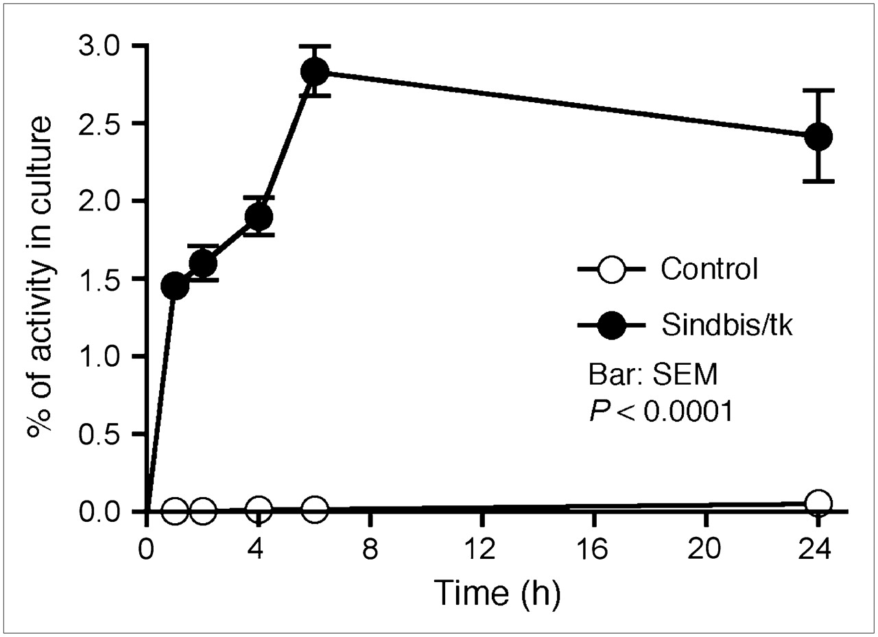
- FIGURE 4.
Sindbis/tk-infected ES-2/Fluc cells took up the HSVtk tracer, 14C-FIAU, specifically and efficiently compared with uninfected control cells. Specific uptakes in cells were determined by a scintillation counter and are presented as percentage cellular uptakes of total radioactivity added into culture.
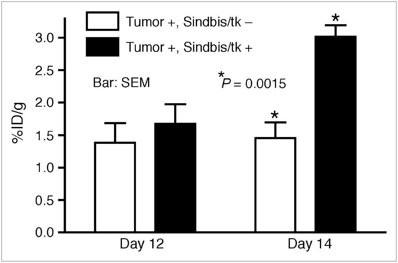
- FIGURE 5.
Quantitative analysis of HSVtk activity in subcutaneous tumors on SCID mice using microPET. BHK subcutaneous tumors were inoculated on right shoulders of SCID mice on day 0. On days 10, 11, and 13, mice received intraperitoneal treatments of Sindbis/tk vectors. Sites of vector treatments were far away from tumor inoculation sites. Untreated tumor-bearing mice were also included as imaging control. Tumor-specific HSVtk activities were measured on days 12 and 14 using 18F-FEAU as tracer. Maximum pixel intensity (%ID/g) in tumor region of each untreated animal (Tumor +, Sindbis/tk –; n = 5) and Sindbis/tk-treated animal (Tumor +, Sindbis/tk +; n = 5) was calculated and analyzed using Student t test (P = 0.0015).
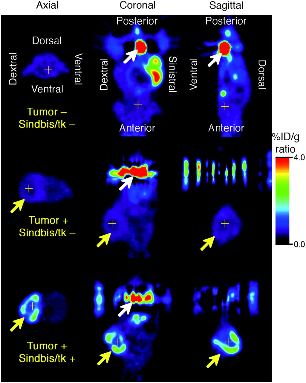
- FIGURE 6.
Noninvasive microPET of HSVtk activity in tumors after Sindbis/tk infection on day 14. BHK subcutaneous tumors were induced on right shoulders of SCID mice as indicated by yellow arrows. Tumor-bearing mice either received no vector treatment (Tumor +, Sindbis/tk –) or received 3 Sindbis/tk treatments via intraperitoneal injection far away from sites of tumor inoculation (Tumor +, Sindbis/tk +). Tumor-free control mice were also included to determine background signals caused by tracer retention within peritoneal cavity (Tumor –, Sindbis/tk –). HSVtk activity was determined after intravenous administration of 18F-FEAU as tracer. Tomographic images are presented in axial, coronal, and sagittal views and crosshairs indicate triangulation points of the 3 views in each set of images. Coronal and sagittal images are shown with animal's head at bottom of image and white arrows indicate activity in urinary bladder. Signal intensity is presented in %ID/g.


