


Article Figures & Data
Figures
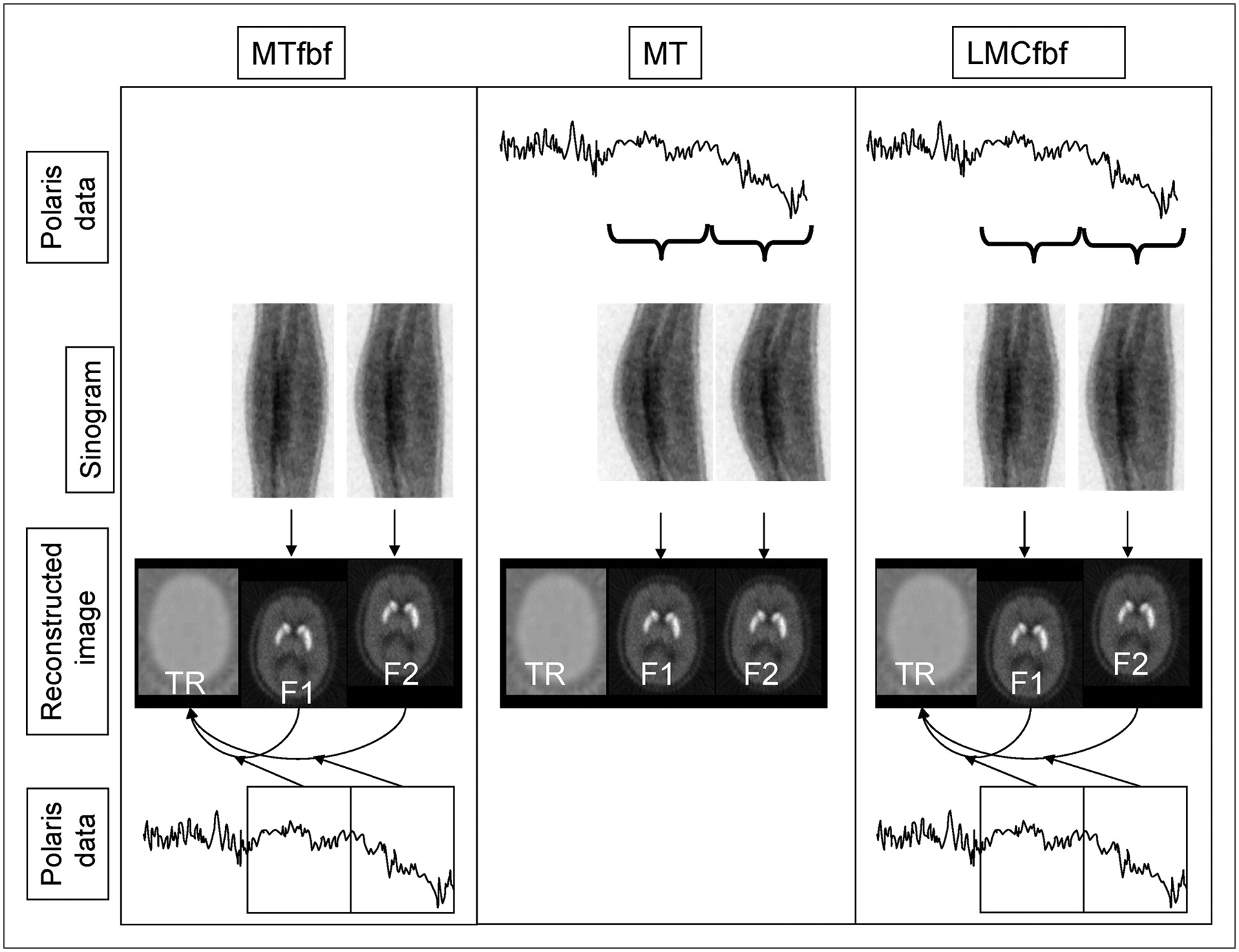
- FIGURE 1.
Different stages involved in realigning emission data with Polaris-based methods. MTfbf is a 1-stage process, where reconstructed frames (F1 and F2) are moved to position of the transmission scan (TR), using the average frame position derived from Polaris data. MT is a 1-stage process in which lines of response (LORs) are moved to the transmission scan position based on Polaris data. LMCfbf is a 2-stage process: First, LORs within a frame are realigned to average position in the frame using Polaris data; then reconstructed frames are moved to transmission scan position using the average frame position derived from Polaris data. For all methods, only 1 Polaris coordinate is shown for simplicity. Full details of the different methods are given in the text.
- FIGURE 2.
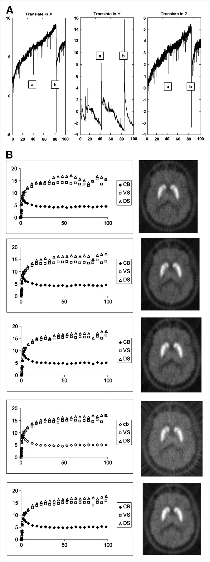
(A) Plots of Polaris translation files (X, left–right; Y, anterior/posterior; Z, dorsal/ventral): x-axis time = min; y-axis from an arbitrary starting point = mm. a and b indicate large movements. (B) Time–activity curve and PET summated images of (from top to bottom): raw PET data, FBF realignment, MTfbf realignment, MT realignment, and LMCfbf realignment. CB = cerebellum; x-axis time = min; y-axis activity = kBq/mL.
- FIGURE 3.
Time–activity curves (left) and RMSEs of motion (right) during scanning for each volunteer. (Left) ▵, DS; □, VS; ♦, cerebellum; y-axis = kBq/mL; x-axis = min. (Right) y-axis = mm; x-axis = min.
- FIGURE 4.
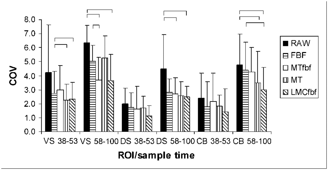
Noise levels (coefficient of variance, COV) in different regions after 5 different analyses: raw data, FBF realignment, and 3 Polaris methods (MTfbf, MT, LMCfbf). ANOVA showed significant effects of region, sample time, and method of analysis, with no interactions (error bars indicate SD; brackets indicate P < 0.05).
- FIGURE 5.
Mean BPs for VS and DS ROIs (brackets indicate paired t test, P < 0.05).
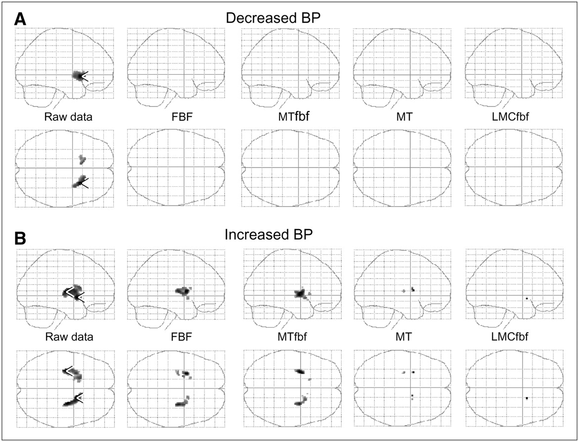
- FIGURE 6.
SPM comparisons of BP between first (A) and second (B) sampling periods. All voxels were significant at P < 0.01, uncorrected. Data are sagittal (top rows) and axial (bottom rows) glass brain views. Arrowheads indicate peak voxels in significant clusters.
- FIGURE 7.
Measured BP in VS and DS ROI as ROI is moved a voxel at a time in the z-axis. Large changes in BP result from small changes in ROI position, demonstrating the susceptibility of measurements to movement.


Tables
VS DS Method 38−53 58−100 38−53 58−100 Raw 1.92 ± 0.71 1.80 ± 0.24 2.33 ± 0.81 2.27 ± 0.23 FBF 1.92 ± 0.68 1.88 ± 0.20 2.28 ± 0.79 2.33 ± 0.17 MTfbf 1.99 ± 0.69 2.03 ± 0.13 2.33 ± 0.80 2.39 ± 0.13 MT 1.86 ± 0.24 1.85 ± 0.13 2.22 ± 0.21 2.22 ± 0.20 LMCfbf 1.94 ± 0.67 1.94 ± 0.10 2.27 ± 0.78 2.30 ± 0.14 VS DS Method Change Variability ICC Change Variability ICC Raw −4.5 ± 14.5 13.5 ± 5.1 0.57 −2.2 ± 4.6 4.2 ± 2.4 0.89 FBF −1.1 ± 9.1 7.3 ± 4.8* 0.75 2.6 ± 5.0 3.6 ± 4.2 0.86 MTfbf 2.6 ± 5.9 5.1 ± 3.6* 0.79 2.9 ± 4.6 3.9 ± 3.8 0.79 MT −0.2 ± 7.0 5.0 ± 4.5*† 0.81 0.8 ± 3.6 2.8 ± 2.2 0.95 LMCfbf 0.6 ± 6.1 4.9 ± 3.3* 0.75 1.2 ± 3.3 2.7 ± 2.1 0.90


Jump to section
Related Articles
Cited By...
- Validation and Evaluation of a Vendor-Provided Head Motion Correction Algorithm on the uMI Panorama PET/CT System
- Dynamic 11C-PiB PET Shows Cerebrospinal Fluid Flow Alterations in Alzheimer Disease and Multiple Sclerosis
- Dynamic 11C-PiB PET shows cerebrospinal fluid flow alterations in Alzheimers disease and multiple sclerosis
- Links between central CB1-receptor availability and peripheral endocannabinoids in patients with first episode psychosis
- An Efficient Approach to Perform MR-Assisted PET Data Optimization in Simultaneous PET/MR Neuroimaging Studies
- Evaluation of Frame-Based and Event-by-Event Motion-Correction Methods for Awake Monkey Brain PET Imaging
- Serotonergic loss in motor circuitries correlates with severity of action-postural tremor in PD
- MRI-Based Nonrigid Motion Correction in Simultaneous PET/MRI
- Serotonin Neuron Loss and Nonmotor Symptoms Continue in Parkinson's Patients Treated with Dopamine Grafts
- Methods for Motion Correction Evaluation Using 18F-FDG Human Brain Scans on a High-Resolution PET Scanner
- Effect of Patient Arm Motion in Whole-Body PET/CT
- Depressive symptoms in PD correlate with higher 5-HTT binding in raphe and limbic structures
- Serotonergic Neurons Mediate Dyskinesia Side Effects in Parkinson's Patients with Neural Transplants
- Increased striatal dopamine (D2/D3) receptor availability and delusions in Alzheimer disease