
Main menu

  • Home
  • Content
    • Current
    • Ahead of print
    • Past Issues
    • JNM Supplement
    • SNMMI Annual Meeting Abstracts
    • Continuing Education
    • JNM Podcasts
  • Subscriptions
    • Subscribers
    • Institutional and Non-member
    • Rates
    • Journal Claims
    • Corporate & Special Sales
  • Authors
    • Submit to JNM
    • Information for Authors
    • Assignment of Copyright
    • AQARA requirements
  • Info
    • Reviewers
    • Permissions
    • Advertisers
  • About
    • About Us
    • Editorial Board
    • Contact Information
  • More
    • Alerts
    • Feedback
    • Help
    • SNMMI Journals
  • SNMMI
    • JNM
    • JNMT
    • SNMMI Journals
    • SNMMI

User menu

  • Subscribe
  • My alerts
  • Log in
  • My Cart

Search

  • Advanced search
Journal of Nuclear Medicine
  • SNMMI
    • JNM
    • JNMT
    • SNMMI Journals
    • SNMMI
  • Subscribe
  • My alerts
  • Log in
  • My Cart
Journal of Nuclear Medicine

Advanced Search

  • Home
  • Content
    • Current
    • Ahead of print
    • Past Issues
    • JNM Supplement
    • SNMMI Annual Meeting Abstracts
    • Continuing Education
    • JNM Podcasts
  • Subscriptions
    • Subscribers
    • Institutional and Non-member
    • Rates
    • Journal Claims
    • Corporate & Special Sales
  • Authors
    • Submit to JNM
    • Information for Authors
    • Assignment of Copyright
    • AQARA requirements
  • Info
    • Reviewers
    • Permissions
    • Advertisers
  • About
    • About Us
    • Editorial Board
    • Contact Information
  • More
    • Alerts
    • Feedback
    • Help
    • SNMMI Journals
  • View or Listen to JNM Podcast
  • Visit JNM on Facebook
  • Join JNM on LinkedIn
  • Follow JNM on Twitter
  • Subscribe to our RSS feeds
OtherBASIC SCIENCE INVESTIGATIONS

Imaging of Tumor Angiogenesis Using 99mTc-Labeled Human Recombinant Anti-ED-B Fibronectin Antibody Fragments

Dietmar Berndorff, Sandra Borkowski, Dieter Moosmayer, Francesca Viti, Beate Müller-Tiemann, Stephanie Sieger, Matthias Friebe, Christoph S. Hilger, Luciano Zardi, Dario Neri and Ludger M. Dinkelborg
Journal of Nuclear Medicine October 2006, 47 (10) 1707-1716;
Dietmar Berndorff
  • Find this author on Google Scholar
  • Find this author on PubMed
  • Search for this author on this site
Sandra Borkowski
  • Find this author on Google Scholar
  • Find this author on PubMed
  • Search for this author on this site
Dieter Moosmayer
  • Find this author on Google Scholar
  • Find this author on PubMed
  • Search for this author on this site
Francesca Viti
  • Find this author on Google Scholar
  • Find this author on PubMed
  • Search for this author on this site
Beate Müller-Tiemann
  • Find this author on Google Scholar
  • Find this author on PubMed
  • Search for this author on this site
Stephanie Sieger
  • Find this author on Google Scholar
  • Find this author on PubMed
  • Search for this author on this site
Matthias Friebe
  • Find this author on Google Scholar
  • Find this author on PubMed
  • Search for this author on this site
Christoph S. Hilger
  • Find this author on Google Scholar
  • Find this author on PubMed
  • Search for this author on this site
Luciano Zardi
  • Find this author on Google Scholar
  • Find this author on PubMed
  • Search for this author on this site
Dario Neri
  • Find this author on Google Scholar
  • Find this author on PubMed
  • Search for this author on this site
Ludger M. Dinkelborg
  • Find this author on Google Scholar
  • Find this author on PubMed
  • Search for this author on this site
  • Article
  • Figures & Data
  • Info & Metrics
  • PDF
Loading

Article Figures & Data

Figures

  • Tables
  • FIGURE 1. 
    • Download figure
    • Open in new tab
    • Download powerpoint
    FIGURE 1. 

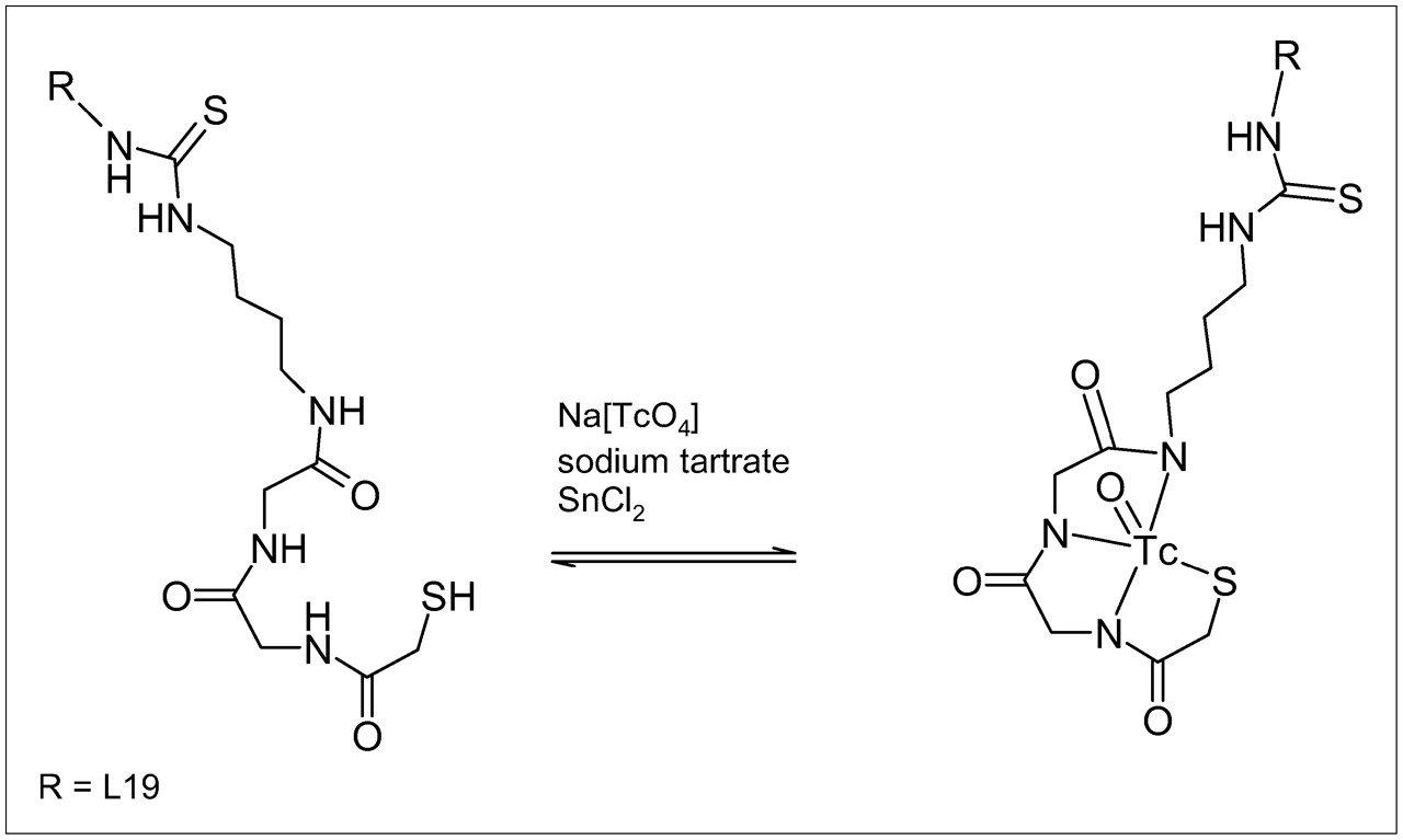
    Synthesis scheme of 99mTc labeling of L19-Hi20.

  • FIGURE 2. 
    • Download figure
    • Open in new tab
    • Download powerpoint
    FIGURE 2. 

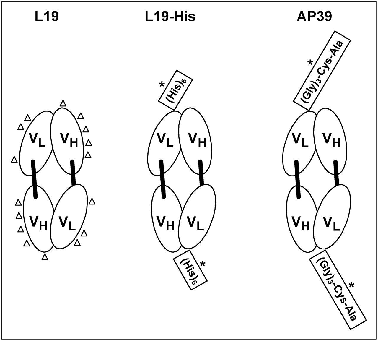
    Schematic drawings show domain structure and potential 99mTc acceptor sites of antibody derivatives that were investigated in this study. Derivatives consist of VL and VH domains of L19 scFv. Using a bifunctional chelator, L19 can be labeled indirectly with 99mTc (potential 99mTc acceptor sites suitable for indirect labeling are indicated by triangles). By insertion of the peptide sequence (His)6 and (Gly)3-Cys-Ala in L19-His and AP39, respectively, these molecules can be labeled directly with 99mTc (potential 99mTc acceptor sites suitable for direct labeling are indicated by asterisks).

  • FIGURE 3. 
    • Download figure
    • Open in new tab
    • Download powerpoint
    FIGURE 3. 

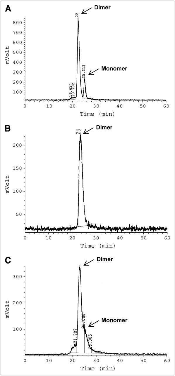
    Representative SE-HPLC chromatograms (radiation detection) of 99mTc-AP39 (A), 99mTc-L19-His (B), and 99mTc-L19-Hi20 (C).

  • FIGURE 4. 
    • Download figure
    • Open in new tab
    • Download powerpoint
    FIGURE 4. 

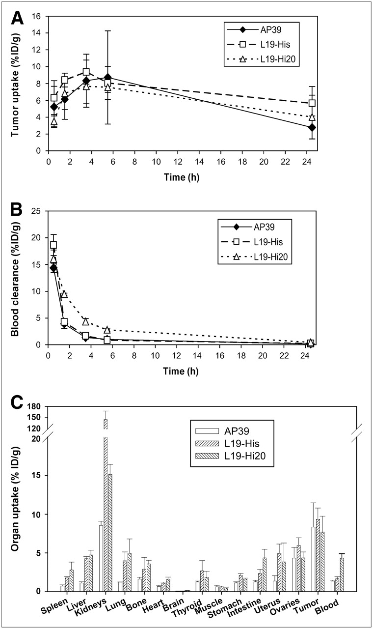
    Comparison of 99mTc-L19 derivatives in F9 tumor–bearing nude mice with regard to tumor uptake up to 24 h after injection (A), blood clearance up to 24 h after injection (B), and organ uptake at 3 h after injection (C). All data are presented as mean ± SD (n = 3).

  • FIGURE 5. 
    • Download figure
    • Open in new tab
    • Download powerpoint
    FIGURE 5. 

    Scintigraphic images (posterior) of 99mTc-AP39 in F9 tumor–bearing mice 3 h (A), 5 h (B), and 24 h (C) after injection.

  • FIGURE 6. 
    • Download figure
    • Open in new tab
    • Download powerpoint
    FIGURE 6. 

    Scintigraphic images (posterior) of 99mTc-L19-His in F9 tumor–bearing mice 5 h (A) and 24 h (B) after injection.

  • FIGURE 7. 
    • Download figure
    • Open in new tab
    • Download powerpoint
    FIGURE 7. 

    Scintigraphic images (posterior) of 99mTc-L19-Hi20 in F9 tumor–bearing mice 5 h (A) and 24 h (B) after injection.

Tables

  • Figures
    • View popup
    TABLE 1

    Biodistribution of 99mTc-AP39 in F9 Tumor–Bearing Mice

    Biodistribution time (h)
    Tissue0.2513524
    Uptake (%ID/g)
     Spleen5.1 ± 0.72.0 ± 0.30.7 ± 0.20.6 ± 0.10.2 ± 0.0
     Liver5.1 ± 0.22.5 ± 0.31.1 ± 0.21.1 ± 0.20.3 ± 0.0
     Kidneys48.2 ± 4.221.8 ± 2.58.6 ± 0.67.9 ± 0.42.6 ± 0.4
     Lung6.9 ± 1.73.1 ± 0.21.2 ± 0.01.1 ± 0.20.8 ± 1.0
     Bone*2.8 ± 0.92.3 ± 0.31.6 ± 0.21.7 ± 0.10.8 ± 0.2
     Heart4.1 ± 1.31.8 ± 0.20.7 ± 0.20.4 ± 0.00.2 ± 0.0
     Brain0.4 ± 0.00.1 ± 0.00.1 ± 0.0NDND
     Thyroid3.4 ± 0.62.0 ± 0.21.3 ± 0.11.3 ± 0.70.8 ± 0.4
     Muscle*0.8 ± 0.10.6 ± 0.00.6 ± 0.20.2 ± 0.00.1 ± 0.0
     Stomach†1.7 ± 0.11.4 ± 0.01.1 ± 0.11.0 ± 0.00.4 ± 0.1
     Intestine‡1.6 ± 0.11.4 ± 0.01.3 ± 0.21.6 ± 0.50.3 ± 0.0
     Uterus2.5 ± 0.82.0 ± 0.51.4 ± 0.72.8 ± 1.21.3 ± 0.7
     Ovaries13.0 ± 1.87.6 ± 2.24.3 ± 1.42.7 ± 0.90.9 ± 0.1
     Tumor5.2 ± 2.56.1 ± 2.08.3 ± 3.28.7 ± 5.52.8 ± 1.4
     Blood14.4 ± 0.93.9 ± 0.81.3 ± 0.11.0 ± 0.10.2 ± 0.0
    Excretion (%ID)
     Urine22.7 ± 3.946.7 ± 4.448.8 ± 6.752.5 ± 2.064.5 ± 6.5
     FecesNDND0.4 ± 0.10.6 ± 0.54.8 ± 1.0
    Tumor/tissue ratio
     T/spleen1.0 ± 0.33.1 ± 0.612.4 ± 7.314.8 ± 11.411.5 ± 4.5
     T/liver1.0 ± 0.42.4 ± 0.57.8 ± 4.08.3 ± 6.28.1 ± 3.6
     T/kidneys0.1 ± 0.00.3 ± 0.11.0 ± 0.41.1 ± 0.81.0 ± 0.4
     T/lung0.8 ± 0.52.0 ± 0.76.8 ± 2.68.3 ± 6.57.5 ± 5.9
     T/muscle6.8 ± 3.410.5 ± 3.213.6 ± 2.638.5 ± 28.925.3 ± 11.2
     T/blood0.4 ± 0.21.6 ± 0.26.4 ± 2.99.0 ± 6.517.2 ± 6.8
     T/intestine3.3 ± 1.34.3 ± 1.36.9 ± 3.66.9 ± 6.98.9 ± 3.7
    • ↵* Tissue aliquot only.

    • ↵† Without content.

    • ↵‡ With content.

    • ND = not detectable; T = tumor.

    • All data are presented as mean ± SD (n = 3).

    • View popup
    TABLE 2

    Biodistribution of 99mTc-L19-His in F9 Tumor–Bearing Mice

    Biodistribution time (h)
    Tissue0.2513524
    Uptake (%ID/g)
     Spleen5.5 ± 0.62.7 ± 0.71.8 ± 0.21.2 ± 0.20.8 ± 0.2
     Liver6.1 ± 0.73.4 ± 0.44.3 ± 0.33.4 ± 0.72.5 ± 0.7
     Kidneys87.5 ± 5.2124.7 ± 6.3145.1 ± 22.3117.0 ± 12.972.4 ± 11.3
     Lung10.4 ± 1.85.2 ± 1.04.0 ± 1.24.1 ± 2.92.0 ± 0.6
     Bone*4.6 ± 0.95.4 ± 1.02.9 ± 1.53.4 ± 0.73.1 ± 0.9
     Heart5.2 ± 0.22.3 ± 0.21.1 ± 0.10.7 ± 0.10.4 ± 0.1
     Brain0.4 ± 0.10.2 ± 0.00.1 ± 0.00.1 ± 0.00.1 ± 0.0
     Thyroid4.9 ± 1.52.9 ± 0.12.7 ± 1.32.7 ± 1.11.1 ± 0.0
     Muscle*0.5 ± 0.20.6 ± 0.10.6 ± 0.10.4 ± 0.10.3 ± 0.1
     Stomach†2.0 ± 0.21.7 ± 0.12.1 ± 0.21.7 ± 0.31.1 ± 0.2
     Intestine‡1.4 ± 0.11.8 ± 0.22.4 ± 0.51.9 ± 0.81.2 ± 0.7
     Uterus3.2 ± 1.74.7 ± 1.34.9 ± 1.22.7 ± 0.84.4 ± 2.4
     Ovaries27.2 ± 18.810.2 ± 2.16.0 ± 1.06.9 ± 1.02.8 ± 0.5
     Tumor6.3 ± 2.08.4 ± 0.89.4 ± 1.48.1 ± 2.05.7 ± 2.0
     Blood18.7 ± 2.04.3 ± 0.11.7 ± 0.30.9 ± 0.10.2 ± 0.1
    Excretion (%ID)
     Urine10.8 ± 3.314.8 ± 5.420.9 ± 5.521.3 ± 4.964.5 ± 6.5
     FecesNDND0.2 ± 0.30.6 ± 0.34.8 ± 1.0
    Tumor/tissue ratio
     T/spleen1.2 ± 0.43.3 ± 1.15.3 ± 0.77.0 ± 2.36.6 ± 1.0
     T/liver1.1 ± 0.42.5 ± 0.52.2 ± 0.52.4 ± 0.62.3 ± 0.2
     T/kidneys0.1 ± 0.00.1 ± 0.00.1 ± 0.00.1 ± 0.00.1 ± 0.0
     T/lung0.6 ± 0.11.7 ± 0.42.5 ± 0.82.5 ± 1.22.9 ± 0.9
     T/muscle12.4 ± 1.415.5 ± 4516.4 ± 4.223.0 ± 9.522.9 ± 6.6
     T/blood0.4 ± 0.12.0 ± 0.25.7 ± 0.59.5 ± 2.623.6 ± 8.3
     T/intestine4.5 ± 1.74.8 ± 0.94.0 ± 0.74.9 ± 2.24.9 ± 1.4
    • ↵* Tissue aliquot only.

    • ↵† Without content.

    • ↵‡ With content.

    • ND = not detectable; T = tumor.

    • All data are presented as mean ± SD (n = 3).

    • View popup
    TABLE 3

    Biodistribution of 99mTc-L19-Hi20 in F9 Tumor–Bearing Mice

    Biodistribution time (h)
    Tissue0.2513524
    Uptake (%ID/g)
     Spleen4.9 ± 0.54.1 ± 0.72.8 ± 1.01.4 ± 0.20.5 ± 0.1
     Liver10.5 ± 2.17.1 ± 0.14.7 ± 0.63.4 ± 0.51.7 ± 0.2
     Kidneys33.5 ± 2.426.6 ± 0.715.2 ± 1.310.9 ± 0.43.0 ± 0.6
     Lung9.4 ± 1.46.2 ± 0.65.0 ± 1.82.6 ± 0.81.3 ± 0.5
     Bone*4.0 ± 0.44.2 ± 0.63.6 ± 0.52.9 ± 0.51.0 ± 0.1
     Heart4.7 ± 1.53.3 ± 0.41.6 ± 0.31.1 ± 0.10.2 ± 0.0
     Brain0.4 ± 0.10.3 ± 0.00.1 ± 0.00.1 ± 0.0ND
     Thyroid3.5 ± 1.44.3 ± 1.21.9 ± 0.83.2 ± 1.10.7 ± 0.1
     Muscle*0.8 ± 0.20.7 ± 0.00.5 ± 0.10.5 ± 0.10.2 ± 0.0
     Stomach†1.9 ± 0.12.5 ± 0.21.6 ± 0.11.5 ± 0.10.6 ± 0.1
     Intestine‡1.6 ± 0.23.3 ± 0.24.4 ± 1.14.2 ± 1.80.9 ± 0.4
     Uterus2.1 ± 0.84.9 ± 1.53.8 ± 2.43.1 ± 0.91.6 ± 0.7
     Ovaries7.6 ± 4.18.4 ± 0.44.4 ± 0.83.6 ± 0.91.0 ± 0.2
     Tumor3.5 ± 0.66.8 ± 1.97.7 ± 2.17.6 ± 0.64.0 ± 2.6
     Blood16.1 ± 1.79.5 ± 0.24.3 ± 0.62.8 ± 0.30.4 ± 0.0
    Excretion (%ID)
     Urine19.1 ± 2.427.1 ± 6.038.8 ± 4.441.8 ± 4.057.8 ± 3.0
     FecesNDND2.6 ± 1.37.3 ± 6.118.6 ± 4.0
    Tumor/tissue ratio
     T/spleen0.7 ± 0.11.7 ± 0.43.1 ± 1.45.4 ± 1.27.7 ± 5.7
     T/liver0.4 ± 0.11.0 ± 0.31.6 ± 0.42.2 ± 0.32.4 ± 1.9
     T/kidneys0.1 ± 0.00.3 ± 0.10.5 ± 0.20.7 ± 0.11.4 ± 1.0
     T/lung0.4 ± 0.01.1 ± 0.21.6 ± 0.53.1 ± 0.74.5 ± 4.9
     T/muscle4.8 ± 1.89.7 ± 3.117.5 ± 8.515.5 ± 2.125.7 ± 13.5
     T/blood0.2 ± 0.10.7 ± 0.21.8 ± 0.52.7 ± 0.49.4 ± 6.4
     T/intestine2.2 ± 0.42.1 ± 0.51.9 ± 0.82.0 ± 0.88.4 ± 2.8
    • ↵* Tissue aliquot only.

    • ↵† Without content.

    • ↵‡ With content.

    • ND = not detectable; T = tumor.

    • All data are presented as mean ± SD (n = 3).

PreviousNext
Back to top

In this issue

Journal of Nuclear Medicine: 47 (10)
Journal of Nuclear Medicine
Vol. 47, Issue 10
October 2006
  • Table of Contents
  • Table of Contents (PDF)
  • About the Cover
  • Index by author
Print
Download PDF
Article Alerts
Sign In to Email Alerts with your Email Address
Email Article

Thank you for your interest in spreading the word on Journal of Nuclear Medicine.

NOTE: We only request your email address so that the person you are recommending the page to knows that you wanted them to see it, and that it is not junk mail. We do not capture any email address.

Enter multiple addresses on separate lines or separate them with commas.
Imaging of Tumor Angiogenesis Using 99mTc-Labeled Human Recombinant Anti-ED-B Fibronectin Antibody Fragments
(Your Name) has sent you a message from Journal of Nuclear Medicine
(Your Name) thought you would like to see the Journal of Nuclear Medicine web site.
Citation Tools
Imaging of Tumor Angiogenesis Using 99mTc-Labeled Human Recombinant Anti-ED-B Fibronectin Antibody Fragments
Dietmar Berndorff, Sandra Borkowski, Dieter Moosmayer, Francesca Viti, Beate Müller-Tiemann, Stephanie Sieger, Matthias Friebe, Christoph S. Hilger, Luciano Zardi, Dario Neri, Ludger M. Dinkelborg
Journal of Nuclear Medicine Oct 2006, 47 (10) 1707-1716;

Citation Manager Formats

  • BibTeX
  • Bookends
  • EasyBib
  • EndNote (tagged)
  • EndNote 8 (xml)
  • Medlars
  • Mendeley
  • Papers
  • RefWorks Tagged
  • Ref Manager
  • RIS
  • Zotero
Share
Imaging of Tumor Angiogenesis Using 99mTc-Labeled Human Recombinant Anti-ED-B Fibronectin Antibody Fragments
Dietmar Berndorff, Sandra Borkowski, Dieter Moosmayer, Francesca Viti, Beate Müller-Tiemann, Stephanie Sieger, Matthias Friebe, Christoph S. Hilger, Luciano Zardi, Dario Neri, Ludger M. Dinkelborg
Journal of Nuclear Medicine Oct 2006, 47 (10) 1707-1716;
Twitter logo Facebook logo LinkedIn logo Mendeley logo
  • Tweet Widget
  • Facebook Like
  • Google Plus One
Bookmark this article

Jump to section

  • Article
    • Abstract
    • MATERIALS AND METHODS
    • RESULTS
    • DISCUSSION
    • CONCLUSION
    • Acknowledgments
    • Footnotes
    • References
  • Figures & Data
  • Info & Metrics
  • PDF

Related Articles

  • This Month in JNM
  • PubMed
  • Google Scholar

Cited By...

  • Prolactin Receptor-Mediated Internalization of Imaging Agents Detects Epithelial Ovarian Cancer with Enhanced Sensitivity and Specificity
  • Targeting of HER2-Expressing Tumors with a Site-Specifically 99mTc-Labeled Recombinant Affibody Molecule, ZHER2:2395, with C-Terminally Engineered Cysteine
  • Antibody Library-based Tumor Endothelial Cells Surface Proteomic Functional Screen Reveals Migration-stimulating Factor as an Anti-angiogenic Target
  • Fibronectin Stimulates Endothelial Cell 18F-FDG Uptake Through Focal Adhesion Kinase-Mediated Phosphatidylinositol 3-Kinase/Akt Signaling
  • Complete eradication of human B-cell lymphoma xenografts using rituximab in combination with the immunocytokine L19-IL2
  • In Vivo Biodistribution, PET Imaging, and Tumor Accumulation of 86Y- and 111In-Antimindin/RG-1, Engineered Antibody Fragments in LNCaP Tumor-Bearing Nude Mice
  • The Extra-domain A of Fibronectin Is a Vascular Marker of Solid Tumors and Metastases
  • Small-Animal PET of Tumor Angiogenesis Using a 76Br-Labeled Human Recombinant Antibody Fragment to the ED-B Domain of Fibronectin
  • Nuclear Imaging Probes: from Bench to Bedside
  • The Antibody-Mediated Targeted Delivery of Interleukin-15 and GM-CSF to the Tumor Neovasculature Inhibits Tumor Growth and Metastasis
  • Human Antibody Against C Domain of Tenascin-C Visualizes Murine Atherosclerotic Plaques Ex Vivo
  • Google Scholar

More in this TOC Section

  • PET/MRI of Hypoxic Atherosclerosis Using 64Cu-ATSM in a Rabbit Model
  • Tumor Uptake of Anti-CD20 Fabs Depends on Tumor Perfusion
  • How Sensitive Is the Upper Gastrointestinal Tract to 90Y Radioembolization? A Histologic and Dosimetric Analysis in a Porcine Model
Show more Basic Science Investigations

Similar Articles

SNMMI

© 2025 SNMMI

Powered by HighWire