


Abstract
The detection of human epidermal growth factor receptor type 2 (HER2) expression in malignant tumors provides important information influencing patient management. Radionuclide in vivo imaging of HER2 may permit the detection of HER2 in both primary tumors and metastases by a single noninvasive procedure. Small (7 kDa) high-affinity anti-HER2 Affibody molecules may be suitable tracers for SPECT visualization of HER2-expressing tumors. The use of generator-produced 99mTc as a label would facilitate the prompt translation of anti-HER2 Affibody molecules into use in clinics. Methods: A C-terminal cysteine was introduced into the Affibody molecule ZHER2:342 to enable site-specific labeling with 99mTc. Two recombinant variants, His6-ZHER2:342-Cys (dissociation constant [KD], 29 pM) and ZHER2:2395-Cys, lacking a His tag (KD, 27 pM), were labeled with 99mTc in yields exceeding 90%. The binding specificity and the cellular processing of Affibody molecules were studied in vitro. Biodistribution and γ-camera imaging studies were performed in mice bearing HER2-expressing xenografts. Results: 99mTc-His6-ZHER2:342-Cys was capable of targeting HER2-expressing SKOV-3 xenografts in SCID mice, but the liver radioactivity uptake was high. A series of comparative biodistribution experiments indicated that the presence of the His tag caused elevated accumulation in the liver. 99mTc-ZHER2:2395-Cys, not containing a His tag, showed low uptake in the liver and high and specific uptake in HER2-expressing xenografts. Four hours after injection, the radioactivity uptake values (percentage of injected activity per gram of tissue [%IA/g]) were 6.9 ± 2.5 (mean ± SD) %IA/g in LS174T xenografts (moderate level of HER2 expression) and 15 ± 3 %IA/g in SKOV-3 xenografts (high level of HER2 expression). The corresponding tumor-to-blood ratios were 88 ± 24 and 121 ± 24, respectively. Both LS174T and SKOV-3 xenografts were clearly visualized with a clinical γ-camera 1 h after injection of 99mTc-ZHER2:2395-Cys. Conclusion: The Affibody molecule 99mTc-ZHER2:2395-Cys is a promising tracer for SPECT visualization of HER2-expressing tumors.
Human epidermal growth factor receptor type 2 (HER2) is a transmembrane protein belonging to the receptor tyrosine kinase superfamily (1). HER2 signaling is associated with increased proliferation and decreased apoptotic capacity (2). Overexpression of HER2 is found in a variety of cancers, for example breast, ovary, colorectal, and urothelial carcinomas, and is associated with more aggressive disease and a poor prognosis (1). This receptor is the target for therapy with the monoclonal antibody trastuzumab (Herceptin; Roche Pharma AG), which interrupts HER2 signaling and prolongs the survival of patients with HER2-expressing breast cancer (3,4). HER2 is, however, overexpressed in only 25%−30% of breast carcinomas. Therefore, the detection of HER2 expression is necessary to identify patients who would benefit from trastuzumab therapy. Both the American Society of Clinical Oncology and the European Group on Tumor Markers have recommended that HER2/c-erbB-2 overexpression should be evaluated for every primary breast cancer (5,6).
Most commonly, HER2 status is determined by immunohistochemical or fluorescent in situ hybridization analysis of biopsy material. Unfortunately, the results of such tests may be adversely affected by tumor sampling errors, discordance in HER2 expression in primary tumors and metastases, and inexperience of laboratories performing analyses (7). The use of radionuclide molecular imaging of HER2 expression may help to avoid these problems associated with biopsies. Moreover, radionuclide imaging may also allow determination of the HER2 expression status in lesions not amenable to biopsy, for example, bone metastases.
Clinical studies on the radionuclide imaging of HER2 were initially performed with 111In-labeled trastuzumab (8,9). However, the detection sensitivity was not optimal. Only 45% of the lesions already detected by routine imaging techniques were detected by the radionuclide scan (9). Low sensitivity is a general problem when full-size IgG antibodies are used as targeting agents because their long residence time in blood causes high radioactivity background, leading to reduced imaging contrast. An analysis of preclinical data showed that smaller tracers provide better imaging contrast and therefore better sensitivity than intact antibodies (10).
The problem of size reduction for a HER2-targeting agent could be solved with Affibody molecules (11). Affibody molecules originate from the 58-amino-acid (7 kDa) cysteine-free scaffold of the staphylococcal protein A–derived Z domain. The scaffold has been subjected to the randomization of 13 surface-exposed residues to generate a library from which high-affinity binders can be selected by phage display. The selection of the Affibody molecule ZHER2:342, which binds to the extracellular domain of HER2 with an affinity of 22 pM, was described earlier (12). High-contrast imaging of HER2-expressing xenografts with indirectly radioiodinated ZHER2:342 was demonstrated (12). The potential of recombinant Affibody molecules for imaging was confirmed in preclinical studies with radioisotopes of bromine (13) and indium in combination with various chelators (14–16).
The spontaneously folding structure of Affibody molecules is amenable to complete peptide synthesis of a well-defined product and site-specific incorporation of various functional groups. Synthetic Affibody molecule ZHER2:342 with DOTA incorporated at the N-terminus has been prepared, labeled with 111In, and characterized in vivo (17). The imaging of HER2-expressing metastases with 111In- and 68Ga-labeled DOTA-ZHER2:342 has been demonstrated in clinical case studies (18).
Generator-produced 99mTc (half-life = 6 h) is an attractive radionuclide for molecular imaging applications because of its photon energy (which is nearly ideal for SPECT), low cost, excellent availability, and low dose burden for the patient. Earlier, mercaptoacetyl-containing synthetic Affibody molecules were labeled with 99mTc and demonstrated high-contrast imaging of HER2-expressing xenografts (19–21).
Mercaptoacetyl is added during chemical peptide synthesis; this process may be applicable only to smaller peptides and monomer Affibody molecules. Therefore, several approaches have been pursued to obtain site-specific labeling of recombinant proteins. We previously reported on the 99mTc labeling of His tag–containing anti-HER2 Affibody dimers with technetium(I) tricarbonyl chemistry (22). Unfortunately, this kind of labeling resulted in high radioactivity uptake in the liver. The absence of cysteine residues in the Affibody scaffold enables the creation of a unique reactive site through the incorporation of a single cysteine. The Cys thiol group, along with 3 amides in the peptide backbone, creates a site-specific N3S chelator for labeling with 99mTc, resulting in a controlled, homogeneous product. This approach has been applied for the labeling of single-chain Fv fragments (23–25). We previously evaluated the labeling of Affibody molecules with CGG and CGGG sequences incorporated at the N terminus of Affibody molecule ZHER2:342 (26). The 99mTc-CGG-ZHER2:342 conjugate was capable of specifically targeting HER2-expressing xenografts in mice and provided a tumor-to-blood ratio of 9.2 (6 h after injection). However, radioactivity uptake in the stomach and salivary glands was higher than that obtained with mercaptoacetyl-containing conjugates, indicating the release of free pertechnetate during catabolism of the conjugate in vivo. Placement of the cysteine residue in a C-terminal position allows for a more stable “GGC”-type chelator, as previously demonstrated for scFv fragments and for technetium-labeled peptide radiopharmaceuticals (27). In a GGC–technetium complex, the N3S ligand set forms a stable chelate with three 5-member rings during complexation with the technetium; however, when the cysteine is located at the N-terminus (CGG), metal complexation through the cysteine yields a 6-member ring in the chelate, which is known to yield a less stable technetium(V) complex (28).
The goal of the study was to evaluate whether the introduction of a single cysteine at the C-terminus of recombinant Affibody molecules would provide stable labeling with 99mTc. An initial characterization was performed with His tag–containing 99mTc-His6-ZHER2:342-Cys (hereafter referred to as 99mTc-H6-ZHER2:342-C) because a His tag facilitates the purification of recombinant proteins. The biodistribution of 99mTc-H6-ZHER2:342-C demonstrated high radioactivity uptake in the liver. Further in vivo studies demonstrated that this elevated liver uptake was associated with the presence of the His tag. An Affibody molecule lacking the His tag, ZHER2:2395-Cys (hereafter referred to as Z2395-C), showed low uptake in the liver and good targeting properties when radiolabeled with 99mTc. γ-camera imaging confirmed that 99mTc-Z2395-C was capable of specific imaging of HER2-expressing xenografts.
MATERIALS AND METHODS
Materials
99mTc was obtained as pertechnetate by elution in an Ultra-TechneKow generator (Tyco) with sterile 0.9% sodium chloride (Mallinckrodt Medical BV). An automated γ-counter with a ∼7.6-cm (3-in) NaI(Tl) detector (1480 WIZARD; Wallac Oy) was used to detect radioactivity. A Cyclone storage phosphor system and OptiQuant image analysis software (both from Perkin-Elmer) were used to measure the radioactivity on instant thin-layer chromatography silica gel–impregnated (ITLC SG) strips. Affibody molecules H6-ZHER2:342-C and Z2395-C were produced and purified as described by Ahlgren et al. (29). These two proteins differ only in the N-terminal sequence (GSSHHHHHHLQVDNKFNK for H6-ZHER2:342-C and AENKFNK for Z2395-C [differences are shown in bold type]).
Labeling Chemistry and In Vitro Characterization
Cysteine-containing HER2-binding Affibody molecules were site-specifically labeled with 99mTc by use of freeze-dried labeling kits each containing 5 mg of sodium α-d-glucoheptonate dihydrate, 100 μg of edetate disodium (EDTA), and 50 μg of tin(II) chloride dihydrate (SnCl2·2H2O) at pH 7.4. The freeze-dried kits were stored at −20°C.
For radiolabeling of 99mTc-H6-ZHER2:342-C, a 99mTc-pertechnetate generator eluate (370 MBq; 350 μL), Affibody molecule H6-ZHER2:342-C dissolved in phosphate-buffered saline (PBS; 200 μg; 91 μL), and 560 μL of saline were added to a freeze-dried kit, and the contents of the vial were swirled. The vial was incubated for 10 min at 100°C and cooled for 15 min at room temperature. The radiochemical purity of 99mTc-H6-ZHER2:342-C was determined by reverse-phase high-pressure liquid chromatography with a water–acetonitrile gradient containing trifluoroacetic acid (supplemental Fig. 1) (supplemental materials are available online only at http://jnm.snmjournals.org). No further purification was needed.
For 99mTc-Z2395-C, the labeling was reoptimized. Radiolabeling was performed by adding the contents of one freeze-dried kit, dissolved in PBS, to 100 μg of Affibody molecule Z2395-C to a total volume of 100 μL. To the reaction solution, 100 μL of a 99mTc-pertechnetate generator eluate (typically, 50 MBq) was added under argon gas, and the solution was incubated at 90°C for 40 min and cooled at room temperature for 15 min. Thereafter, 1 μL samples were taken for analysis of the labeling yield by ITLC SG with PBS as the mobile phase and for analysis of reduced hydrolyzed technetium colloid levels by ITLC SG with pyridine:acetic acid:water (5:3:1.5) as the mobile phase. No further purification was needed. For in vitro and in vivo experiments, the labeling solution was diluted with PBS.
To test the labeling stability in vitro, two 5 μL samples of 99mTc-Z2395-C were mixed with 100 μL of mouse plasma. After incubation at 37°C for 2 h, 20 μL of each sample was electrophoresed on a sodium dodecyl sulfate–polyacrylamide gel along with a sample of free 99mTcO4− diluted in PBS.
The in vitro binding specificity of 99mTc-Z2395-C was verified in HER2-expressing SKOV-3 ovarian carcinoma cells by receptor presaturation with unlabeled Affibody molecules as described by Ahlgren et al. (29). Antigen-binding capacity after radiolabeling was studied as described by Tran et al. (26). Cellular retention and internalization of radioactivity were evaluated as described by Ahlgren et al. (29).
Animal Studies
The animal experiments were planned and performed in accordance with national legislation on the protection of laboratory animals. The animal study plans were approved by the local ethics committee for animal research. Biodistribution studies with non–tumor-bearing mice were performed with female immunocompetent NMRI mice.
In experiments with tumor-bearing mice, female outbred BALB/c nu/nu or SCID mice were used. Xenografts of HER2-expressing LS174T colon adenocarcinoma cells and SKOV-3 cells were subcutaneously implanted in the right hind leg. For xenografts in BALB/c nu/nu mice, 106 LS174T cells or 107 SKOV-3 cells were used, whereas in SCID mice, 5 × 10⁶ SKOV-3 cells were used. As negative controls for use in the imaging experiment, mice were given xenografts of 107 non–HER2-expressing A431 cells. At the time of the biodistribution experiment, the average (mean ± SD) tumor sizes were 0.3 ± 0.15 g for LS174T xenografts in BALB/c nu/nu mice, 0.4 ± 0.1 g for SKOV-3 xenografts in BALB/c nu/nu mice, and 1.0 ± 0.5 g for SKOV-3 xenografts in SCID mice.
For biodistribution studies with SKOV-3 xenograft–bearing SCID mice, animals were randomized into groups of 3 and injected intravenously with 60 ng of 99mTc-H6-ZHER2:342-C conjugate (94 kBq) in 100 μL of PBS. For studies with xenograft-bearing BALB/c nu/nu mice, animals were randomized into groups of 4 and injected intravenously with 1 μg of conjugate (50 kBq) in 100 μL of PBS. At predesignated time points, the mice were euthanized by anesthesia overdosing. Blood was collected by heart puncture. Organ samples were collected in preweighed plastic vials. All tissue uptake values were calculated as the percentage of injected activity (%IA) per gram of tissue (%IA/g), except for the thyroid, gastrointestinal tract, and carcass, where the tissue uptake values were calculated as the %IA per whole sample. The radioactivity in the gastrointestinal tract and its contents was used as a measure of hepatobiliary excretion.
The biodistributions of 99mTc-Z2395-C and 99mTc-H6-ZHER2:342-C in NMRI mice were compared at 4 h after injection to verify that the elevated uptake in the liver was attributable to the His tag.
The biodistribution of 99mTc-Z2395-C in BALB/c mice bearing LS174T xenografts was measured at 0.5, 1, 4, and 6 h after injection, and in BALB/c nu/nu mice bearing SKOV-3 xenografts at 4 h after injection (Table 3). To verify the specificity of HER2 targeting, HER2 was presaturated in one group of mice bearing LS174T xenografts and in another group of mice bearing SKOV-3 xenografts by subcutaneous injection with 600 μg of unlabeled His6-ZHER2:342 45 min before injection of 99mTc-Z2395-C. The animals were euthanized 4 h after injection, and uptake in tumors was measured.
For γ-camera imaging, two groups of LS174T xenograft–bearing mice were injected with 1.5 MBq of 99mTc-Z2395-C (amount of protein, 3 μg). In a blocking experiment, one animal from each group was preinjected with unlabeled Affibody molecules as described earlier. One group of animals was euthanized 1 h after injection, and the other group was euthanized 4 h after injection. For imaging of SKOV-3 xenograft–bearing mice, 4 animals were injected with 1.5 MBq of 99mTc-Z2395-C (amount of protein, 3 μg). Two of them were euthanized 1 h after injection, and the other two were euthanized 4 h after injection. As a negative control, two animals bearing non–HER2-expressing A431 xenografts were injected with the same amount of 99mTc-Z2395-C as the SKOV-3 xenograft–bearing mice and euthanized 4 h after injection. All animals were euthanized by overdose with ketamine hydrochloride (Ketalar; Pfizer) and xylazine hydrochloride (Rompun; Bayer). After euthanasia, the urinary bladders were dissected. Imaging was performed at the Department of Nuclear Medicine at Uppsala University Hospital with an e.Cam γ-camera (Siemens Medical Systems, Inc.) equipped with a low-energy, high-resolution collimator. Acquisition of static images was performed with a 256 × 256 matrix and a zoom factor of 3.2. The energy window settings were 140 keV and 15%. The images were evaluated with Osiris 4.19 software (Digital Imaging Group, University Hospital of Geneva).
RESULTS
Analysis of Peptides
Data concerning the characterization of recombinant Affibody molecules were reported by Ahlgren et al. (29). All purified proteins demonstrated a single peak on liquid chromatography–mass spectrometry with a mass coinciding with the theoretically calculated value ± 1.5 Da, which is within the accuracy of the instrument. The introduction of a cysteine at the C terminus did not have an effect on the binding to HER2 because the apparent dissociation constants (KD) of 29 pM for H6-ZHER2:342-C and 27 pM for Z2395-C did not deviate from the KD for parental Affibody molecule ZHER2:342 (22 pM) and were within the accuracy of the measurement method.
Labeling Chemistry and In Vitro Characterization
The radiochemical purity determined by reverse-phase high-pressure liquid chromatography for 99mTc-H6-ZHER2:342-C was 93%. For 99mTc-Z2395-C samples, labeling yields determined by ITLC were greater than 97%, and the levels of reduced hydrolyzed technetium colloids were less than 1%. A specific radioactivity of 3.5 GBq/μmol was achieved for 99mTc-Z2395-C. Phosphorimaging analysis of the radioactivity distribution on a sodium dodecyl sulfate–polyacrylamide gel showed that after incubation at 37°C for 2 h in mouse plasma, 96% ± 0.2% of the radioactivity was associated with 99mTc-Z2395-C and the rest of the radioactivity matched free 99mTcO4−. For biologic experiments, all 99mTc conjugates were used without additional purification.
In the in vitro specificity analysis, presaturation of HER2 in SKOV-3 cells with nonlabeled Affibody molecules reduced the cell-bound radioactivity from 30.3% ± 0.9% to 0.9% ± 0.1% (P < 10−6) after 1 h of incubation at 37°C (Fig. 1A). This finding indicated that the binding of 99mTc-Z2395-C to HER2-expressing cells was specific. The antigen-binding capacity after radiolabeling was 86% ± 1.6% when tested with SKOV-3 cells. The cellular retention and internalization analyses of 99mTc-Z2395-C indicated that the conjugate was efficiently retained by HER2-expressing cells (Fig. 1B). The cellular retention of the total radioactivity was good, with 68% ± 1% being cell associated after 24 h of incubation at 37°C. The internalization of 99mTc-Z2395-C was relatively slow, with less than 11% internalized radioactivity after 24 h. Most of the radioconjugate was released within the first 0.5 h, with a slow increase of released radioactivity up to 8 h. Thereafter, the levels were almost stable up to 24 h.
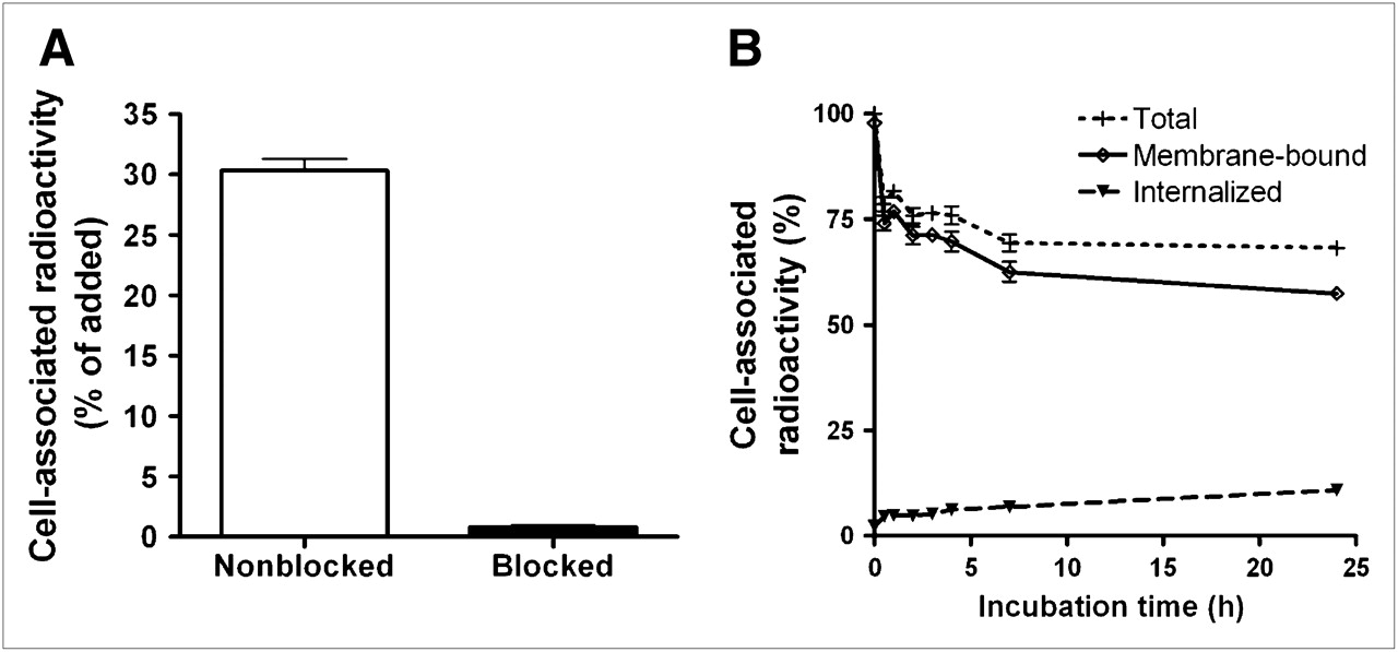
(A) In vitro specificity analysis. One group of culture dishes with SKOV-3 cells was pretreated with saturating amounts of nonlabeled His6-ZHER2:342 before incubation with 99mTc-ZHER2:2395-C. Cell-associated radioactivity was calculated as percentage of total added radioactivity. (B) Cell-associated radioactivity as function of time after interrupted incubation of SKOV-3 cells with 99mTc-ZHER2:2395-C. Cell-associated radioactivity at time zero after interrupted incubation was considered to be 100%. Radioactivity that was removed from cells by treatment with 4 M urea solution in 0.2 M glycine buffer (pH 2.0) was considered to be membrane bound, and remainder was considered to be internalized. Data are presented as mean ± SD (n = 3). Error bars may not be visible because they are smaller than the symbols.
Animal Studies
The biodistribution data for 99mTc-H6-ZHER2:342-C in SCID mice are shown in Table 1. The conjugate showed rapid tumor targeting and rapid clearance from a majority of nontarget organs. Kidney uptake was high because of renal excretion and reabsorption; this pattern is typical for radiometal-labeled Affibody molecules. Liver uptake was also high (about 20 %IA/g); this finding is unusual for ZHER2:342 conjugates. To examine whether the atypical liver uptake was attributable to the physiologic features of the mouse strain used (SCID), the biodistribution of a different ZHER2:342-based tracer, 111In-tetraazacyclododecanetetraacetic acid (DOTA)-ZHER2:342, was investigated in SCID mice bearing SKOV-3 xenografts (Supplemental Table 1). The biodistribution data for 111In-DOTA-ZHER2:342 in SCID mice showed a high level of agreement with previously published data on biodistribution in BALB/c nu/nu mice (17).
Biodistribution of 99mTc-H6-ZHER2:342-C in SCID Mice Bearing SKOV-3 Tumors
A comparison of the biodistributions of the His tag–containing conjugate, 99mTc-H6-ZHER2:342-C, and a corresponding conjugate lacking the His tag, 99mTc-Z2395-C, is shown in Figure 2. The general biodistributions for both conjugates were similar, except that the His tag–containing conjugate showed elevated (nearly 5-fold) uptake of radioactivity in the liver 4 h after injection (P < 10−4). This finding is a strong indication that the His tag inherently increases liver uptake and, thus, that His tag–containing conjugates are less suitable for future imaging applications. Therefore, only 99mTc-Z2395-C was evaluated further.
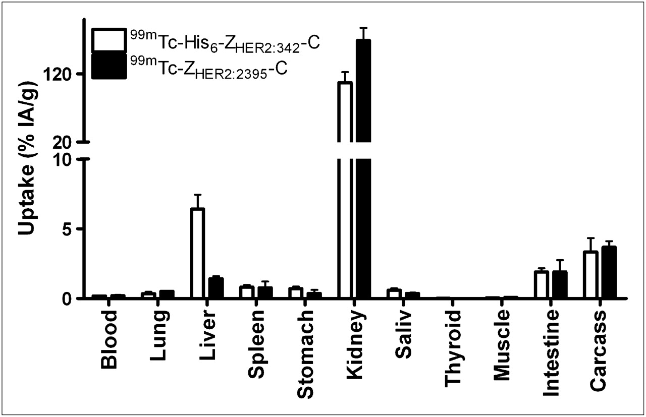
Biodistributions of radiolabeled Affibody molecules 99mTc-H6-ZHER2:342-C, containing His tag, and 99mTc-ZHER2:2395-C, lacking His tag, in NMRI (normal) mice 4 h after injection. Data are expressed as %IA per whole sample for gastrointestinal tract and contents, thyroid, and carcass and as %IA/g for other tissues and are presented as mean ± SD for 4 animals. Saliv = salivary gland.
The biodistribution data for 99mTc-Z2395-C in nude mice bearing LS174T xenografts at 0.5, 1, 2, 4, and 6 h after injection are shown in Tables 2 and 3. Besides the kidneys, the only site with high uptake was the tumor. Tumor uptake was high (7 ± 2 %IA/g) as early as 0.5 h after injection, and it did not change significantly during the experiment. The uptake in all other organs and tissues was low. Generally, tumor-to-organ ratios increased between 0.5 and 6 h because of a decrease in radioactivity in nontarget organs.
Uptake of 99mTc-ZHER2:2395-C in BALB/c nu/nu Mice Bearing Xenografts of LS174T or SKOV-3 Tumors
Tumor-to-Organ Ratios for 99mTc-ZHER2:2395-C in BALB/c nu/nu Mice Bearing Xenografts of LS174T or SKOV-3 Tumors
The biodistribution data for 99mTc-Z2395-C in nude mice bearing SKOV-3 xenografts at 4 h after injection are also shown in Tables 2 and 3. The uptake in these xenografts, with high HER2 levels of expression, was also high (15 ± 3 %IA/g), and the tumor-to-blood ratio was 121 ± 24.
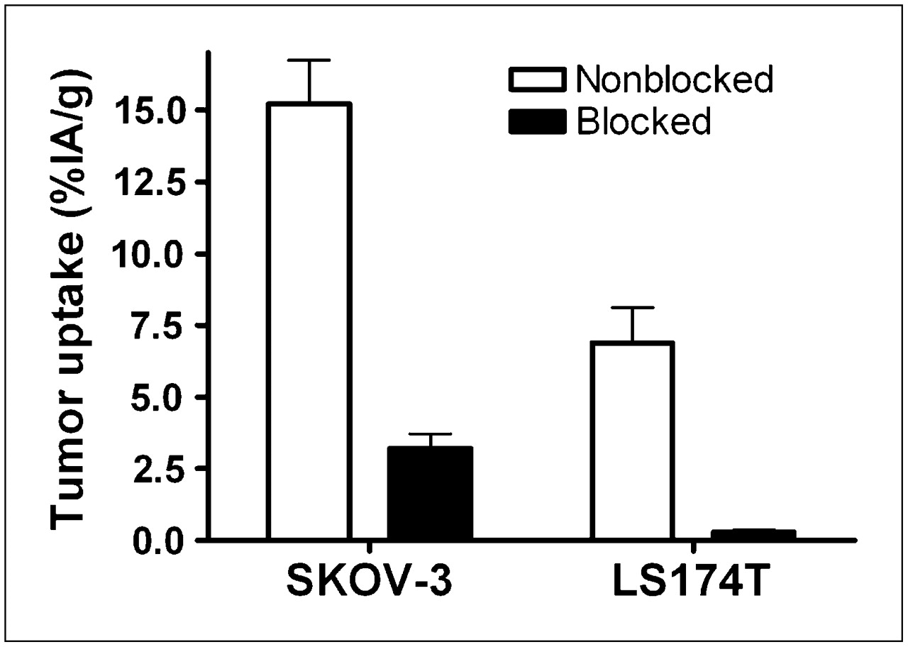
The results of the in vivo specificity analysis shown in Figure 3 demonstrated the specificity of uptake. Presaturation of receptors led to a significant decrease in radioactivity uptake in tumors for both LS174T (P < 0.005) and SKOV-3 (P < 0.0005) xenografts.

Specificity of 99mTc-ZHER2:2395-C tumor uptake in SKOV-3 and LS174T xenografts 4 h after injection. To saturate HER2 in tumors, one group of animals for each tumor model was preinjected with 600 μg of nonlabeled His6-ZHER2:342 45 min before injection of radiolabeled conjugate (designated as blocked). All animals were injected with 1 μg of labeled Affibody molecule. Data are expressed as %IA/g and are presented as mean ± SD for 4 animals.
γ-Camera Imaging
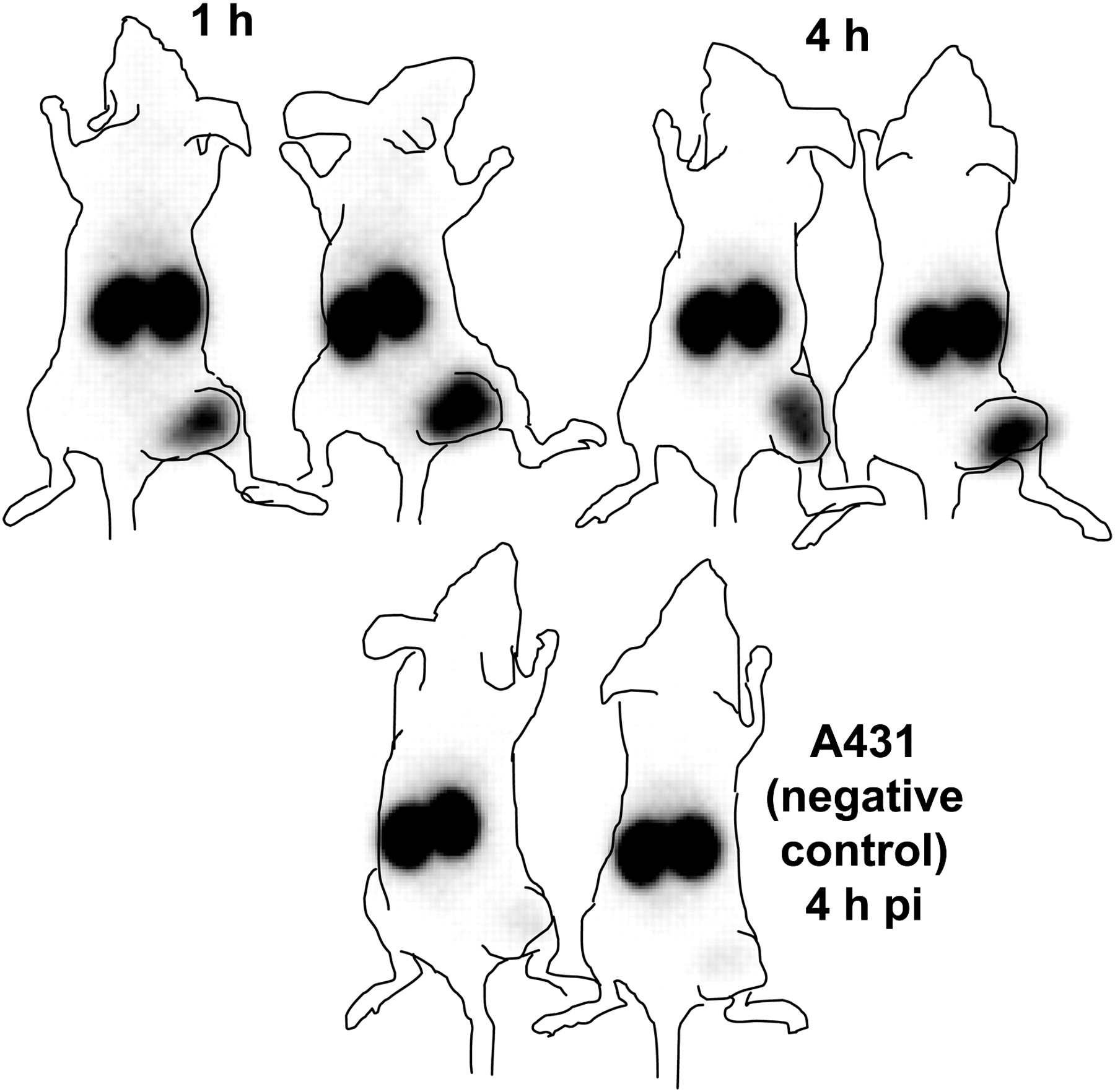
Images acquired 1 and 4 h after the intravenous injection of 99mTc-Z2395-C into nude mice bearing subcutaneous LS174T or SKOV-3 xenografts confirmed that tumor visualization was feasible as early as 1 h after injection (Figs. 4 and 5). The renal route of elimination of the conjugate led to substantial kidney retention on the images as well. Other organs and tissues were not visualized. The rapid clearance from the blood and nontarget organs allowed for increased contrast at the later time point (4 h after injection). The tumor-to-thigh (contralateral) ratios were 10.0 ± 0.4 (average ± maximum error) for LS174T xenografts and 34 ± 5 for SKOV-3 xenografts 4 h after injection. LS174T xenografts were not visualized in mice preinjected with unlabeled Affibody molecules to saturate HER2. Therefore, the specificity of HER2 imaging of 99mTc-Z2395-C was confirmed in this experiment. The absence of uptake in HER2-negative A431 xenografts also confirmed the specificity of HER2 targeting.
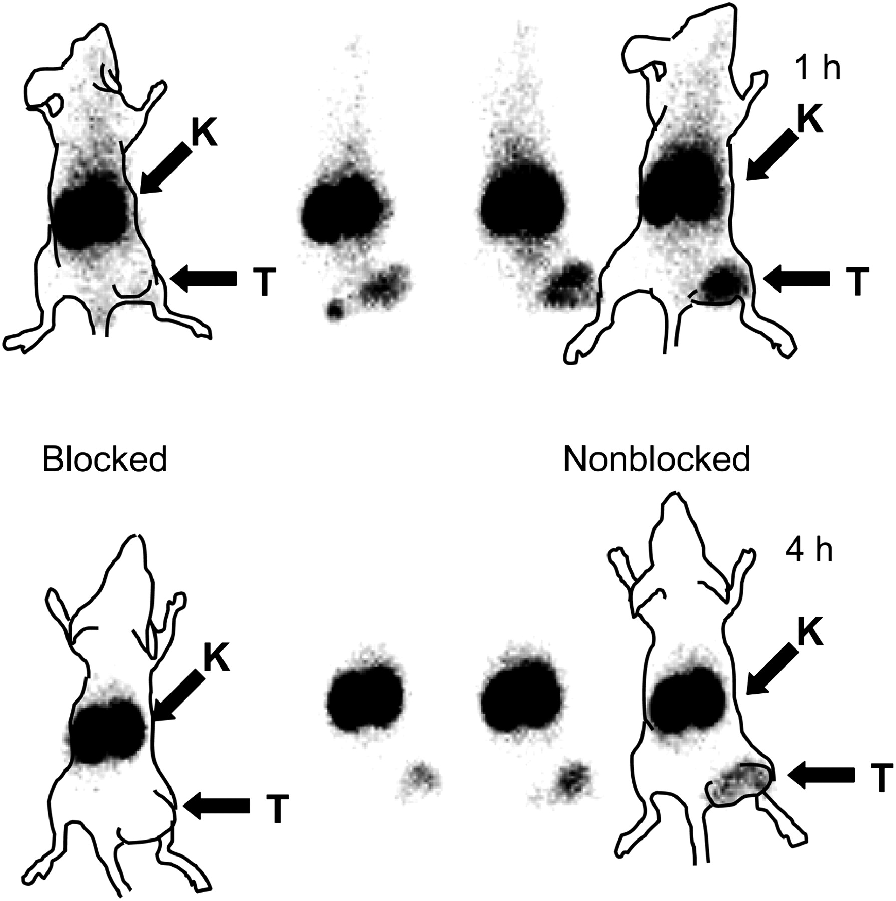
Imaging of HER2 expression in LS174T xenografts in BALB/c nu/nu mice with 99mTc-ZHER2:2395-C. In blocking experiment, HER2 was saturated by preinjection of 600 μg of nonlabeled Affibody molecule. Planar γ-camera images were acquired 1 and 4 h after administration of 99mTc-ZHER2:2395-C. Tumors (right hind leg) were clearly visualized without blocking but were not seen in blocking experiment, indicating specific HER2 binding to 99mTc-ZHER2:2395-C. Animal contours were derived from digital photographs and superimposed over γ-camera images to facilitate interpretation. K = kidney; T = tumor.

Imaging of HER2 expression in SKOV-3 and A431 xenografts in BALB/c nu/nu mice with 99mTc-ZHER2:2395-C. A431 tumors were used as negative controls because they are considered to be non–HER2-expressing tumors. Planar γ-camera images were acquired 1 and 4 h after administration of 99mTc-ZHER2:2395-C in mice with SKOV-3 xenografts and 4 h after administration in mice with A431 xenografts. Tumors (right hind leg) were clearly visualized in SKOV-3 xenografts but were not seen in negative controls, indicating specific HER2 binding to 99mTc-ZHER2:2395-C. Animal contours were derived from digital photographs and superimposed over γ-camera images to facilitate interpretation. pi = after injection.
DISCUSSION
Affibody molecules were proven earlier to be promising tracers for in vivo molecular imaging (11,30). The use of 99mTc as a label would facilitate their rapid implementation in clinical practice because of the favorable properties of this nuclide and its ready availability. The thiophilic nature of technetium(V) enables the site-specific labeling of thiol-containing proteins (25), providing a well-defined, homogeneous product. The use of cysteine-based peptide chelators would enable the recombinant production of Affibody molecule–based tracers in a single process with high reproducibility. We previously demonstrated the feasibility of using an N-terminal cysteine to label an anti-HER2 Affibody molecule (26). However, elevated uptake in the stomach and salivary glands (3–4 %IA/g 4 h after injection) indicated a certain degree of catabolic instability of the label in vivo. Literature data suggested that using a C-terminal cysteine to label scFv fragments provided catabolically more stable labels (14,25). The biodistribution of Affibody molecules and thereby their imaging contrast are influenced by the nature of the chelator–metal complex (15,19–21,26,29). Therefore, an in vivo evaluation was necessary for the development of this type of labeling for Affibody molecules.
The introduction of a C-terminal cysteine did not affect the affinity of ZHER2:342 for HER2 appreciably and enabled efficient labeling. The initial His tag–containing tracer, 99mTc-H6-ZHER2:342-C, showed reduced radioactivity uptake in the stomach of tumor-bearing SCID mice (Table 1) relative to literature data on N-terminal cysteine–containing 99mTc-CGG-ZHER2:342. This finding indicated superior stability of the cysteine-containing chelator in the C-terminal position rather than the N-terminal position. However, 99mTc-H6-ZHER2:342-C yielded high liver uptake. To verify that the SCID animal model was not the cause of the high liver uptake, a comparison between SCID and BALB/c nu/nu mice was performed. The biodistribution of synthetic 111In-DOTA-ZHER2:342 in SCID mice bearing SKOV-3 tumors was compared with the previously described biodistribution of the same conjugate in BALB/c nu/nu mice (17). The results obtained in the two animal models showed a high level of agreement (Supplemental Table 1). The liver uptake of 111In-DOTA-ZHER2:342 in SCID mice was somewhat elevated in comparison with the uptake in BALB/c nu/nu mice, but not to such an extent that it could be considered the reason for the high liver uptake of all Affibody molecules, including 99mTc-H6-ZHER2:342-C, in this animal model. An alternative hypothesis for the high liver uptake was the presence of a His tag in the conjugate. Elevated liver uptake of a His tag–containing conjugate was detected for H6-ZHER2:342-C labeled with 111In when a maleimido derivative of DOTA was used (29). Therefore, a variant of HER2-binding Affibody molecule ZHER2:342 lacking the His tag but with a C-terminal cysteine, Z2395-C, was evaluated. A comparison of the biodistributions of the His tag–containing conjugate, 99mTc-H6-ZHER2:342-C, and the corresponding variant lacking the His tag, 99mTc-Z2395-C, confirmed the negative effect of the His tag on liver uptake (Fig. 2).
99mTc-Z2395-C showed adequate stability both in vitro and in vivo. Characterization in vitro with HER2-expressing SKOV-3 cells showed that both binding specificity (Fig. 1A) and a high degree of binding capacity were retained after labeling. The cellular retention of 99mTc-Z2395-C in SKOV-3 cells was good throughout the experiment (24 h), mainly because of strong membrane binding rather than intracellular trapping (Fig. 1B). In vivo, the specificity was confirmed by a blocking experiment, in which presaturation of HER2 in xenografts of two cell lines reduced the tumor accumulation of 99mTc-Z2395-C (Fig. 3). In LS174T-bearing and SKOV-3–bearing mice a prominent uptake in tumors but very low uptake in normal tissues was shown, resulting in high tumor-to-background ratios. A comparison of the biodistributions of 99mTc-Z2395-C and 99mTc-CGG-ZHER2:342 (26) in SKOV-3–bearing mice 4 h after injection revealed that the radioactivity uptake of 99mTc-Z2395-C in the stomach and thyroid was strongly reduced—8-fold in the stomach and nearly 40-fold in the thyroid. These numbers indicate that the stability of the 99mTc-labeled Affibody molecule conjugate is improved when the cysteine is placed in a C-terminal position rather than in an N-terminal position. The only organ with high 99mTc-Z2395-C radioactivity uptake was the kidney. High accumulation in the kidneys is expected for small targeting proteins (<60 kDa) because of reabsorption in the proximal tubules (31). Anatomically, the kidneys are well separated from the main metastatic sites for breast cancer, which is the type of cancer for which HER2 imaging primarily will have a clinical application. Therefore, it should be possible to visualize tumors with SPECT/CT without interference from the high accumulation of radioactivity in the kidneys.
99mTc-Z2395-C showed rapid tumor targeting and rapid clearance from nontarget tissues, thereby enabling high-contrast imaging as early as 1 h after injection (Figs. 4 and 5). The tumor-to-blood ratios increased at the later time points, indicating that optimal contrast enhancement could be achieved once the tracer was allowed to clear from nontarget tissues. The characterization of 99mTc-Z2395-C revealed the excellent targeting properties of this tracer.
The requirement of high specific radioactivity is not as crucial for HER2-imaging agents as it is for many other tracers because of the very high level of HER2 overexpression in malignant tumors. For example, an immunohistochemical score of 3+ corresponds to ∼2.3 million of these receptors per cell (32). Moreover, the presence of shed HER2 can alter the blood kinetics and biodistribution of HER2-targeting agents if the specific radioactivity is too high. The preliminary results of a clinical study on the imaging of HER2 expression in breast cancer with 89Zr-trastuzumab (33) showed that for satisfactory imaging, the mass of radiolabeled trastuzumab should be at least 50 mg (∼300 nmol) for trastuzumab-naive patients. Preclinical data demonstrated that the levels of uptake of ZHER2:342 in SKOV-3 xenografts do not differ significantly for injected masses ranging from 0.1 to 10 μg (14 pmol−1.4 nmol) per mouse (34). Therefore, the results of biodistribution experiments with slightly different masses of injected Affibody molecules in mice bearing SKOV-3 xenografts may be directly compared.
The use of another 99mTc-labeled anti-HER2 tracer, a 99mTc-hydrazinonicotinamide-trastuzumab Fab fragment, was previously reported (35). This tracer yielded a tumor-to-blood ratio of 3.2 ± 0.68 at 24 h after injection in a BT-474 xenograft model with a high level of HER2 expression. In comparison, the smaller 99mTc-Z2395-C provided contrast nearly 40 times higher as early as 4 h after injection when evaluated in SKOV-3 xenografts, which have a HER2 expression level similar to that in BT-474 xenografts (36). The improved contrast is due to higher tumor uptake and to more rapid blood clearance of Affibody molecules than Fab fragments. In comparison with 99mTc-CGG-ZHER2:342, 99mTc-Z2395-C showed not only improved catabolic stability of the label but also a lower level of hepatobiliary excretion—an advantage for the imaging of extrahepatic abdominal metastases. These data might indicate that hepatobiliary excretion is determined not only by the overall charge and lipophilicity of proteins but also by the regional distribution of amino acids. In comparison with synthetic mercaptoacetyl-containing anti-HER2 Affibody conjugates (19–21), Z2395-C showed better affinity, which may have been attributable to the better spatial separation of the chelator and the binding site of the Affibody molecule. This characteristic translated into higher tumor uptake of 99mTc-Z2395-C and a higher tumor-to-blood ratio. The combination of high affinity, high label stability, and favorable biodistribution properties renders 99mTc-Z2395-C the best preclinically tested 99mTc-labeled HER2-imaging tracer to date. The introduction of a C-terminal cysteine to create a technetium chelator also provides more flexibility in the manufacturing of Affibody molecules than can be achieved with mercaptoacetyl derivatives because a species with only native amino acids can be produced in a recombinant fashion.
CONCLUSION
The introduction of a C-terminal cysteine provided a unique site for the labeling of recombinant Affibody molecules with 99mTc. The tracer 99mTc-Z2395-C, not containing a His tag, showed efficient targeting of HER2-expressing xenografts and rapid clearance from all normal tissues, except the kidneys. Clear and specific visualization of HER2 expression in a murine xenograft model with 99mTc-Z2395-C is possible within a few hours after injection, and the tumor-to-blood ratio (121 ± 24 at 4 h after injection in SKOV-3 xenografts) is better for 99mTc-Z2395-C than for any other 99mTc-labeled HER2-targeting agent tested. In conclusion, 99mTc-Z2395-C is a promising SPECT tracer for the detection of HER2 expression in malignant tumors.
Acknowledgments
This research was financially supported in part by a grant from the Swedish Cancer Society (Cancerfonden) and a research scholarship from the Schuberts Fund.
Footnotes
-
COPYRIGHT © 2009 by the Society of Nuclear Medicine, Inc.
References
- Received for publication August 12, 2008.
- Accepted for publication February 5, 2009.
In this issue


Jump to section
Related Articles
Cited By...
- Preclinical and clinical applications of specific molecular imaging for HER2-positive breast cancer
- 188Re-ZHER2:V2, a Promising Affibody-Based Targeting Agent Against HER2-Expressing Tumors: Preclinical Assessment
- HER2-Positive Tumors Imaged Within 1 Hour Using a Site-Specifically 11C-Labeled Sel-Tagged Affibody Molecule
- Molecular Design and Optimization of 99mTc-Labeled Recombinant Affibody Molecules Improves Their Biodistribution and Imaging Properties
- Targeting of HER2-Expressing Tumors Using 111In-ABY-025, a Second-Generation Affibody Molecule with a Fundamentally Reengineered Scaffold
- Targeting Prostate Cancer Cells In Vivo Using a Rapidly Internalizing Novel Human Single-Chain Antibody Fragment