
Main menu

  • Home
  • Content
    • Current
    • Ahead of print
    • Past Issues
    • JNM Supplement
    • SNMMI Annual Meeting Abstracts
    • Continuing Education
    • JNM Podcasts
  • Subscriptions
    • Subscribers
    • Institutional and Non-member
    • Rates
    • Journal Claims
    • Corporate & Special Sales
  • Authors
    • Submit to JNM
    • Information for Authors
    • Assignment of Copyright
    • AQARA requirements
  • Info
    • Reviewers
    • Permissions
    • Advertisers
  • About
    • About Us
    • Editorial Board
    • Contact Information
  • More
    • Alerts
    • Feedback
    • Help
    • SNMMI Journals
  • SNMMI
    • JNM
    • JNMT
    • SNMMI Journals
    • SNMMI

User menu

  • Subscribe
  • My alerts
  • Log in
  • My Cart

Search

  • Advanced search
Journal of Nuclear Medicine
  • SNMMI
    • JNM
    • JNMT
    • SNMMI Journals
    • SNMMI
  • Subscribe
  • My alerts
  • Log in
  • My Cart
Journal of Nuclear Medicine

Advanced Search

  • Home
  • Content
    • Current
    • Ahead of print
    • Past Issues
    • JNM Supplement
    • SNMMI Annual Meeting Abstracts
    • Continuing Education
    • JNM Podcasts
  • Subscriptions
    • Subscribers
    • Institutional and Non-member
    • Rates
    • Journal Claims
    • Corporate & Special Sales
  • Authors
    • Submit to JNM
    • Information for Authors
    • Assignment of Copyright
    • AQARA requirements
  • Info
    • Reviewers
    • Permissions
    • Advertisers
  • About
    • About Us
    • Editorial Board
    • Contact Information
  • More
    • Alerts
    • Feedback
    • Help
    • SNMMI Journals
  • View or Listen to JNM Podcast
  • Visit JNM on Facebook
  • Join JNM on LinkedIn
  • Follow JNM on Twitter
  • Subscribe to our RSS feeds
OtherBasic Science Investigations

64Cu-Labeled Folate-Conjugated Shell Cross-Linked Nanoparticles for Tumor Imaging and Radiotherapy: Synthesis, Radiolabeling, and Biologic Evaluation

Raffaella Rossin, Dipanjan Pan, Kai Qi, Jeffrey L. Turner, Xiankai Sun, Karen L. Wooley and Michael J. Welch
Journal of Nuclear Medicine July 2005, 46 (7) 1210-1218;
Raffaella Rossin
  • Find this author on Google Scholar
  • Find this author on PubMed
  • Search for this author on this site
Dipanjan Pan
  • Find this author on Google Scholar
  • Find this author on PubMed
  • Search for this author on this site
Kai Qi
  • Find this author on Google Scholar
  • Find this author on PubMed
  • Search for this author on this site
Jeffrey L. Turner
  • Find this author on Google Scholar
  • Find this author on PubMed
  • Search for this author on this site
Xiankai Sun
  • Find this author on Google Scholar
  • Find this author on PubMed
  • Search for this author on this site
Karen L. Wooley
  • Find this author on Google Scholar
  • Find this author on PubMed
  • Search for this author on this site
Michael J. Welch
  • Find this author on Google Scholar
  • Find this author on PubMed
  • Search for this author on this site
  • Article
  • Figures & Data
  • Info & Metrics
  • PDF
Loading

Article Figures & Data

Figures

  • Tables
  • FIGURE 1.
    • Download figure
    • Open in new tab
    • Download powerpoint
    FIGURE 1.

    Preparation of SCKs functionalized with folate for targeting and labeled with FTSC or TETA for fluorescence or radionuclide detection, respectively. Reagents and conditions: (i) polymer micelle formation: tetrahydrofuran, followed by controlled addition of water and dialysis against water; (ii) shell crosslinking: block copolymer micelles, 2,2′-(ethylenedioxy)-bis(ethylamine), 1-(3′-dimethylaminopropyl)-3-ethylcarbodiimide (EDC) methiodide, room temperature (RT), followed by dialysis against water; (iii) shell functionalization with folate-PEG1,600-amine (PEG1,600 is a poly(ethylene glycol) spacer with a molecular weight of 1,600 Da): nonfunctionalized SCKs, folate-PEG1,600-amine, EDC methiodide, RT, followed by dialysis against sodium phosphate-buffered saline at pH 7.3; (iv) shell functionalization with fluorescein thiosemicarbazide (FTSC): SCK-folate, FTSC, EDC methiodide, RT, followed by dialysis against sodium phosphate-buffered saline at pH 7.3; (v) shell functionalization with TETA-amine: SCK-folate, sulfo-NHS (s-NHS), EDC, 4°C, 2 h, followed by Centricon separation, TETA-amine, 4°C, overnight.
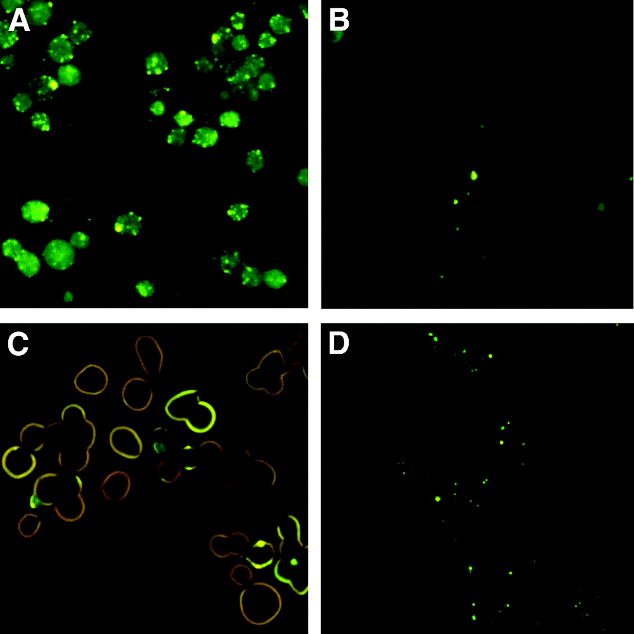
  • FIGURE 2.
    • Download figure
    • Open in new tab
    • Download powerpoint
    FIGURE 2.

    Fluorescence micrographs of KB cells incubated for 4 h at 37°C (A and B) and 4°C (C and D) with FTSC and folate-conjugated SCKs (10 μg FTSC-SCK-folate per 33-mm culture dish, plated with 3 × 10⁵ cells per dish 24 h before assay). Incubation at 4°C led to surface uptake of FTSC-SCK-folate onto KB cells (C), whereas fluorescent endocytotic vesicles are visible inside the cells after 4 h of incubation at 37°C (A). At both incubation temperatures, excess folic acid (1 mmol/L) competitively inhibited FTSC-SCK-folate cell uptake (B and D).
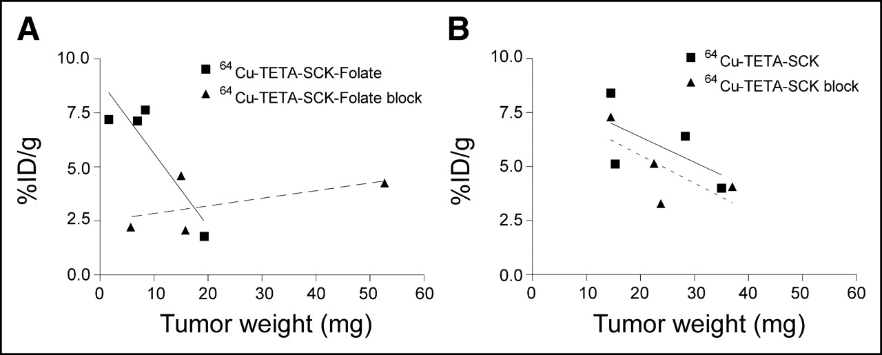
  • FIGURE 3.
    • Download figure
    • Open in new tab
    • Download powerpoint
    FIGURE 3.

    Dependence of tumor uptake on tumor size 4 h after injection of 64Cu-labeled folated (A) and nonfolated (B) SCKs in KB xenograft-bearing mice. Data are presented as %ID/g. Within each experimental group, small tumors exhibited enhanced nanoparticle uptake. Coadministration of excess folic acid led to competitive block of 64Cu-TETA-SCK-folate tumor uptake (A, dashed line), whereas it had no noticeable effect on 64Cu-TETA-SCK tumor uptake (B, dashed line).
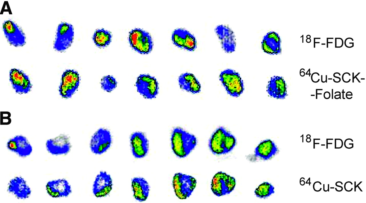
  • FIGURE 4.
    • Download figure
    • Open in new tab
    • Download powerpoint
    FIGURE 4.

    Dual-tracer autoradiographs demonstrate regional concentration of 64Cu-labeled folated (A) or nonfolated (B) SCKs in mice bearing 0.3- to 0.6-g KB xenografts. Tumors were resected and sectioned into 1-mm slices 1 h after 18F-FDG administration and 24 h after administration of 64Cu-radiolabeled SCKs. Images of 18F-FDG distribution were collected shortly after tumor resection, whereas distribution of 64Cu-labeled SCKs in the same slices was imaged with a 20-h delay. Both tumors exhibit scattered necrotic regions (no radiotracer uptake).

  • FIGURE 5.
    • Download figure
    • Open in new tab
    • Download powerpoint
    FIGURE 5.

    Cross-sectional slice from a tumor generated by subcutaneous inoculation of a nu/nu athymic mouse with 1 × 10⁶ human KB cells. Tumor mass is enclosed in a capsule (C) and it is characterized by scattered necrotic regions (N) surrounded by viable tissues (V). (hematoxylin–eosin, ×40)

Tables

  • Figures
    • View popup
    TABLE 1

    Biodistribution Data of 64Cu-TETA-SCK-Folate in KB Tumor Cell Xenograft-Bearing Athymic Nude Female Mice (n = 4)

    Biodistribution%ID/g%ID/organ
    10 min1 h4 h24 h10 min1 h4 h24 h
    Blood2.1 ± 0.22.1 ± 0.22.5 ± 0.32.6 ± 0.63.1 ± 0.23.4 ± 0.24.2 ± 0.34.3 ± 0.9
    Lung39.7 ± 5.110.9 ± 3.814.4 ± 5.910.8 ± 1.45.1 ± 0.91.5 ± 0.22.1 ± 0.91.5 ± 0.2
    Liver56.0 ± 7.138.4 ± 7.933.8 ± 2.621.5 ± 2.952.3 ± 5.641.7 ± 3.034.1 ± 2.525.2 ± 2.9
    Spleen18.3 ± 6.310.8 ± 3.28.8 ± 0.54.7 ± 1.01.5 ± 0.41.0 ± 0.30.8 ± 0.20.5 ± 0.2
    Kidney7.2 ± 1.68.1 ± 1.87.9 ± 0.88.5 ± 1.31.2 ± 0.31.3 ± 0.21.4 ± 0.21.5 ± 0.4
    Muscle0.8 ± 0.10.8 ± 0.20.9 ± 0.21.0 ± 0.17.2 ± 0.57.5 ± 1.38.6 ± 1.79.5 ± 1.3
    Heart2.3 ± 0.22.7 ± 0.53.7 ± 0.34.3 ± 0.50.2 ± 0.00.3 ± 0.10.4 ± 0.00.5 ± 0.0
    Tumor3.4 ± 1.22.3 ± 0.75.9 ± 2.82.9 ± 3.10.04 ± 0.020.12 ± 0.020.04 ± 0.020.09 ± 0.07
    • Data are presented as percentage injected dose per gram (%ID/g) and percentage injected dose per organ (%ID/organ) ± SD.

    • View popup
    TABLE 2

    Biodistribution Data of 64Cu-TETA-SCK in KB Tumor Cell Xenograft-Bearing Athymic Nude Female Mice (n = 4)

    Biodistribution%ID/g%ID/organ
    10 min1 h4 h24 h10 min1 h4 h24 h
    Blood3.3 ± 0.32.4 ± 0.72.6 ± 0.83.1 ± 0.35.1 ± 0.43.7 ± 0.83.8 ± 1.34.8 ± 0.2
    Lung32.5 ± 14.015.0 ± 4.113.8 ± 6.612.7 ± 1.84.6 ± 1.81.9 ± 0.31.8 ± 0.91.8 ± 0.1
    Liver45.7 ± 3.535.3 ± 8.831.8 ± 11.926.9 ± 3.945.6 ± 2.534.3 ± 7.327.3 ± 10.226.1 ± 1.0
    Spleen11.5 ± 2.58.2 ± 3.44.7 ± 2.04.5 ± 0.61.0 ± 0.20.8 ± 0.30.4 ± 0.20.4 ± 0.1
    Kidney9.0 ± 0.68.6 ± 2.09.5 ± 0.810.8 ± 1.11.5 ± 0.01.4 ± 0.41.4 ± 0.31.7 ± 0.1
    Muscle1.1 ± 0.10.9 ± 0.20.9 ± 0.21.4 ± 0.210.1 ± 0.28.4 ± 2.07.6 ± 1.913.1 ± 2.6
    Heart3.7 ± 0.14.0 ± 1.34.7 ± 1.56.2 ± 0.40.4 ± 0.00.5 ± 0.20.4 ± 0.20.6 ± 0.0
    Tumor2.2 ± 0.33.2 ± 0.76.0 ± 1.95.6 ± 0.90.04 ± 0.040.17 ± 0.070.13 ± 0.040.18 ± 0.11
    • Data are presented as percentage injected dose per gram (%ID/g) and percentage injected dose per organ (%ID/organ) ± SD.

PreviousNext
Back to top

In this issue

Journal of Nuclear Medicine: 46 (7)
Journal of Nuclear Medicine
Vol. 46, Issue 7
July 1, 2005
  • Table of Contents
  • About the Cover
  • Index by author
Print
Download PDF
Article Alerts
Sign In to Email Alerts with your Email Address
Email Article

Thank you for your interest in spreading the word on Journal of Nuclear Medicine.

NOTE: We only request your email address so that the person you are recommending the page to knows that you wanted them to see it, and that it is not junk mail. We do not capture any email address.

Enter multiple addresses on separate lines or separate them with commas.
64Cu-Labeled Folate-Conjugated Shell Cross-Linked Nanoparticles for Tumor Imaging and Radiotherapy: Synthesis, Radiolabeling, and Biologic Evaluation
(Your Name) has sent you a message from Journal of Nuclear Medicine
(Your Name) thought you would like to see the Journal of Nuclear Medicine web site.
Citation Tools
64Cu-Labeled Folate-Conjugated Shell Cross-Linked Nanoparticles for Tumor Imaging and Radiotherapy: Synthesis, Radiolabeling, and Biologic Evaluation
Raffaella Rossin, Dipanjan Pan, Kai Qi, Jeffrey L. Turner, Xiankai Sun, Karen L. Wooley, Michael J. Welch
Journal of Nuclear Medicine Jul 2005, 46 (7) 1210-1218;

Citation Manager Formats

  • BibTeX
  • Bookends
  • EasyBib
  • EndNote (tagged)
  • EndNote 8 (xml)
  • Medlars
  • Mendeley
  • Papers
  • RefWorks Tagged
  • Ref Manager
  • RIS
  • Zotero
Share
64Cu-Labeled Folate-Conjugated Shell Cross-Linked Nanoparticles for Tumor Imaging and Radiotherapy: Synthesis, Radiolabeling, and Biologic Evaluation
Raffaella Rossin, Dipanjan Pan, Kai Qi, Jeffrey L. Turner, Xiankai Sun, Karen L. Wooley, Michael J. Welch
Journal of Nuclear Medicine Jul 2005, 46 (7) 1210-1218;
Twitter logo Facebook logo LinkedIn logo Mendeley logo
  • Tweet Widget
  • Facebook Like
  • Google Plus One
Bookmark this article

Jump to section

  • Article
    • Abstract
    • MATERIALS AND METHODS
    • RESULTS
    • DISCUSSION
    • CONCLUSION
    • Acknowledgments
    • Footnotes
    • REFERENCES
  • Figures & Data
  • Info & Metrics
  • PDF

Related Articles

  • THIS MONTH IN JNM
  • PubMed
  • Google Scholar

Cited By...

  • Bombesin Antagonist-Based Radioligands for Translational Nuclear Imaging of Gastrin-Releasing Peptide Receptor-Positive Tumors
  • The Advantages of Nanoparticles for PET
  • In Vivo Imaging of 64Cu-Labeled Polymer Nanoparticles Targeted to the Lung Endothelium
  • Google Scholar

More in this TOC Section

  • 11C-Methionine PET of Myocardial Inflammation in a Rat Model of Experimental Autoimmune Myocarditis
  • Counting Rate Characteristics and Image Distortion in Preclinical PET Imaging During Radiopharmaceutical Therapy
  • Design and Fabrication of Kidney Phantoms for Internal Radiation Dosimetry Using 3D Printing Technology
Show more Basic Science Investigations

Similar Articles

SNMMI

© 2025 SNMMI

Powered by HighWire