
Main menu

  • Home
  • Content
    • Current
    • Ahead of print
    • Past Issues
    • JNM Supplement
    • SNMMI Annual Meeting Abstracts
    • Continuing Education
    • JNM Podcasts
  • Subscriptions
    • Subscribers
    • Institutional and Non-member
    • Rates
    • Journal Claims
    • Corporate & Special Sales
  • Authors
    • Submit to JNM
    • Information for Authors
    • Assignment of Copyright
    • AQARA requirements
  • Info
    • Reviewers
    • Permissions
    • Advertisers
  • About
    • About Us
    • Editorial Board
    • Contact Information
  • More
    • Alerts
    • Feedback
    • Help
    • SNMMI Journals
  • SNMMI
    • JNM
    • JNMT
    • SNMMI Journals
    • SNMMI

User menu

  • Subscribe
  • My alerts
  • Log in
  • My Cart

Search

  • Advanced search
Journal of Nuclear Medicine
  • SNMMI
    • JNM
    • JNMT
    • SNMMI Journals
    • SNMMI
  • Subscribe
  • My alerts
  • Log in
  • My Cart
Journal of Nuclear Medicine

Advanced Search

  • Home
  • Content
    • Current
    • Ahead of print
    • Past Issues
    • JNM Supplement
    • SNMMI Annual Meeting Abstracts
    • Continuing Education
    • JNM Podcasts
  • Subscriptions
    • Subscribers
    • Institutional and Non-member
    • Rates
    • Journal Claims
    • Corporate & Special Sales
  • Authors
    • Submit to JNM
    • Information for Authors
    • Assignment of Copyright
    • AQARA requirements
  • Info
    • Reviewers
    • Permissions
    • Advertisers
  • About
    • About Us
    • Editorial Board
    • Contact Information
  • More
    • Alerts
    • Feedback
    • Help
    • SNMMI Journals
  • View or Listen to JNM Podcast
  • Visit JNM on Facebook
  • Join JNM on LinkedIn
  • Follow JNM on Twitter
  • Subscribe to our RSS feeds
OtherClinical Investigations

Detection of Klatskin’s Tumor in Extrahepatic Bile Duct Strictures Using Delayed 18F-FDG PET/CT: Preliminary Results for 22 Patient Studies

Michael J. Reinhardt, Holger Strunk, Thomas Gerhardt, Roland Roedel, Ursula Jaeger, Jan Bucerius, Tilman Sauerbruch, Hans-Juergen Biersack and Franz L. Dumoulin
Journal of Nuclear Medicine July 2005, 46 (7) 1158-1163;
Michael J. Reinhardt
  • Find this author on Google Scholar
  • Find this author on PubMed
  • Search for this author on this site
Holger Strunk
  • Find this author on Google Scholar
  • Find this author on PubMed
  • Search for this author on this site
Thomas Gerhardt
  • Find this author on Google Scholar
  • Find this author on PubMed
  • Search for this author on this site
Roland Roedel
  • Find this author on Google Scholar
  • Find this author on PubMed
  • Search for this author on this site
Ursula Jaeger
  • Find this author on Google Scholar
  • Find this author on PubMed
  • Search for this author on this site
Jan Bucerius
  • Find this author on Google Scholar
  • Find this author on PubMed
  • Search for this author on this site
Tilman Sauerbruch
  • Find this author on Google Scholar
  • Find this author on PubMed
  • Search for this author on this site
Hans-Juergen Biersack
  • Find this author on Google Scholar
  • Find this author on PubMed
  • Search for this author on this site
Franz L. Dumoulin
  • Find this author on Google Scholar
  • Find this author on PubMed
  • Search for this author on this site
  • Article
  • Figures & Data
  • Info & Metrics
  • PDF
Loading

Article Figures & Data

Figures

  • Tables
  • FIGURE 1.
    • Download figure
    • Open in new tab
    • Download powerpoint
    FIGURE 1.

    18F-FDG PET/CT of patient with no evidence of malignancy after PDT. (A) Native CT image shows discrete hypodense areas in liver segments 4B and 5. (B) Fused image (CT, 50%; PET, 50%) enables anatomic correlation of glucose metabolism. (C) 18F-FDG PET image shows 75% and 100% isocontour areas used for SUV analysis (SUVmax = 3.2 and SUV75% = 2.5). Note atrophy of left liver lobe.

  • FIGURE 2.
    • Download figure
    • Open in new tab
    • Download powerpoint
    FIGURE 2.

    18F-FDG PET/CT of patient with residual Klatskin’s tumor after PDT and Yamakawa stent implantation. (A) Native CT image shows large hypodense area in liver hilus. (B) Fused image (CT, 50%; PET, 50%) enables anatomic correlation of increased glucose metabolism. (C) 18F-FDG PET image shows 75% and 100% isocontour areas used for SUV analysis (SUVmax, 5.0, and SUV75%, 4.1).

  • FIGURE 3.
    • Download figure
    • Open in new tab
    • Download powerpoint
    FIGURE 3.

    Box plot analysis of perihilar SUVmax in 22 patient studies with and without cholangiocarcinoma. ○ = outliers; * = extreme cases; CC = cholangiocarcinoma.

  • FIGURE 4.
    • Download figure
    • Open in new tab
    • Download powerpoint
    FIGURE 4.

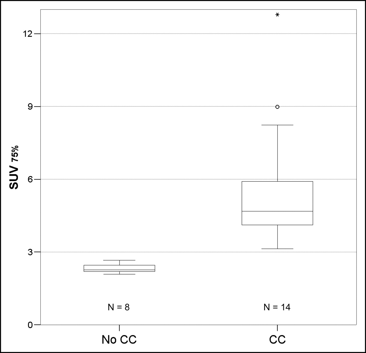
    Box plot analysis of perihilar SUV75% in 22 patient studies with and without cholangiocarcinoma. ○ = outliers; * = extreme cases; CC = cholangiocarcinoma.

Tables

  • Figures
    • View popup
    TABLE 1

    Characteristics, 18F-FDG PET/CT Indications, Perihilar SUV, Histology, and Outcome of 20 Patients with Extrahepatic Bile Duct Strictures

    Patient no.Age (y)SexIndication for 18F-FDG PET/CTPerihilar SUVmaxPerihilar SUV75%HistologyOutcome
    163FStaging before surgery3.93.1CCCurative resection
    246MStaging before surgery5.24.3CCCurative resection
    368MStaging before surgery6.15.1CCCurative resection
    466FStaging before surgery6.65.6CCCurative resection
    571MStaging before PDT5.24.2CCStable disease
    675MStaging before PDT6.35.2CCStable disease
    776MStaging before PDT9.88.2CCStable disease
    Tx control after PDT11.09.0CCDisease progress on CT 5 mo later
    877FTx control after PDT15.811.8CCDisease progress on CT 6 mo later
    976MTx control after PDT5.24.3CCStable disease for 6 mo
    Follow-up after 6 mo7.15.9CCDisease progress on CT 7 mo later
    1061FTx control after PDT3.93.2CCDisease progress on CT 9 mo later
    1164FTx control after PDT5.04.1CCDisease progress on CT 14 mo later
    1278FExclusion of CC*4.23.5CCCurative resection
    1364MExclusion of CC*3.22.7No CC12-mo follow-up negative
    1482FExclusion of CC*2.72.2No CC18-mo follow-up negative
    1559MExclusion of CC*3.02.2No CC20-mo follow-up negative
    1663MExclusion of CC*2.92.3No CC21-mo follow-up negative
    1760MPSC, exclusion of CC*3.12.4No CC18-mo follow-up negative
    1835FPSC, exclusion of CC*2.72.2No CC19-mo follow-up negative
    1937FPSC, exclusion of CC*2.52.1No CC20-mo follow-up negative
    2071FTx control after PDT of CC3.02.5No CC18-mo follow-up negative
    • ↵* To avoid surgery when 18F-FDG PET/CT gave no evidence of malignancy.

    • CC = cholangiocarcinoma; Tx = therapy.

PreviousNext
Back to top

In this issue

Journal of Nuclear Medicine: 46 (7)
Journal of Nuclear Medicine
Vol. 46, Issue 7
July 1, 2005
  • Table of Contents
  • About the Cover
  • Index by author
Print
Download PDF
Article Alerts
Sign In to Email Alerts with your Email Address
Email Article

Thank you for your interest in spreading the word on Journal of Nuclear Medicine.

NOTE: We only request your email address so that the person you are recommending the page to knows that you wanted them to see it, and that it is not junk mail. We do not capture any email address.

Enter multiple addresses on separate lines or separate them with commas.
Detection of Klatskin’s Tumor in Extrahepatic Bile Duct Strictures Using Delayed 18F-FDG PET/CT: Preliminary Results for 22 Patient Studies
(Your Name) has sent you a message from Journal of Nuclear Medicine
(Your Name) thought you would like to see the Journal of Nuclear Medicine web site.
Citation Tools
Detection of Klatskin’s Tumor in Extrahepatic Bile Duct Strictures Using Delayed 18F-FDG PET/CT: Preliminary Results for 22 Patient Studies
Michael J. Reinhardt, Holger Strunk, Thomas Gerhardt, Roland Roedel, Ursula Jaeger, Jan Bucerius, Tilman Sauerbruch, Hans-Juergen Biersack, Franz L. Dumoulin
Journal of Nuclear Medicine Jul 2005, 46 (7) 1158-1163;

Citation Manager Formats

  • BibTeX
  • Bookends
  • EasyBib
  • EndNote (tagged)
  • EndNote 8 (xml)
  • Medlars
  • Mendeley
  • Papers
  • RefWorks Tagged
  • Ref Manager
  • RIS
  • Zotero
Share
Detection of Klatskin’s Tumor in Extrahepatic Bile Duct Strictures Using Delayed 18F-FDG PET/CT: Preliminary Results for 22 Patient Studies
Michael J. Reinhardt, Holger Strunk, Thomas Gerhardt, Roland Roedel, Ursula Jaeger, Jan Bucerius, Tilman Sauerbruch, Hans-Juergen Biersack, Franz L. Dumoulin
Journal of Nuclear Medicine Jul 2005, 46 (7) 1158-1163;
Twitter logo Facebook logo LinkedIn logo Mendeley logo
  • Tweet Widget
  • Facebook Like
  • Google Plus One
Bookmark this article

Jump to section

  • Article
    • Abstract
    • MATERIALS AND METHODS
    • RESULTS
    • DISCUSSION
    • CONCLUSION
    • Acknowledgments
    • Footnotes
    • REFERENCES
  • Figures & Data
  • Info & Metrics
  • PDF

Related Articles

  • THIS MONTH IN JNM
  • PubMed
  • Google Scholar

Cited By...

  • Cholangiocarcinoma: Risk Factors, Environmental Influences and Oncogenesis
  • Cholestasis From Malignant Melanoma
  • Dual-Time-Point 18F-FDG PET for the Evaluation of Gallbladder Carcinoma
  • Google Scholar

More in this TOC Section

  • Feasibility of Ultra-Low-Activity 18F-FDG PET/CT Imaging Using a Long–Axial-Field-of-View PET/CT System
  • Cardiac Presynaptic Sympathetic Nervous Function Evaluated by Cardiac PET in Patients with Chronotropic Incompetence Without Heart Failure
  • Validation and Evaluation of a Vendor-Provided Head Motion Correction Algorithm on the uMI Panorama PET/CT System
Show more Clinical Investigations

Similar Articles

SNMMI

© 2025 SNMMI

Powered by HighWire