
Main menu

  • Home
  • Content
    • Current
    • Ahead of print
    • Past Issues
    • JNM Supplement
    • SNMMI Annual Meeting Abstracts
    • Continuing Education
    • JNM Podcasts
  • Subscriptions
    • Subscribers
    • Institutional and Non-member
    • Rates
    • Journal Claims
    • Corporate & Special Sales
  • Authors
    • Submit to JNM
    • Information for Authors
    • Assignment of Copyright
    • AQARA requirements
  • Info
    • Reviewers
    • Permissions
    • Advertisers
  • About
    • About Us
    • Editorial Board
    • Contact Information
  • More
    • Alerts
    • Feedback
    • Help
    • SNMMI Journals
  • SNMMI
    • JNM
    • JNMT
    • SNMMI Journals
    • SNMMI

User menu

  • Subscribe
  • My alerts
  • Log in
  • My Cart

Search

  • Advanced search
Journal of Nuclear Medicine
  • SNMMI
    • JNM
    • JNMT
    • SNMMI Journals
    • SNMMI
  • Subscribe
  • My alerts
  • Log in
  • My Cart
Journal of Nuclear Medicine

Advanced Search

  • Home
  • Content
    • Current
    • Ahead of print
    • Past Issues
    • JNM Supplement
    • SNMMI Annual Meeting Abstracts
    • Continuing Education
    • JNM Podcasts
  • Subscriptions
    • Subscribers
    • Institutional and Non-member
    • Rates
    • Journal Claims
    • Corporate & Special Sales
  • Authors
    • Submit to JNM
    • Information for Authors
    • Assignment of Copyright
    • AQARA requirements
  • Info
    • Reviewers
    • Permissions
    • Advertisers
  • About
    • About Us
    • Editorial Board
    • Contact Information
  • More
    • Alerts
    • Feedback
    • Help
    • SNMMI Journals
  • View or Listen to JNM Podcast
  • Visit JNM on Facebook
  • Join JNM on LinkedIn
  • Follow JNM on Twitter
  • Subscribe to our RSS feeds
OtherClinical Investigations

5-HT1A Receptors Are Reduced in Temporal Lobe Epilepsy After Partial-Volume Correction

Giampiero Giovacchini, Maria T. Toczek, Robert Bonwetsch, Anto Bagic, Lixin Lang, Charles Fraser, Pat Reeves-Tyer, Peter Herscovitch, William C. Eckelman, Richard E. Carson and William H. Theodore
Journal of Nuclear Medicine July 2005, 46 (7) 1128-1135;
Giampiero Giovacchini
  • Find this author on Google Scholar
  • Find this author on PubMed
  • Search for this author on this site
Maria T. Toczek
  • Find this author on Google Scholar
  • Find this author on PubMed
  • Search for this author on this site
Robert Bonwetsch
  • Find this author on Google Scholar
  • Find this author on PubMed
  • Search for this author on this site
Anto Bagic
  • Find this author on Google Scholar
  • Find this author on PubMed
  • Search for this author on this site
Lixin Lang
  • Find this author on Google Scholar
  • Find this author on PubMed
  • Search for this author on this site
Charles Fraser
  • Find this author on Google Scholar
  • Find this author on PubMed
  • Search for this author on this site
Pat Reeves-Tyer
  • Find this author on Google Scholar
  • Find this author on PubMed
  • Search for this author on this site
Peter Herscovitch
  • Find this author on Google Scholar
  • Find this author on PubMed
  • Search for this author on this site
William C. Eckelman
  • Find this author on Google Scholar
  • Find this author on PubMed
  • Search for this author on this site
Richard E. Carson
  • Find this author on Google Scholar
  • Find this author on PubMed
  • Search for this author on this site
William H. Theodore
  • Find this author on Google Scholar
  • Find this author on PubMed
  • Search for this author on this site
  • Article
  • Figures & Data
  • Info & Metrics
  • PDF
Loading

Article Figures & Data

Figures

  • Tables
  • FIGURE 1.
    • Download figure
    • Open in new tab
    • Download powerpoint
    FIGURE 1.

    Coronal T2-weighted (A, top) and T1-weighed (A, bottom) MR images of patient with TLE with a right mesial temporal focus and coregistered 18F-FCWAY distribution volume (V/f1, B) and CMRglu (C) images before (top) and after (bottom) PVC. Coronal images are at level of posterior hippocampus, anterior to pons. V/f1 and CMRglu images are scaled to a maximum of 100 mL/mL and 10 mg/min/100 g, respectively.

  • FIGURE 2.
    • Download figure
    • Open in new tab
    • Download powerpoint
    FIGURE 2.

    Relationship between percentage increase in 18F-FCWAY distribution volume (V/f1, mL/mL) and CMRglu (mg/min/100 g) in TLE patients. Each point represents mean ROI value. Regression equation was V = 21.5 + 0.90 CMRglu; r2 = 0.95; P < 0.001. Dashed line represents identity line.
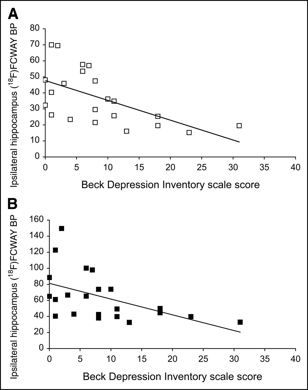
  • FIGURE 3.
    • Download figure
    • Open in new tab
    • Download powerpoint
    FIGURE 3.

    Relationship between 18F-FCWAY BP (mL/mL) in hippocampus ipsilateral to seizure focus and BDI scale score in TLE patients before (A) and after (B) PVC. Regression equations were y = 47.6 –1.24x; r2 = 0.35, P = 0.004 (A) and y = 81.2 –1.97x; r2 = 0.26, P = 0.016 (B).

Tables

  • Figures
    • View popup
    TABLE 1

    BP Values (mL/mL) in Control Subjects and Patients with TLE Before and After PVC

    RegionBefore PVCAfter PVC
    ControlsPatientsPControlsPatientsP
    I. Superior temporal45 ± 1532 ± 12∗88 ± 2576 ± 32
    C. Superior temporal46 ± 1336 ± 1484 ± 2373 ± 27
    I. Middle temporal51 ± 1737 ± 12∗95 ± 2879 ± 29
    C. Middle temporal52 ± 1341 ± 15∗91 ± 2377 ± 25
    I. Inferior temporal52 ± 1838 ± 12∗100 ± 2978 ± 28
    C. Inferior temporal54 ± 1646 ± 1697 ± 2590 ± 32
    I. Fusiform gyrus73 ± 2245 ± 15‡126 ± 3480 ± 24‡
    C. Fusiform gyrus71 ± 2355 ± 19∗120 ± 3296 ± 32
    I. Parahippocampus90 ± 2647 ± 16‡144 ± 3883 ± 27‡
    C. Parahippocampus86 ± 2359 ± 17‡144 ± 41102 ± 30∗
    I. Hippocampus88 ± 2137 ± 17‡127 ± 2864 ± 31‡
    C. Hippocampus85 ± 2458 ± 16‡124 ± 3497 ± 30∗
    I. Amygdala54 ± 1433 ± 15‡72 ± 1650 ± 20∗
    C. Amygdala53 ± 1442 ± 16∗75 ± 1660 ± 22
    I. Frontal35 ± 1131 ± 1168 ± 1766 ± 24
    C. Frontal34 ± 1030 ± 1165 ± 1663 ± 23
    I. Parietal32 ± 1026 ± 963 ± 1764 ± 22
    C. Parietal31 ± 1027 ± 1162 ± 1866 ± 24
    I. Insula56 ± 1638 ± 13‡95 ± 2763 ± 21‡
    C. Insula55 ± 1540 ± 14∗95 ± 2766 ± 21‡
    I. Occipital19 ± 617 ± 639 ± 1044 ± 14
    C. Occipital18 ± 417 ± 637 ± 846 ± 17
    Raphe54 ± 1338 ± 13∗N/AN/AN/A
    • ↵* Uncorrected P < 0.05, controls vs. patients, unpaired t test.

    • ↵‡ P < 0.05 after correction for multiple region comparisons, controls vs. patients, unpaired t test.

    • I. = ipsilateral to seizure focus; C. = contralateral to seizure focus; N/A = not available.

    • View popup
    TABLE 2

    AIs of 18F-FCWAY BP and CMRglu Before and After PVC

    Region18F-FCWAY BPCMRglu
    Before PVCAfter PVCBefore PVCAfter PVC
    ControlsPatientsPControlsPatientsPPatientsPPatientsP
    Superior temporal−3 ± 8−8 ± 124 ± 93 ± 30−7 ± 131 ± 28
    Middle temporal−5 ± 12−7 ± 113 ± 82 ± 28−11 ± 14−6 ± 26
    Inferior temporal−3 ± 13−14 ± 13∗2 ± 11−12 ± 19∗−12 ± 13−11 ± 22
    Fusiform gyrus3 ± 11−17 ± 21∗4 ± 13−15 ± 23∗−12 ± 14−11 ± 20
    Parahippocampus4 ± 5−21 ± 16‡1 ± 8−20 ± 16∗−8 ± 12‡−8 ± 14§
    Hippocampus4 ± 7−42 ± 26‡4 ± 8−40 ± 25‡−14 ± 14‡−13 ± 16‡
    Amygdala2 ± 15−20 ± 20∗−4 ± 12−17 ± 19∗−10 ± 14§−9 ± 14
    Frontal3 ± 41 ± 74 ± 34 ± 15−2 ± 30 ± 9
    Parietal1 ± 5−2 ± 132 ± 4−3 ± 14−2 ± 4−3 ± 5
    Insula1 ± 6−7 ± 11∗−1 ± 4−5 ± 14−6 ± 6−5 ± 8
    Occipital2 ± 9−2 ± 62 ± 12−2 ± 11−2 ± 3−2 ± 11
    • ↵* Uncorrected P < 0.05, controls vs. patients, unpaired t test.

    • ↵‡ P < 0.05 after correction for multiple comparisons, controls vs. patients, unpaired t test.

    • ↵‡ P < 0.05 after correction for multiple region comparisons, 18F-FCWAY vs. 18F-FDG in patient group, paired t test.

    • ↵§ Uncorrected P < 0.05, 18F-FCWAY vs. 18F-FDG in patient group, paired t test.

PreviousNext
Back to top

In this issue

Journal of Nuclear Medicine: 46 (7)
Journal of Nuclear Medicine
Vol. 46, Issue 7
July 1, 2005
  • Table of Contents
  • About the Cover
  • Index by author
Print
Download PDF
Article Alerts
Sign In to Email Alerts with your Email Address
Email Article

Thank you for your interest in spreading the word on Journal of Nuclear Medicine.

NOTE: We only request your email address so that the person you are recommending the page to knows that you wanted them to see it, and that it is not junk mail. We do not capture any email address.

Enter multiple addresses on separate lines or separate them with commas.
5-HT1A Receptors Are Reduced in Temporal Lobe Epilepsy After Partial-Volume Correction
(Your Name) has sent you a message from Journal of Nuclear Medicine
(Your Name) thought you would like to see the Journal of Nuclear Medicine web site.
Citation Tools
5-HT1A Receptors Are Reduced in Temporal Lobe Epilepsy After Partial-Volume Correction
Giampiero Giovacchini, Maria T. Toczek, Robert Bonwetsch, Anto Bagic, Lixin Lang, Charles Fraser, Pat Reeves-Tyer, Peter Herscovitch, William C. Eckelman, Richard E. Carson, William H. Theodore
Journal of Nuclear Medicine Jul 2005, 46 (7) 1128-1135;

Citation Manager Formats

  • BibTeX
  • Bookends
  • EasyBib
  • EndNote (tagged)
  • EndNote 8 (xml)
  • Medlars
  • Mendeley
  • Papers
  • RefWorks Tagged
  • Ref Manager
  • RIS
  • Zotero
Share
5-HT1A Receptors Are Reduced in Temporal Lobe Epilepsy After Partial-Volume Correction
Giampiero Giovacchini, Maria T. Toczek, Robert Bonwetsch, Anto Bagic, Lixin Lang, Charles Fraser, Pat Reeves-Tyer, Peter Herscovitch, William C. Eckelman, Richard E. Carson, William H. Theodore
Journal of Nuclear Medicine Jul 2005, 46 (7) 1128-1135;
Twitter logo Facebook logo LinkedIn logo Mendeley logo
  • Tweet Widget
  • Facebook Like
  • Google Plus One
Bookmark this article

Jump to section

  • Article
    • Abstract
    • MATERIALS AND METHODS
    • RESULTS
    • DISCUSSION
    • CONCLUSION
    • Acknowledgments
    • Footnotes
    • REFERENCES
  • Figures & Data
  • Info & Metrics
  • PDF

Related Articles

  • THIS MONTH IN JNM
  • PubMed
  • Google Scholar

Cited By...

  • Temporal lobe epilepsy and affective disorders: the role of the subgenual anterior cingulate cortex
  • The 5-HT1A receptor and 5-HT transporter in temporal lobe epilepsy
  • PET of Serotonin 1A Receptors and Cerebral Glucose Metabolism for Temporal Lobectomy
  • Using Cerebral White Matter for Estimation of Nondisplaceable Binding of 5-HT1A Receptors in Temporal Lobe Epilepsy
  • Decreased GABA-A binding on FMZ-PET in succinic semialdehyde dehydrogenase deficiency
  • Hippocampal volume and depression: insights from epilepsy surgery
  • MRI-Based Correction for Partial-Volume Effect Improves Detectability of Intractable Epileptogenic Foci on 123I-Iomazenil Brain SPECT Images
  • Striatal D2 Receptor Availability After Shunting in Idiopathic Normal Pressure Hydrocephalus
  • Disulfiram Inhibits Defluorination of 18F-FCWAY, Reduces Bone Radioactivity, and Enhances Visualization of Radioligand Binding to Serotonin 5-HT1A Receptors in Human Brain
  • Prognosis of children with partial epilepsy: MRI and serial 18FDG-PET
  • Hippocampal 1H-MRSI correlates with severity of depression symptoms in temporal lobe epilepsy
  • Synthesis and Biologic Evaluation of a Novel Serotonin 5-HT1A Receptor Radioligand, 18F-Labeled Mefway, in Rodents and Imaging by PET in a Nonhuman Primate
  • Google Scholar

More in this TOC Section

  • Cardiac Presynaptic Sympathetic Nervous Function Evaluated by Cardiac PET in Patients with Chronotropic Incompetence Without Heart Failure
  • Validation and Evaluation of a Vendor-Provided Head Motion Correction Algorithm on the uMI Panorama PET/CT System
  • Prognostic Role of 68Ga-PSMA11 PET–Based Response in Patients with Prostate Cancer Undergoing Taxane-Based Chemotherapy
Show more Clinical Investigations

Similar Articles

SNMMI

© 2025 SNMMI

Powered by HighWire