


Article Figures & Data
Figures
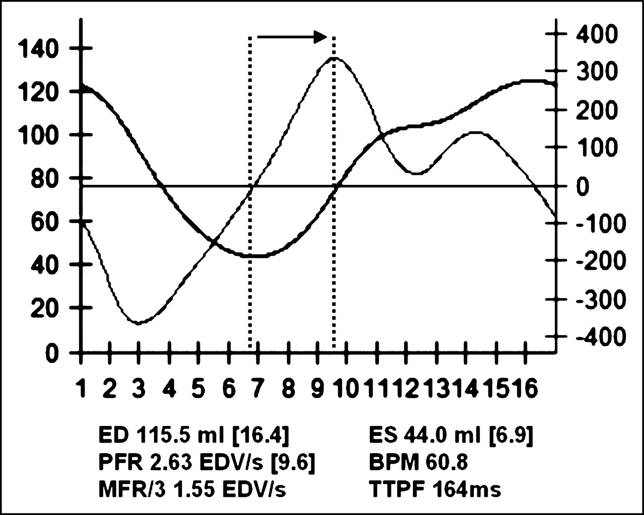
- FIGURE 1.
Example of a patient’s volume and filling curves over time in 16-frame gated MPS. Numbers in brackets represent exact frame numbers from which parameters are derived. Arrow shows TTPF, defined by time from ES to greatest filling rate in early diastole. Peak filling is normalized to EDV. ED = end diastole; ES = end systole; BPM = beats per minute HR; MFR/3 = mean filling rate over first third of diastole.
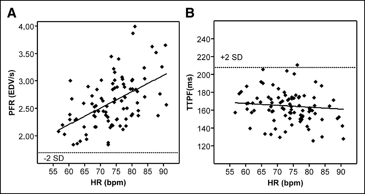
- FIGURE 2.
Scatter plots of relationship between PFR and HR (A) and TTPF and HR (B). Dotted line indicates 2-SD threshold. Solid line is regression line. In A, correlation coefficient (r) is 0.514, P = 0.01. In B, TTPF shows no correlation with HR.
- FIGURE 3.
Scatter plots of relationship between PFR and EF (%) (A) and TTPF and EF (%) (B). Dotted line indicates 2-SD threshold. Solid line is regression line. In A, correlation coefficient (r) is 0.529, P = 0.01. In B, TTPF shows no correlation with EF (%).
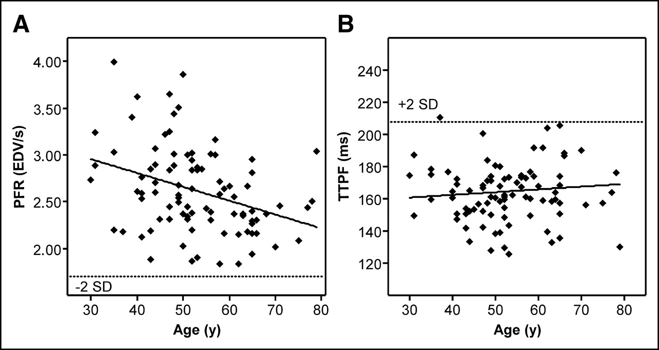
- FIGURE 4.
Scatter plots of relationship between PFR and age (A) and TTPF and age (B). Dotted line indicates 2-SD threshold. Solid line is regression line. In A, correlation coefficient (r) is −0.348, P = 0.01. In B, TTPF shows no correlation with age.


Tables
Characteristic Derivation group Validation group Overall P value Males* (%) 41/50 (82) 30/40 (75) 71/90 (79) NS Age (y) 53.3 ± 11.4 52.5 ± 10.3 52.9 ± 10.9 NS Resting HR 62.7 ± 8.2 61.3 ± 8.2 62.1 ± 8.2 NS Resting SBP 126.0 ± 16.1 130.4 ± 17.2 127.9 ± 16.6 NS Resting DBP 77.0 ± 8.3 76.8 ± 8.1 76.9 ± 8.1 NS MPHR (%) 95.6 ± 5.9 94.1 ± 6.1 94.9 ± 6.0 NS (+) Clinical response 0/50 2/40 2/90 NS (+) ECG response 2/50 2/40 4/90 NS ↵* Values in parentheses are percentages.
NS = not significant; SBP = systolic blood pressure (mm Hg); DBP = diastolic blood pressure (mm Hg).
Parameter Derivation group Validation group Overall P value HR (bpm) 73.6 ± 8.0 73.8 ± 8.0 73.7 ± 8.0 NS LVEF (%) 63.0 ± 5.4 64.6 ± 5.8 63.7 ± 5.6 NS EDV (mL) 104.2 ± 19.7 107.9 ± 21.7 105.9 ± 20.6 NS ESV (mL) 39.1 ± 11.5 38.9 ± 12.1 39.0 ± 11.7 NS PFR (EDV/s) 2.55 ± 0.42 2.70 ± 0.50 2.62 ± 0.46 NS TTPF (ms) 166.4 ± 25.1 162.3 ± 16.5 164.6 ± 21.7 NS HR = HR during poststress gated MPS acquisition; NS = not significant.
Parameter Time interval P value 0–30 min (n = 14) 30–45 min (n = 33) >45 min (n = 43) PFR (EDV/s) 2.59 ± 0.48 2.67 ± 0.48 2.58 ± 0.45 NS TTPF (ms) 169.8 ± 17.4 165.0 ± 27.9 162.6 ± 17.4 NS LVEF (%) 62.2 ± 5.2 64.9 ± 6.4 63.3 ± 4.9 NS NS = not significant.
Parameter Age group <50 y (n = 36) 50–59 y (n = 29) ≥60 y (n = 25) HR (bpm) 76.4 ± 7.6 72.0 ± 7.2 71.6 ± 8.5 LVEF (%) 62.5 ± 5.8 63.7 ± 6.4 65.6 ± 3.6 EDV (mL) 106.2 ± 22.0 110.3 ± 19.6 100.2 ± 18.9 ESV (mL) 40.4 ± 12.2 40.8 ± 12.6 34.8 ± 9.1 PFR* (EDV/s) 2.81 ± 0.49 2.58 ± 0.45 2.37 ± 0.29 TTPF (ms) 163.1 ± 17.1 161.6 ± 15.8 170.2 ± 31.4 ↵* P < 0.005; across 3 groups and age group <50 y vs. ≥60 y.
HR = HR during poststress gated MPS acquisition.
Parameter Men (n = 71) Women (n = 19) P value Age (y) 53.2 ± 10.7 51.8 ± 11.5 NS HR (bpm) 73.3 ± 7.8 75.0 ± 8.7 NS LVEF (%) 62.9 ± 5.1 66.8 ± 6.3 <0.01 EDV (mL) 109.7 ± 19.7 91.4 ± 17.2 <0.001 ESV (mL) 41.2 ± 11.1 30.5 ± 10.5 <0.001 PFR (EDV/s) 2.53 ± 0.40 2.95 ± 0.54 <0.001 TTPF (ms) 165.2 ± 23.1 162.3 ± 15.9 NS NS = not significant; HR = HR during poststress gated MPS acquisition.
Variable β-Coefficient 95% CI for β (lower bound − upper bound) P value Model for PFR R = 0.779; R2 = 0.607; adjusted R2 = 0.589; SEE = 0.296 Age −0.405 (−0.023) − (−0.011) <0.001 Male sex −0.188 (−0.372) − (−0.051) <0.05 LVEF 0.484 (0.027) − (0.053) <0.001 HR 0.274 (0.007) − (0.024) <0.001 Model for TTPF R = 0.120; R2 = 0.014; adjusted R2 = −0.032; SEE = 22.059 Age 0.059 (−0.344) − (0.581) 0.613 Male sex 0.057 (8.958) − (14.946) 0.620 LVEF 0.041 (−0.808) − (1.130) 0.742 HR −0.074 (−0.843) − (0.438) 0.531 95% CI = 95% confidence interval; R = ρ-coefficient.
In this issue


Jump to section
Related Articles
Cited By...
- Quantitative Clinical Nuclear Cardiology, Part 1: Established Applications
- Presence of Postsystolic Shortening Increases the Likelihood of Coronary Artery Disease: A Rest Electrocardiography-Gated Myocardial Perfusion SPECT Study
- Automated Segmentation of Routine Clinical Cardiac Magnetic Resonance Imaging for Assessment of Left Ventricular Diastolic Dysfunction
- Automatic Global and Regional Phase Analysis from Gated Myocardial Perfusion SPECT Imaging: Application to the Characterization of Ventricular Contraction in Patients with Left Bundle Branch Block
- Diastolic Filling Parameters Derived from Myocardial Perfusion Imaging Can Predict Left Ventricular End-Diastolic Pressure at Subsequent Cardiac Catheterization