


Article Figures & Data
Figures
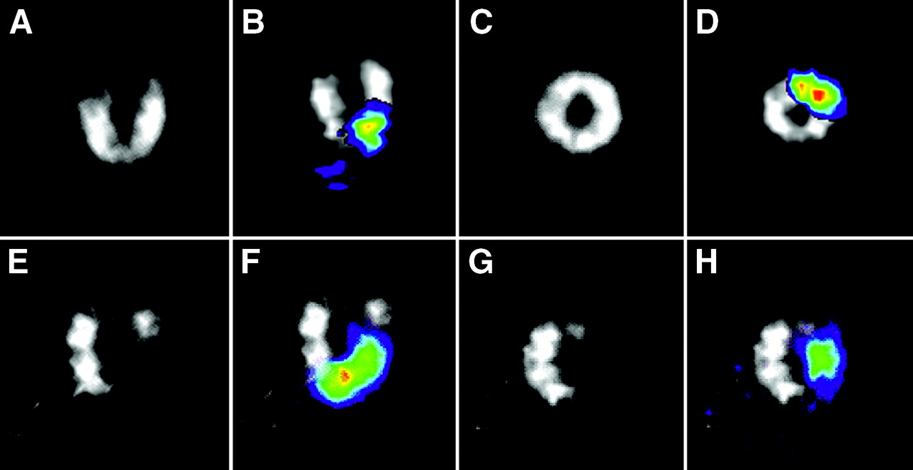
- FIGURE 1.
Cardiac long- and short-axis SPECT images of normal (A and C) and infarcted (E and G) heart using perfusion tracer 99mTc-sestamibi. 111In signal (color) was overlaid on gray-scale 99mTc-sestamibi images for normal (B and D) and infarcted (F and H) heart.
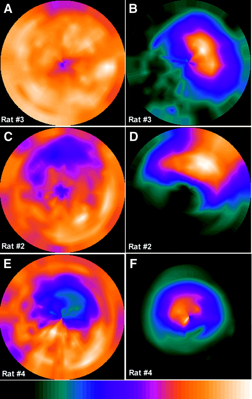
- FIGURE 2.
Bull’s-eye plots of 99mTc-sestamibi signal (A, C, and E) and 111In signal (B, D, and F) from normal rat (rat 3) and 2 rats with different infarction sizes (rats 2 and 4). Color bar represents transition from weak to strong signal intensity from left to right.
- FIGURE 3.
Time course of background 111In signal intensity in region of lungs and thorax in rats 3 (top) and 4 (bottom). Each image was normalized to give same maximum count to show relative washout of tracer from thoracic cavity. Signal detected in thoracic region in rat 3 in first image (acquired within 2 h after injection) was likely due to some leakage during injection of cells.
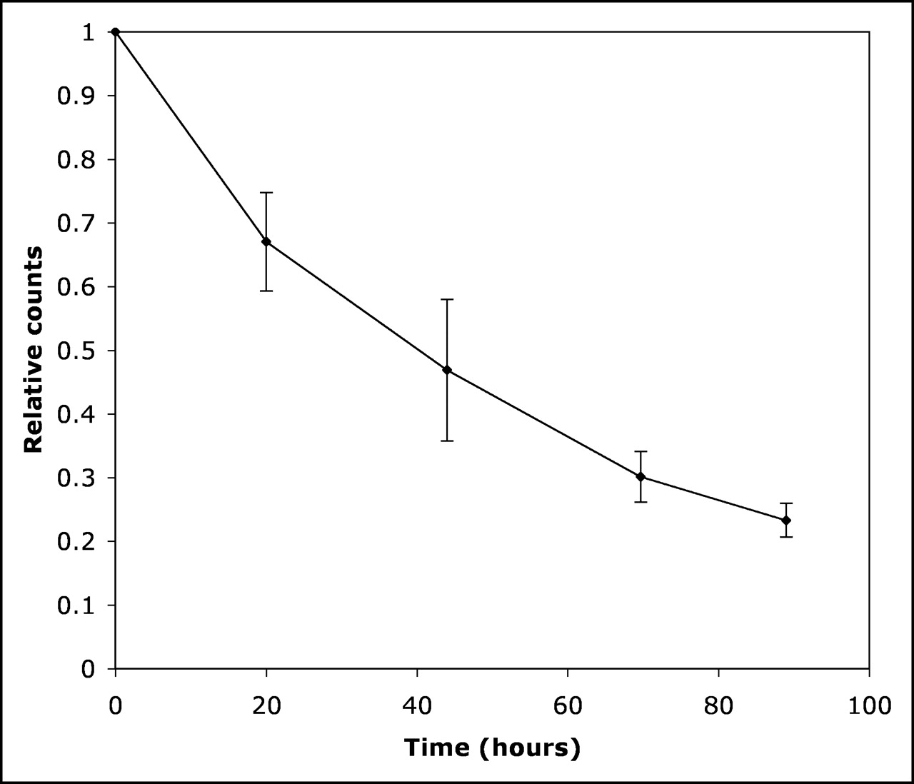
- FIGURE 4.
Time course of 111In signal intensity in hearts from 6 animals. Curve was corrected for radioactive decay of 111In. Error bars represent 1 SD.
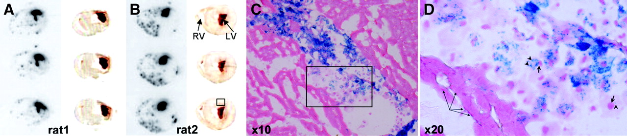
- FIGURE 5.
(A and B) Autoradiographs (left) of 3 adjacent slices and corresponding photographs (right). Region of intense 111In uptake (boxed area) was identified on each slice. (C and D) Histologic analysis of adjacent tissue slices reveals injected cells localized in region outlined in box. SPIO particles (arrowheads) are stained blue, whereas nuclei (short arrows) are counterstained red. Magnified view (D) of boxed area in C shows intensely SPIO-labeled cells (e.g., 1 in center) and slightly labeled cells (e.g., 1 in right corner, which contained only 1 blue dot; the cell is round, as delineated by pink-stained cytoplasm, and has a large nucleus). Unique morphology distinguishes injected cells from surrounding myocardium. Bundle of muscle fibers (long arrows) is also seen. LV = left ventricle; RV = right ventricle.

In this issue


Jump to section
Related Articles
Cited By...
- Perspectives on Assessment of Stem Cell Therapy in Stroke by 18F-FDG PET
- 99mTc-Based Imaging of Transplanted Neural Stem Cells and Progenitor Cells
- Current Perspectives on Imaging Cardiac Stem Cell Therapy
- Noninvasive Quantification and Optimization of Acute Cell Retention by In Vivo Positron Emission Tomography After Intramyocardial Cardiac-Derived Stem Cell Delivery
- In Vivo SPECT Quantification of Transplanted Cell Survival After Engraftment Using 111In-Tropolone in Infarcted Canine Myocardium
- Small-Animal SPECT and SPECT/CT: Important Tools for Preclinical Investigation
- Imaging of Gene Expression in Live Pancreatic Islet Cell Lines Using Dual-Isotope SPECT
- In vivo imaging of T cell delivery to tumors after adoptive transfer therapy
- Intramyocardial Implantation of Bone Marrow-Derived Stem Cells Enhances Perfusion in Chronic Myocardial Infarction: Dependency on Initial Perfusion Depth and Follow-up Assessed by Gated Pinhole SPECT
- Imaging Stem Cells Implanted in Infarcted Myocardium