


Article Figures & Data
Figures
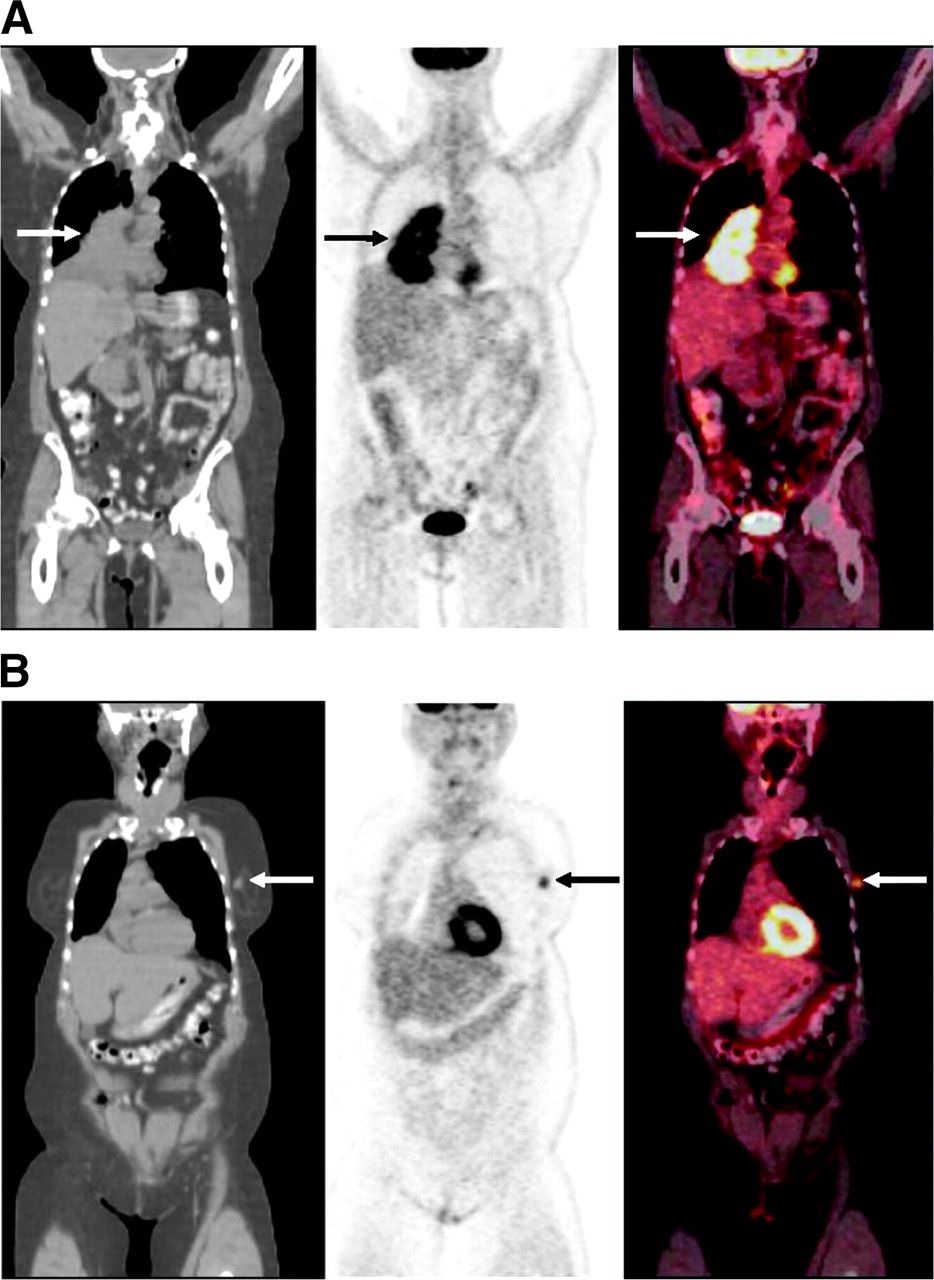
- FIGURE 1.
Coronal PET/CT images of 73-y-old woman with recently diagnosed cancer in right lung. (A) Images show large 18F-FDG-avid mass in right lung (arrow), consistent with patient’s known lung cancer. Biopsy revealed small cell lung cancer. (B) Anterior slices from same PET/CT study showed 18F-FDG-avid nodule in left breast (arrow), highly suggestive of malignancy. Pathology revealed infiltrating ductal carcinoma. Thus, this case was true-positive for an additional primary malignancy.
- FIGURE 2.
Coronal PET/CT images of 73-y-old woman with non-small cell right-upper-lobe lung cancer recently diagnosed by biopsy. (A) Lung mass shows intensely increased metabolic activity, compatible with the known lung cancer (arrow). (B) Additional slices from same PET/CT study showed increased activity in a right thyroid nodule (arrow). Fine-needle aspiration biopsy showed benign adenomatoid nodule. This case was thus falsely positive for an additional primary. Mildly increased activity seen in right axilla (arrowhead) was likely due to minimal tracer infiltration after right antecubital injection of tracer.

Tables
Parameter Patients Lesions (n) Histologic findings n % Total 1,912 100 Suspected second primary 79 4.1 81 Pathologically proven malignant 22* 1.2 24* Lung 7* 8* Adenocarcinoma (6), squamous cell carcinoma (1), poorly differentiated non-small cell carcinoma (1) Thyroid 6* 6* Papillary carcinoma Colon 4 4 Adenocarcinoma (3), lymphoma (1) Breast 2 2 Infiltrating ductal carcinoma Esophagus 2 2 Squamous cell carcinoma Bile duct† 1 1 Adenocarcinoma Head and neck (not thyroid) 1 1 Squamous cell carcinoma (oropharynx) Pathologically proven benign 10 0.5 10 Thyroid 5 5 Adenomatoid nodule Uterus 2 2 Normal epithelium Head and neck (not thyroid) 2 2 Reactive lymph nodes (1), lymphoid hyperplasia (1) Lung 1 1 Benign respiratory epithelium Benign by follow-up (8–23 mo) 8 0.4 8 Head and neck (not thyroid) 2 2 Breast 2 2 Colon 2 2 Thyroid 1 1 Other site‡ 1 1 Not yet confirmed 39 2.0 39 Thyroid 17 17 Head and neck (not thyroid) 5 5 Lung 3 3 Uterus 3 3 Breast 3 3 Colon 2 2 Kidney 2 2 Other sites§ 4 4 ↵* Two lesions in lung and 1 in thyroid were found in 1 patient.
↵† Pancreatic cancer was suspected on PET/CT and bile duct cancer was confirmed by operation.
↵‡ At junction of right adrenal, inferior vena cava, and liver; assumed to be focal brown fat uptake.
↵§ Stomach, mediastinum, adrenal gland, and prostate.
Patient no. Age (y) Sex Known or suspected primary Histology of known primary Suspected additional primary Histology of additional primary Comments 1 66 F Melanoma Malignant melanoma Thyroid Papillary thyroid carcinoma 2 54 F GIST (duodenum) Malignant GIST Thyroid Papillary thyroid carcinoma 3 67 F Melanoma Malignant melanoma Thyroid Papillary thyroid carcinoma 4 68 M Colon (1 site) Moderately differentiated adenocarcinoma Colon (adjacent separate site) Moderately differentiated adenocarcinoma 5 72 M Esophagus Squamous cell carcinoma Lung Adenocarcinoma 6 73 F Lung Small cell carcinoma Breast Infiltrating ductal carcinoma 7 78 F Breast NA (operated 1991) Colon Low-grade B-cell lymphoma 8 58 F Breast NA (operated 1998) Lung Squamous cell carcinoma 9 59 M Groin tumor, kidney Merkel cell carcinoma, renal cell cercinoma Lung (2 noncontiguous lesions), thyroid Lung: poorly differentiated adenocarcinoma, thyroid: papillary thyroid carcinoma Two different types of lung tumor by immunostaining 10 80 F Lymphoma Diffuse B-cell lymphoma Thyroid Papillary thyroid carcinoma 11 70 M Bile duct Moderately differentiated adenocarcinoma Lung Adenocarcinoma Spiculated lung nodule suggestive of additional primary 12 79 M Colon NA (operated 2001) Lung Adenocarcinoma Metastasis was ruled out by immunohistochemistry 13 76 M Pharynx Squamous cell carcinoma Esophagus Squamous cell carcinoma 14 60 F Uterus (endometrial) NA (operated 1975) Lung Poorly differentiated carcinoma 15 57 M Lymphoma High-grade B-cell lymphoma Colon Adenocarcinoma 16 74 M Lung Poorly differentiated non-small cell carcinoma Colon Moderately differentiated adenocarcinoma 17 80 F Colon Moderately differentiated adenocarcinoma Lung Moderately differentiated adenocarcinoma Metastasis was ruled out by immunohistochemistry 18 62 F Breast Infiltrating lobular carcinoma Pancreas Adenocarcinoma Metastasis was ruled out by immunohistochemistry 19 62 M Colon Moderately differentiated adenocarcinoma Oropharynx Squamous cell carcinoma 20 66 F Lung NA Breast Infiltrating well to moderately differentiated mammary carcinoma The breast lesion was typical breast cancer 21 59 M Lymphoma Follicular lymphoma Thyroid Papillary thyroid carcinoma 22 81 F Tonsil Squamous cell carcinoma Esophagus Squamous cell carcinoma GIST = gastrointestinal stromal tumor; NA = not applicable.
In this issue


Jump to section
Related Articles
Cited By...
- Detection of Additional Primary Neoplasms on 18F-Fluciclovine PET/CT in Patients with Primary Prostate Cancer
- Multiple primary tumours: challenges and approaches, a review
- Assessment of incidental and clinically unsuspected fluorodeoxyglucose-avid foci detected on oncological positron emission tomography/CT
- The role of the breast radiologist in evaluation of breast incidentalomas detected on 18-fludeoxyglucose positron emission tomography/CT
- Incidental findings on positron emission tomography/CT scans performed in the investigation of lung cancer
- Significance of incidental focal uptake in prostate on 18-fluoro-2-deoxyglucose positron emission tomography CT images
- Incidental findings in imaging diagnostic tests: a systematic review
- Comparison of Whole-Body PET/CT, Dedicated High-Resolution Head and Neck PET/CT, and Contrast-Enhanced CT in Preoperative Staging of Clinically M0 Squamous Cell Carcinoma of the Head and Neck
- Prospective Evaluation of Whole-Body Cancer Screening With Multiple Modalities Including [18F]Fluorodeoxyglucose Positron Emission Tomography in a Healthy Population: A Preliminary Report
- Incidental Detection of Concurrent Extramedullary Plasmacytoma and Amyloidoma of the Nasopharynx on [18F]Fluorodeoxyglucose Positron Emission Tomography/Computed Tomography
- Detection of extrapulmonary lesions with integrated PET/CT in the staging of lung cancer
- Role of Nuclear Medicine in the Management of Cutaneous Malignant Melanoma
- Focal Thyroid Lesions Incidentally Identified by Integrated 18F-FDG PET/CT: Clinical Significance and Improved Characterization