
Main menu

  • Home
  • Content
    • Current
    • Ahead of print
    • Past Issues
    • JNM Supplement
    • SNMMI Annual Meeting Abstracts
    • Continuing Education
    • JNM Podcasts
  • Subscriptions
    • Subscribers
    • Institutional and Non-member
    • Rates
    • Journal Claims
    • Corporate & Special Sales
  • Authors
    • Submit to JNM
    • Information for Authors
    • Assignment of Copyright
    • AQARA requirements
  • Info
    • Reviewers
    • Permissions
    • Advertisers
  • About
    • About Us
    • Editorial Board
    • Contact Information
  • More
    • Alerts
    • Feedback
    • Help
    • SNMMI Journals
  • SNMMI
    • JNM
    • JNMT
    • SNMMI Journals
    • SNMMI

User menu

  • Subscribe
  • My alerts
  • Log in
  • My Cart

Search

  • Advanced search
Journal of Nuclear Medicine
  • SNMMI
    • JNM
    • JNMT
    • SNMMI Journals
    • SNMMI
  • Subscribe
  • My alerts
  • Log in
  • My Cart
Journal of Nuclear Medicine

Advanced Search

  • Home
  • Content
    • Current
    • Ahead of print
    • Past Issues
    • JNM Supplement
    • SNMMI Annual Meeting Abstracts
    • Continuing Education
    • JNM Podcasts
  • Subscriptions
    • Subscribers
    • Institutional and Non-member
    • Rates
    • Journal Claims
    • Corporate & Special Sales
  • Authors
    • Submit to JNM
    • Information for Authors
    • Assignment of Copyright
    • AQARA requirements
  • Info
    • Reviewers
    • Permissions
    • Advertisers
  • About
    • About Us
    • Editorial Board
    • Contact Information
  • More
    • Alerts
    • Feedback
    • Help
    • SNMMI Journals
  • View or Listen to JNM Podcast
  • Visit JNM on Facebook
  • Join JNM on LinkedIn
  • Follow JNM on Twitter
  • Subscribe to our RSS feeds
OtherBasic Science Investigations

Biodistribution and Radiation Dosimetry of 11C-WAY100,635 in Humans

Ramin V. Parsey, Marie-Jose Belanger, Gregory M. Sullivan, Norman R. Simpson, Michael G. Stabin, Ronald Van Heertum and J. John Mann
Journal of Nuclear Medicine April 2005, 46 (4) 614-619;
Ramin V. Parsey
  • Find this author on Google Scholar
  • Find this author on PubMed
  • Search for this author on this site
Marie-Jose Belanger
  • Find this author on Google Scholar
  • Find this author on PubMed
  • Search for this author on this site
Gregory M. Sullivan
  • Find this author on Google Scholar
  • Find this author on PubMed
  • Search for this author on this site
Norman R. Simpson
  • Find this author on Google Scholar
  • Find this author on PubMed
  • Search for this author on this site
Michael G. Stabin
  • Find this author on Google Scholar
  • Find this author on PubMed
  • Search for this author on this site
Ronald Van Heertum
  • Find this author on Google Scholar
  • Find this author on PubMed
  • Search for this author on this site
J. John Mann
  • Find this author on Google Scholar
  • Find this author on PubMed
  • Search for this author on this site
  • Article
  • Figures & Data
  • Info & Metrics
  • PDF
Loading

Article Figures & Data

Figures

  • Tables
  • FIGURE 1.
    • Download figure
    • Open in new tab
    • Download powerpoint
    FIGURE 1.

    Representative whole-body scan of a woman after bolus injection of 11C-WAY100,635. Acquisitions from frame 1 (0–6 min) (A and E), frame 2 (10–16 min) (B and F), frame 3 (20–30 min) (C and G), and frame 5 (56–72 min) (D and H) in coronal (A–D) and transaxial (E–H) planes sometimes spanned 2 bed positions (D). Time–activity curves were obtained for each ROI at each bed position.
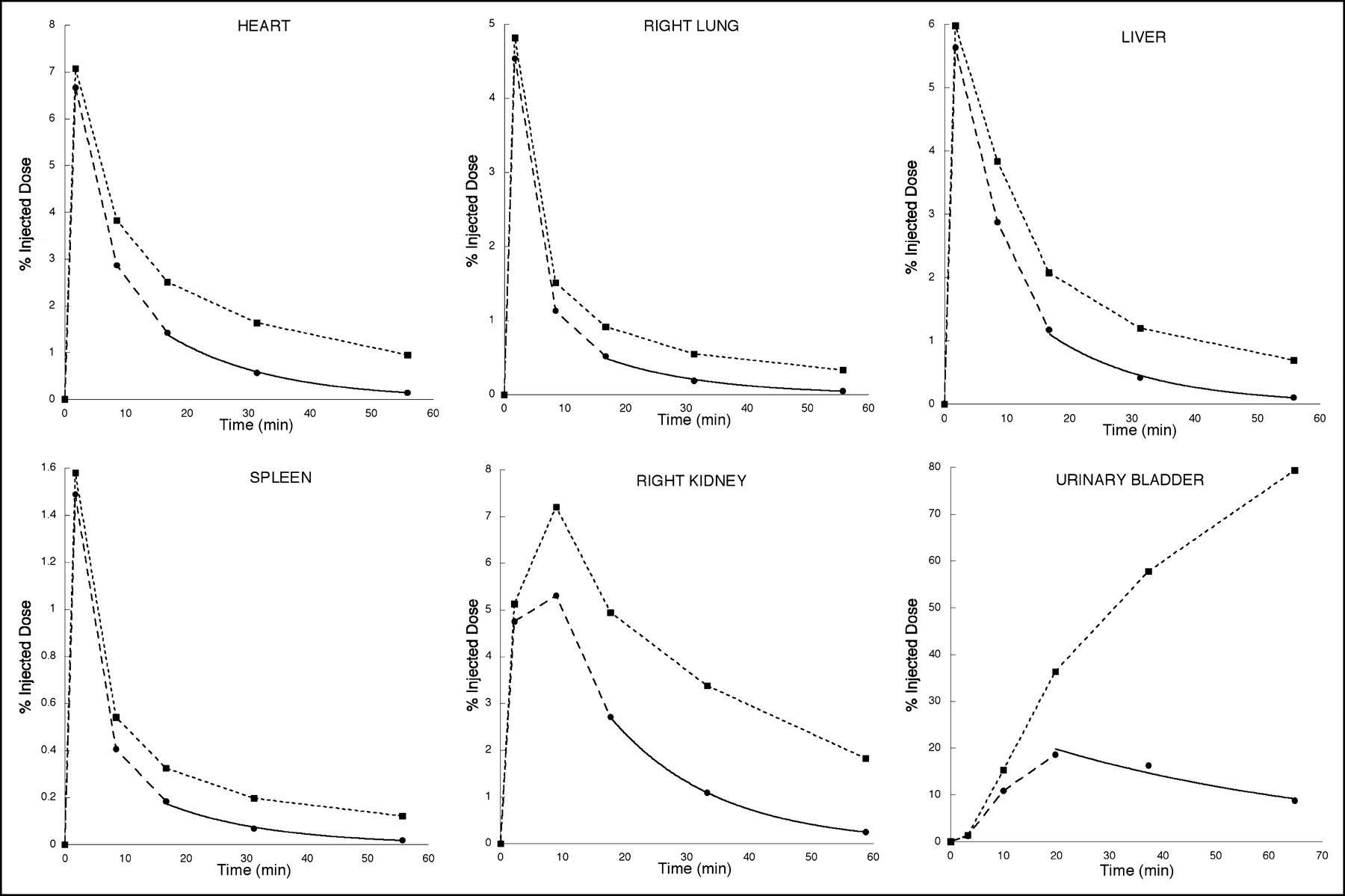
  • FIGURE 2.
    • Download figure
    • Open in new tab
    • Download powerpoint
    FIGURE 2.

    Time–activity curves generated from ROIs shown in Figure 1. Initial fast distribution of radiotracer throughout most of the body, with slower accumulation in kidneys, is followed by accumulation in urinary bladder, suggesting that major route of elimination is urinary system. Solid circles indicate activity in ROI as percentage of injected dose. Area under curve is determined by trapezoidal integration of first 3 time points through origin and integration of single exponential fit through final 3 time points. Time points are different in different organs as organs occupy different bed positions. Solid squares and dashed lines indicate decay-corrected time–activity curve.
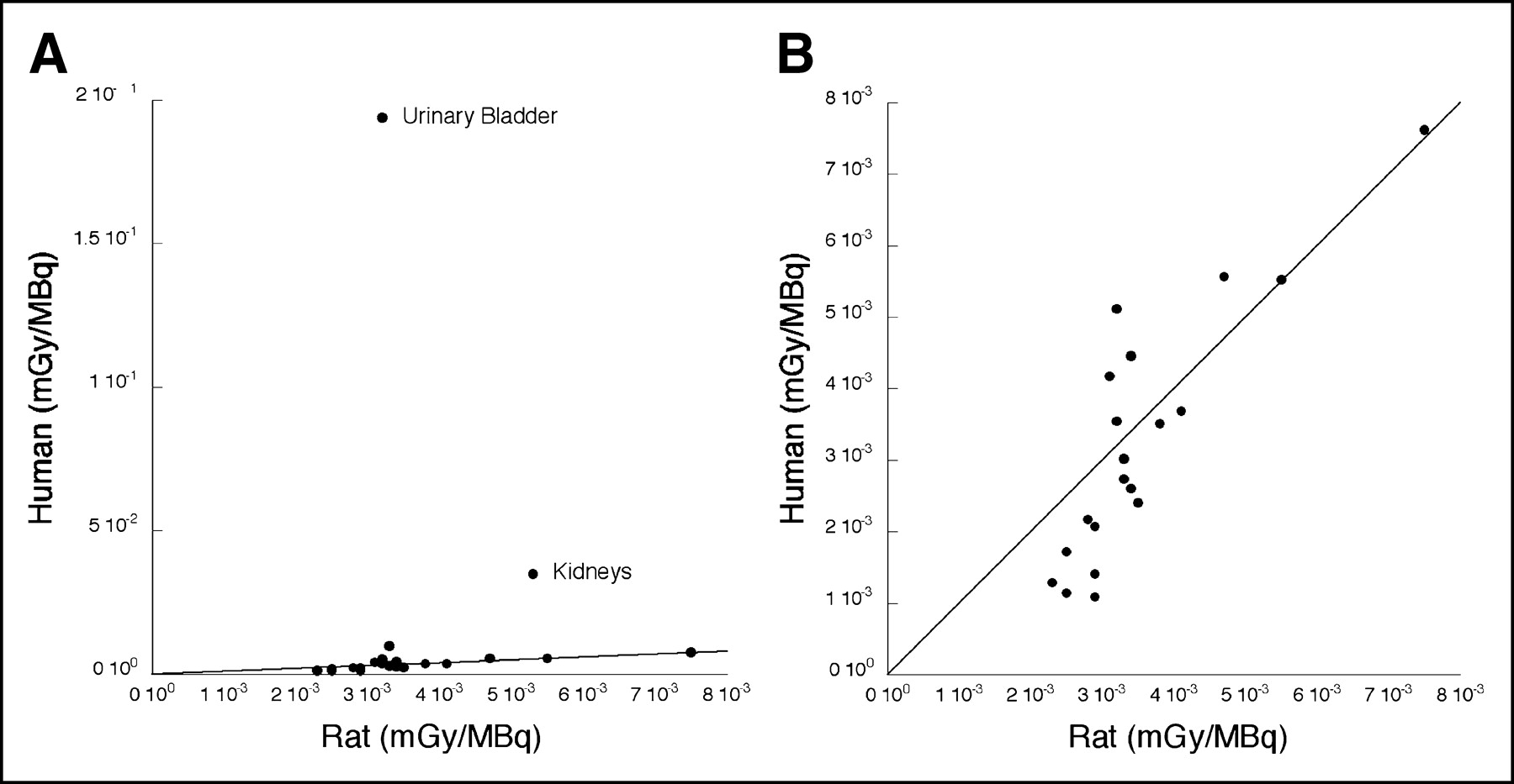
  • FIGURE 3.
    • Download figure
    • Open in new tab
    • Download powerpoint
    FIGURE 3.

    Comparison of radiation dose estimate from in vivo human PET studies and ex vivo rat biodistribution and dosimetry studies with all data points (A) and without urinary bladder and kidneys (B). Solid line in both plots is identity.

Tables

  • Figures
    • View popup
    TABLE 1

    Individual and Mean Residence Times for 11C-WAY100,635

    RegionMale 1Male 2Male 3Female 1Female 2Female 3Male average (±SD)Female average (±SD)Total average (±SD)*
    Urinary bladder2.63E−012.20E−012.60E−012.71E−012.25E−012.13E−012.48E−01 ± 2.41E−022.36E−01 ± 3.08E−022.42E−01 ± 2.55E−02
    Kidneys3.92E−023.49E−023.72E−022.43E−024.10E−024.25E−023.71E−02 ± 2.15E−033.59E−02 ± 1.01E−023.65E−02 ± 6.56E−03
    Liver2.78E−023.57E−023.66E−023.47E−023.57E−023.57E−023.33E−02 ± 4.86E−033.54E−02 ± 5.70E−043.44E−02 ± 3.29E−03
    Brain1.41E−021.06E−021.13E−021.26E−022.26E−021.39E−021.20E−02 ± 1.87E−031.64E−02 ± 5.46E−031.42E−02 ± 4.36E−03
    Lungs1.48E−021.36E−026.72E−031.25E−021.96E−028.20E−031.17E−02 ± 4.37E−031.35E−02 ± 5.76E−031.26E−02 ± 4.67E−03
    Heart7.60E−036.26E−039.21E−036.56E−031.32E−028.23E−037.69E−03 ± 1.48E−039.32E−03 ± 3.45E−038.51E−03 ± 2.53E−03
    Spleen3.62E−031.85E−032.37E−032.12E−033.86E−031.27E−032.61E−03 ± 9.07E−042.42E−03 ± 1.32E−032.52E−03 ± 1.02E−03
    Stomach wall7.99E−042.40E−032.13E−031.51E−032.88E−034.40E−031.78E−03 ± 8.58E−042.93E−03 ± 1.45E−032.35E−03 ± 1.24E−03
    Gallbladder1.78E−041.02E−032.32E−049.25E−042.23E−042.52E−044.77E−04 ± 4.72E−044.67E−04 ± 3.97E−044.72E−04 ± 3.90E−04
    • ↵* Descending order.

    • Data are in hours.

    • View popup
    TABLE 2

    Mean Radiation Absorbed Dose Estimates for 11C-WAY100,635

    RegionMale average (±SD)Female average (±SD)Total average (±SD)*
    Urinary bladder wall1.67E−01 ± 1.59E−022.20E−01 ± 2.87E−021.94E−01 ± 3.57E−02
    Kidneys3.39E−02 ± 1.85E−033.57E−02 ± 9.76E−033.48E−02 ± 6.36E−03
    Uterus9.81E−03 ± 9.48E−049.81E−03 ± 9.48E−04
    Liver6.38E−03 ± 8.12E−048.86E−03 ± 2.51E−047.62E−03 ± 1.46E−03
    Spleen5.16E−03 ± 1.31E−035.98E−03 ± 2.37E−035.57E−03 ± 1.77E−03
    Ovaries5.52E−03 ± 3.84E−045.52E−03 ± 3.84E−04
    Lower large intestine wall4.56E−03 ± 1.62E−045.68E−03 ± 4.20E−045.12E−03 ± 6.78E−04
    Lungs3.65E−03 ± 1.09E−035.26E−03 ± 1.84E−034.46E−03 ± 1.61E−03
    Heart wall3.54E−03 ± 3.65E−044.82E−03 ± 1.32E−034.18E−03 ± 1.11E−03
    Brain2.88E−03 ± 4.07E−044.49E−03 ± 1.42E−033.69E−03 ± 1.28E−03
    Stomach wall2.34E−03 ± 9.75E−044.75E−03 ± 2.17E−033.55E−03 ± 2.00E−03
    Gallbladder wall3.23E−03 ± 1.22E−033.80E−03 ± 8.74E−043.52E−03 ± 9.98E−04
    Testes3.19E−03 ± 7.23E−053.19E−03 ± 7.23E−05
    Small intestine2.65E−03 ± 5.13E−053.38E−03 ± 1.17E−043.02E−03 ± 4.08E−04
    Upper large intestine wall2.37E−03 ± 9.17E−053.10E−03 ± 1.31E−042.74E−03 ± 4.14E−04
    Adrenals2.26E−03 ± 1.38E−042.95E−03 ± 3.97E−042.61E−03 ± 4.59E−04
    Pancreas2.13E−03 ± 1.53E−042.69E−03 ± 3.32E−042.41E−03 ± 3.85E−04
    Muscle1.95E−03 ± 7.00E−052.40E−03 ± 8.62E−052.17E−03 ± 2.55E−04
    Red marrow1.82E−03 ± 9.54E−052.33E−03 ± 1.29E−042.07E−03 ± 2.95E−04
    Bone surfaces1.50E−03 ± 1.30E−041.94E−03 ± 1.47E−041.72E−03 ± 2.71E−04
    Thymus1.22E−03 ± 1.39E−041.61E−03 ± 1.74E−041.41E−03 ± 2.54E−04
    Skin1.15E−03 ± 9.87E−051.43E−03 ± 1.10E−041.29E−03 ± 1.83E−04
    Breasts9.85E−04 ± 1.34E−041.31E−03 ± 1.31E−041.15E−03 ± 2.12E−04
    Thyroid9.88E−04 ± 1.57E−041.19E−03 ± 1.50E−041.09E−03 ± 1.77E−04
    Total body2.31E−03 ± 5.20E−052.99E−03 ± 8.19E−052.65E−03 ± 3.77E−04
    Effective dose equivalent1.52E−02 ± 1.10E−031.95E−02 ± 1.25E−031.74E−02 ± 2.56E−03
    Effective dose1.22E−02 ± 7.57E−041.60E−02 ± 1.43E−031.41E−02 ± 2.35E−03
    • ↵* Descending order.

PreviousNext
Back to top

In this issue

Journal of Nuclear Medicine: 46 (4)
Journal of Nuclear Medicine
Vol. 46, Issue 4
April 1, 2005
  • Table of Contents
  • About the Cover
  • Index by author
Print
Download PDF
Article Alerts
Sign In to Email Alerts with your Email Address
Email Article

Thank you for your interest in spreading the word on Journal of Nuclear Medicine.

NOTE: We only request your email address so that the person you are recommending the page to knows that you wanted them to see it, and that it is not junk mail. We do not capture any email address.

Enter multiple addresses on separate lines or separate them with commas.
Biodistribution and Radiation Dosimetry of 11C-WAY100,635 in Humans
(Your Name) has sent you a message from Journal of Nuclear Medicine
(Your Name) thought you would like to see the Journal of Nuclear Medicine web site.
Citation Tools
Biodistribution and Radiation Dosimetry of 11C-WAY100,635 in Humans
Ramin V. Parsey, Marie-Jose Belanger, Gregory M. Sullivan, Norman R. Simpson, Michael G. Stabin, Ronald Van Heertum, J. John Mann
Journal of Nuclear Medicine Apr 2005, 46 (4) 614-619;

Citation Manager Formats

  • BibTeX
  • Bookends
  • EasyBib
  • EndNote (tagged)
  • EndNote 8 (xml)
  • Medlars
  • Mendeley
  • Papers
  • RefWorks Tagged
  • Ref Manager
  • RIS
  • Zotero
Share
Biodistribution and Radiation Dosimetry of 11C-WAY100,635 in Humans
Ramin V. Parsey, Marie-Jose Belanger, Gregory M. Sullivan, Norman R. Simpson, Michael G. Stabin, Ronald Van Heertum, J. John Mann
Journal of Nuclear Medicine Apr 2005, 46 (4) 614-619;
Twitter logo Facebook logo LinkedIn logo Mendeley logo
  • Tweet Widget
  • Facebook Like
  • Google Plus One
Bookmark this article

Jump to section

  • Article
    • Abstract
    • MATERIALS AND METHODS
    • RESULTS
    • DISCUSSION
    • CONCLUSION
    • Acknowledgments
    • Footnotes
    • REFERENCES
  • Figures & Data
  • Info & Metrics
  • PDF

Related Articles

  • THIS MONTH IN JNM
  • PubMed
  • Google Scholar

Cited By...

  • 11C Dosimetry Scans Should Be Abandoned
  • Internal Dose Assessment of (-)-18F-Flubatine, Comparing Animal Model Datasets of Mice and Piglets with First-in-Human Results
  • Dynamic PET Denoising with HYPR Processing
  • 1-11C-Methyl-4-Piperidinyl-N-Butyrate Radiation Dosimetry in Humans by Dynamic Organ-Specific Evaluation
  • Whole-Body Biodistribution and Estimation of Radiation-Absorbed Doses of the Dopamine D1 Receptor Radioligand 11C-NNC 112 in Humans
  • Google Scholar

More in this TOC Section

  • Tumor-Specific Binding of Radiolabeled PEGylated GIRLRG Peptide: A Novel Agent for Targeting Cancers
  • PET/MRI of Hypoxic Atherosclerosis Using 64Cu-ATSM in a Rabbit Model
  • Tumor Uptake of Anti-CD20 Fabs Depends on Tumor Perfusion
Show more Basic Science Investigations

Similar Articles

SNMMI

© 2025 SNMMI

Powered by HighWire