


Article Figures & Data
Figures
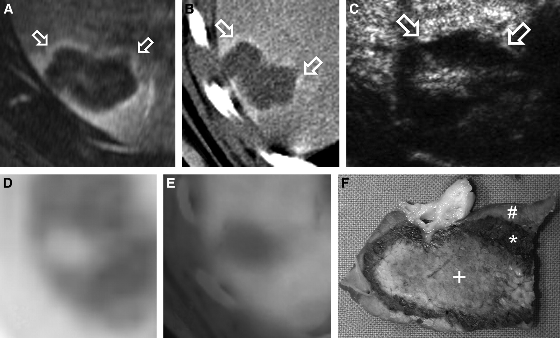
- FIGURE 1.
Transversal images immediately after RF-ablation. Rim-like increase of contrast enhancement in arterial phase (arrows) was detected on MRI (A), CT (B), and US (C). Functional data provided by PET (D) and PET/CT (E) demonstrated an area of decreased 18F-FDG uptake surrounded by homogeneous tracer distribution. Macroscopic tissue sample generated after sacrificing 1 animal (F) shows central necrosis (+), adjacent rim of blood-filled sinusoids (*), and macroscopically normal liver parenchyma (#). Note blurred margins of lesion on PET (D) leading to slight underestimation of lesion size.
- FIGURE 2.
Qualitative image analysis according to 3-point scale revealed increased contrast enhancement in periphery of induced necrosis when compared with normal liver parenchyma with all morphologic imaging procedures. On PET, no increase in glucose metabolism was found, leading to a score of 1 in all 19 lesions.
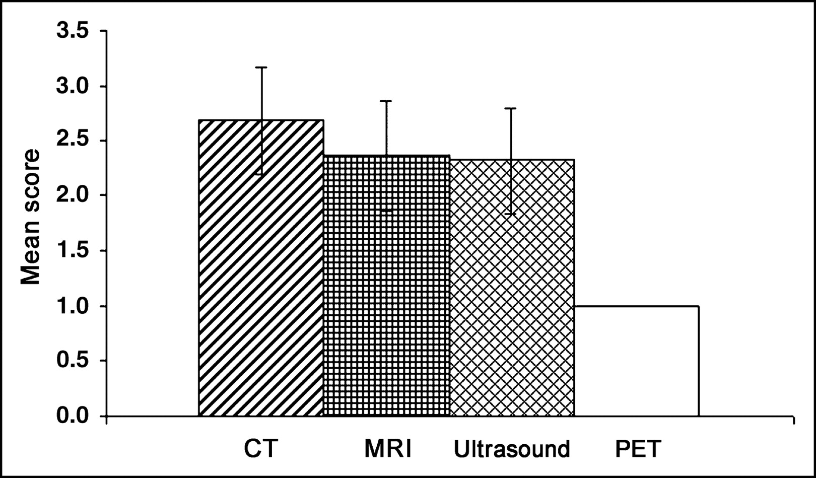
- FIGURE 3.
Quantitative image analysis by determination of ratio of contrast enhancement of lesion periphery/contrast enhancement of normal liver parenchyma for CT and MRI as well as ratio of activity concentration of lesion periphery/activity concentration of normal liver parenchyma on PET. Due to rim-like increase of contrast enhancement in periphery of necrosis, ratios are elevated for morphologic imaging procedures, whereas homogeneous tracer distribution on PET led to a mean ratio of 1.05 for functional data analysis.
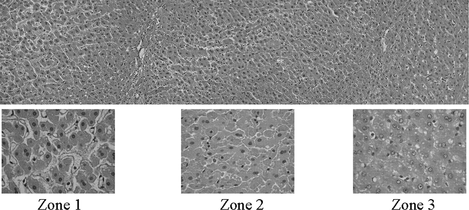
- FIGURE 4.
Fragmentation of liver plates were visible in center of lesion (zone 1), demonstrating early signs of necrosis. No blood cells were detected in zone 1 due to occlusion of sinusoids caused by edema of hepatocytes. This edema, furthermore, led to widening of space of Disse. In transitional zone (zone 2), sinusoids were engorged with blood caused by outflow obstruction. Peripheral zone (zone 3) is characterized by unspecific vacuolar transformation of cytoplasm of hepatocytes as sign of reactive changes.


Tables
- TABLE 1
Mean Maximal Diameters (Large-Axis Measurement) and Mean Minimal Diameters (Small-Axis Measurement) of RF-Induced Necrotic Zone as Determined by Different Imaging Procedures
Imaging procedure Measurement 1 Measurement 2 Maximal diameter (cm) Minimal diameter (cm) Maximal diameter (cm) Minimal diameter (cm) US 3.7 ± 0.7 2.5 ± 0.6 3.6 ± 0.6 2.4 ± 0.4 CT 3.8 ± 0.7 2.5 ± 0.5 3.8 ± 0.7 2.5 ± 0.5 MRI 3.8 ± 0.9 2.5 ± 0.6 4.1 ± 0.9 2.7 ± 0.7 PET 3.4 ± 0.8 2.3 ± 0.5 — — PET/CT 3.8 ± 0.7 2.5 ± 0.3 — — For US, CT, and MRI, measurement 1 represents determination of lesion size in venous phase, and measurement 2 represents lesion size in arterial phase. For PET and PET/CT, only 1 measurement was performed (measurement 1). Measurements are expressed as mean ± SD.
In this issue


Jump to section
Related Articles
Cited By...
- Iodine quantification with dual-energy CT: phantom study and preliminary experience with VX2 residual tumour in rabbits after radiofrequency ablation
- Oncologic PET/MRI, Part 1: Tumors of the Brain, Head and Neck, Chest, Abdomen, and Pelvis
- Monitoring and Predicting Response to Therapy with 18F-FDG PET in Colorectal Cancer: A Systematic Review
- Real-Time Iterative Monitoring of Radiofrequency Ablation Tumor Therapy with 15O-Water PET Imaging
- Morphologic and Functional Changes in Nontumorous Liver Tissue After Radiofrequency Ablation in an In Vivo Model: Comparison of 18F-FDG PET/CT, MRI, Ultrasound, and CT
- Evaluation of Image Registration in PET/CT of the Liver and Recommendations for Optimized Imaging
- Can PET/CT Replace Separate Diagnostic CT for Cancer Imaging? Optimizing CT Protocols for Imaging Cancers of the Chest and Abdomen