


Article Figures & Data
Figures
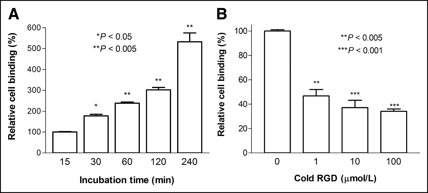
- FIGURE 1.
Endothelial cell binding of 125I-c(RGD(I)yV). (A) Incubation time-dependent increase of 125I-c(RGD(I)yV) binding to HUVEC cells. Results are expressed as percent uptake relative to 15-min value. (B) Dose-dependent inhibition of cell binding by excess nonradiolabeled c(RGD(I)yV). Results are expressed as percent uptake relative to sample without nonradiolabeled c(RGD(I)yV). All results are mean ± SD of duplicates.
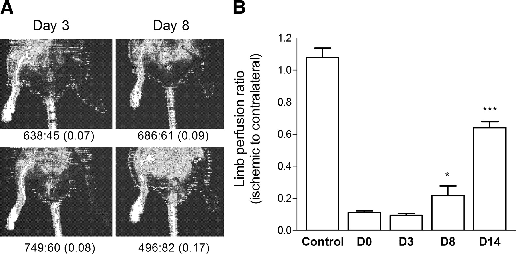
- FIGURE 2.
Hindimb blood flow monitored with laser Doppler flowmetry. (A) Doppler images of representative animals on days 3 and 8 of ischemia. Right:left hindlimb flow measurements (perfusion ratios in parentheses) are denoted below each image. (B) Temporal change of right-to-left hindlimb perfusion ratios. There was severe hypoperfusion in left hindlimb immediately after surgery (D0) and on day 3 (D3), which gradually improved on day 8 (D8) and day 14 (D14). Data are shown as mean ± SD of left-to-right ratios. *P < 0.05; ***P < 0.0001 compared with immediate postoperative ratios.
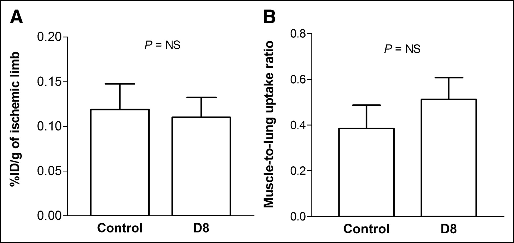
- FIGURE 3.
125I-c(RGD(I)yV) uptake in ischemic hindlimbs according to periods of ischemia. 125I-c(RGD(I)yV) uptake in ischemic hindlimb muscles is expressed as %ID/g (A) and muscle-to-lung uptake ratios (B). Data are expressed as mean ± SD of 6 animals for each ischemia period and 7 animals for control.
- FIGURE 4.
Uptake of control peptide (125I-GfVGV). Uptake in muscle tissue of ischemic hindlimb and hindlimb of control animals is expressed as %ID/g (A) and muscle-to-lung uptake ratios (B). Data are shown as mean ± SD of 5 ischemic hindlimb models and 4 control animals.
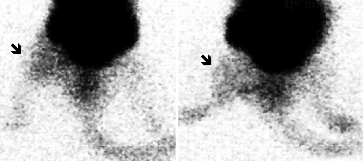
- FIGURE 5.
Scintigraphic 123I-c(RGD(I)yV) images in hindlimb ischemic models on day 3 demonstrate higher radioactivity in ischemic left (arrows) compared with contralateral hindlimbs.
- FIGURE 6.
Immunohistochemical detection of αv integrin. Hematoxylin and eosin staining (A, C, E, G) and αv integrin immunostaining (B, D, F, H) of hindlimb muscle tissue microsections. Tissues are from normal control animals (A and B) and mice at 3 d (C and D), 8 d (E and F), and 14 d (G and H) of ischemia. Length of bar indicates 400 μm.
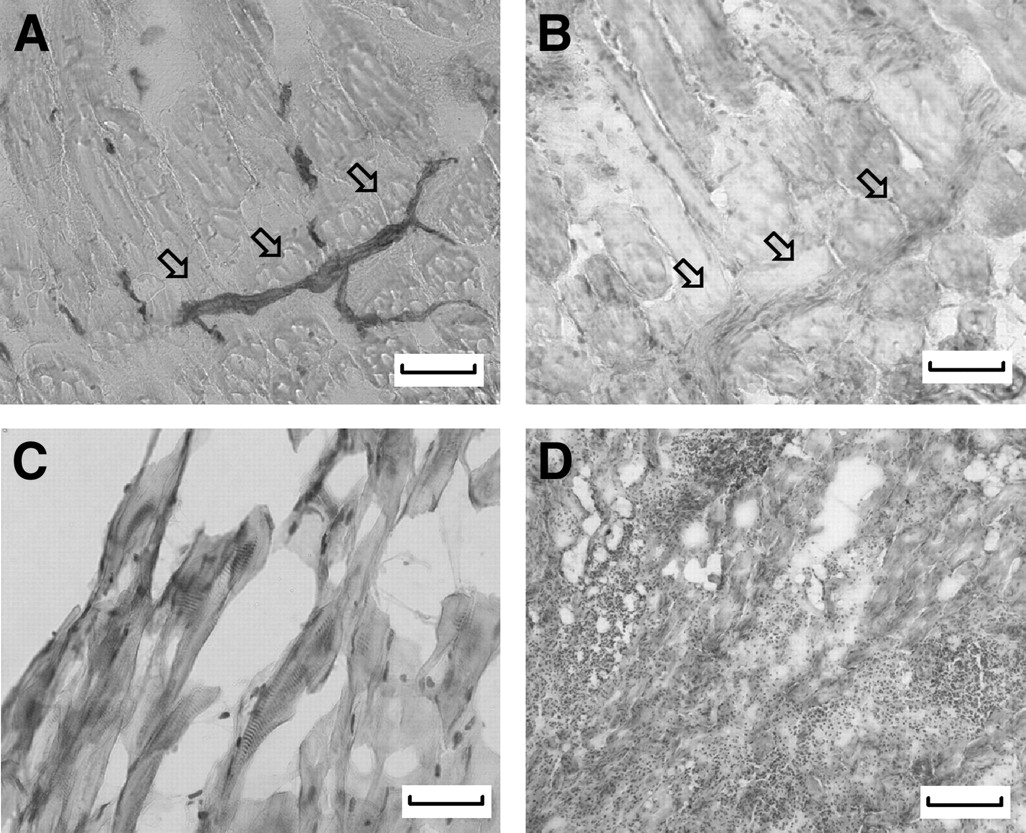
- FIGURE 7.
Histologic localization of αv integrin staining. Alkaline phosphatase staining (A) demonstrates localization of αv integrin immunoreactivity (B) to vascular endothelial cells. High magnification shows αv integrin immunostaining localized to skeletal muscle cells (C). Hematoxylin and eosin counterstaining shows lack of αv immunostaining in inflammatory cell infiltration sites (D). Length of bar indicates 200 μm for A, B, and D and 100 μm for C.


Tables
Tissue Control (n = 8) Hindlimb ischemia model 3 d (n = 6) 8 d (n = 6) 14 d (n = 6) Blood 0.41 ± 0.20 0.39 ± 0.09 0.28 ± 0.07 0.31 ± 0.10 Heart 0.18 ± 0.09 0.20 ± 0.04 0.12 ± 0.03 0.13 ± 0.04 Lung 0.43 ± 0.19 0.38 ± 0.15 0.29 ± 0.08 0.30 ± 0.09 Liver 0.68 ± 0.37 0.99 ± 0.24 1.05 ± 0.33 0.60 ± 0.23 Spleen 0.29 ± 0.12 0.32 ± 0.07 0.23 ± 0.04 0.23 ± 0.12 Pancreas 0.16 ± 0.09 0.24 ± 0.06 0.14 ± 0.06 0.15 ± 0.04 Kidneys 0.85 ± 0.27 1.02 ± 0.31 0.84 ± 0.19 0.63 ± 0.27 Hindlimb muscle* 0.16 ± 0.05 0.85 ± 0.76† 0.43 ± 0.23† 0.43 ± 0.28†
In this issue


Jump to section
Related Articles
Cited By...
- Radiotracer Imaging of Peripheral Vascular Disease
- State-of-the-Art Methods for Evaluation of Angiogenesis and Tissue Vascularization: A Scientific Statement From the American Heart Association
- Radiotracer Imaging of Peripheral Vascular Disease
- Approaches to Multimodality Imaging of Angiogenesis
- Targeted Imaging Offers Advantages Over Physiological Imaging for Evaluation of Angiogenic Therapy
- Radionuclide Imaging: A Molecular Key to the Atherosclerotic Plaque
- Preparation of a Promising Angiogenesis PET Imaging Agent: 68Ga-Labeled c(RGDyK)-Isothiocyanatobenzyl-1,4,7-Triazacyclononane-1,4,7-Triacetic Acid and Feasibility Studies in Mice
- Monitoring of the Biological Response to Murine Hindlimb Ischemia With 64Cu-Labeled Vascular Endothelial Growth Factor-121 Positron Emission Tomography