


Article Figures & Data
Figures
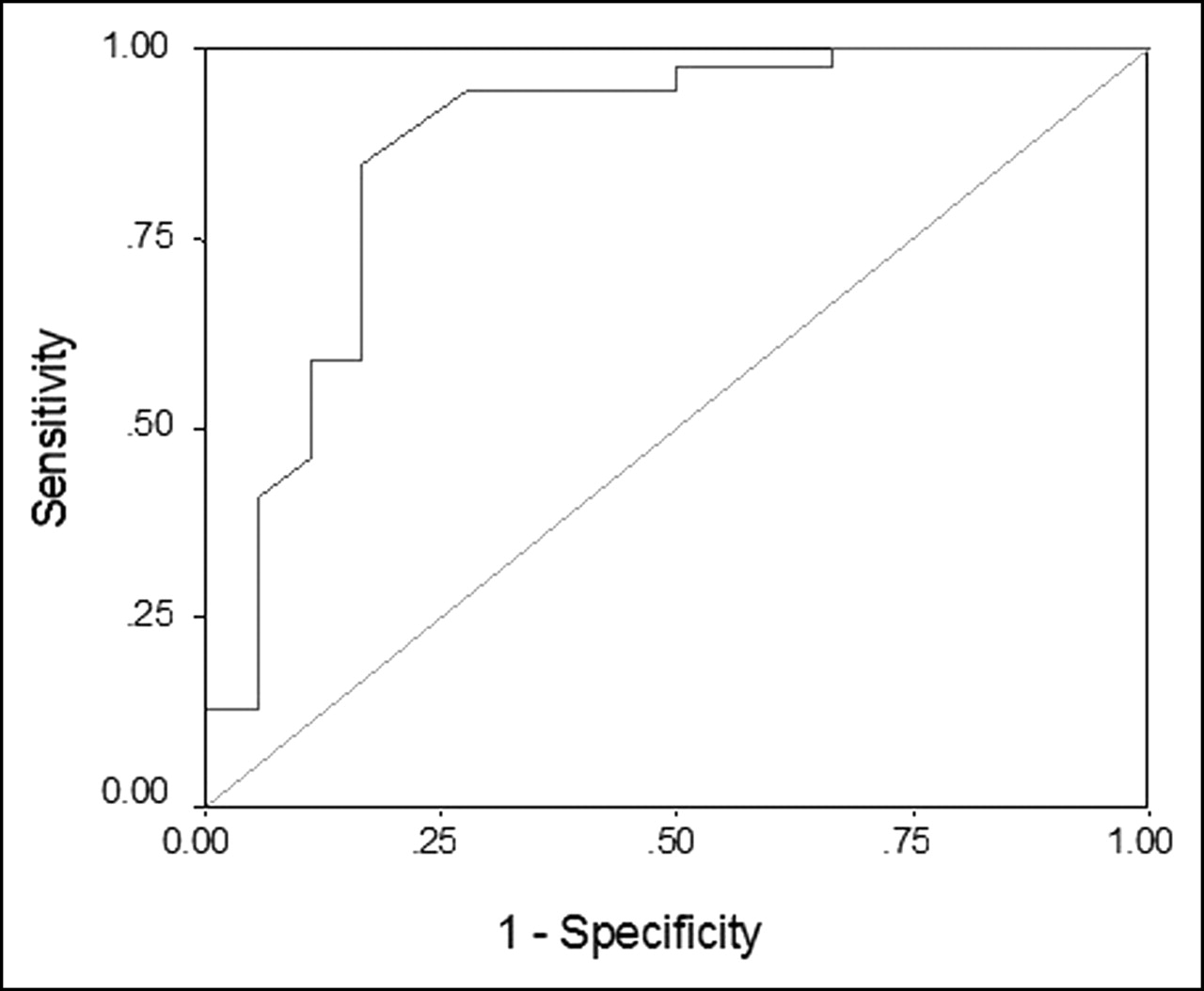
- FIGURE 1.
ROC curve for percent change in average SUVs.
Tables
Sample Average SUV at time: % Change in average SUV Histopathologic findings Tumor grade Size (cm) 1 2 1 1.2 1.4 16.7 IDC Low 0.2 2 1.5 1.7 13.3 IDC High 1.5 3 0.7 0.7 0 IDC + ILC Moderate 3.5 4 0.6 0.7 16.7 IDC Not known 1.0 5 1.1 0.9 −18.2 IDC Moderate 0.2 6 3.0 3.3 10.0 IDC High 2.0 7 2.1 2.3 9.5 IDC Moderate 2.4 8 2.1 2.2 4.8 IDC + ILC Low 4.0 9 1.4 1.6 14.3 IDC Moderate 0.5 10 1.9 2.3 21.1 IDC High Not known 11 1.8 1.9 5.6 ILC + IDC Moderate 1.5 12 0.8 0.9 12.5 IDC Low Not known 13 7.4 9.1 23.0 IDC High Not known 14 2.3 2.5 8.7 IDC Moderate 2.9 15 1.8 2.0 11.1 IDC Moderate 1.9 16 0.9 0.9 0 IDC Moderate 1.6 17 2.0 2.3 15.0 IDC Low 3.5 18 2.7 3.5 29.6 IDC + ILC High 2.4 19 0.6 0.8 33.3 IDC Low 1.6 20 11.7 15.7 34.2 IDC High 2.6 21 0.7 0.8 14.3 IDC Low 1.4 22 2.9 3.2 10.3 IDC Moderate 2.1 23 15.3 20.0 30.7 IDC High 2.8 24 5.7 5.9 3.5 IDC High 1.9 25 0.7 0.7 0 IDC + ILC Low 0.3 26 6.0 6.9 15.0 Medullary carcinoma High 1.8 27 5.2 5.8 11.5 IDC Moderate 2.5 28 2.2 2.4 9.1 IDC High 2.2 29 1.5 1.7 13.3 IDC Moderate 1.2 30 0.4 0.4 0 IDC Moderate 0.4 31 2.5 2.6 4.0 IDC High 0.5 32 2.5 2.7 8.0 IDC Low Not known 33 1.7 1.9 11.8 IDC + ILC Low Not known 34 1.4 1.7 21.4 IDC Moderate 0.9 35 0.8 1.0 25 IDC High Not known 36 2.7 3.6 33.3 IDC Moderate 1.0 37 6.2 7.7 24.2 IDC Moderate 4.0 38 3.0 3.2 6.7 IDC Moderate Not known 39 3.3 3.0 −9.1 Adenocarcinoma Not known Not known Mean 2.88 3.38 12.7 SD 3.04 3.98 11.4 IDC = invasive ductal carcinoma; ILC = invasive lobular carcinoma.
Sample Average SUV at time: % Change in average SUV Histopathologic findings 1 2 1 1.7 1.4 −0.176 No tumor, Bx Rx + 2 1.1 0.8 −0.273 Benign, Bx Rx + 3 1.5 1.9 0.267 No tumor, Bx Rx +, Ch Inf 4 1.3 1.1 −0.154 No tumor, Bx Rx + 5 1.0 0.7 −0.300 No tumor 6 1.3 1.2 −0.077 No tumor, Bx Rx + 7 1.0 0.8 −0.200 No tumor, Bx Rx + 8 1.0 1.1 0.100 Bx Rx +, proliferative fibroblasts 9 1.6 1.6 0 No tumor, Bx Rx + 10 1.5 1.3 −0.133 No tumor, Bx Rx + 11 1.0 0.8 −0.200 No tumor, Bx Rx + 12 0.8 0.6 −0.250 No tumor, Bx Rx + 13 1.4 1.3 −0.071 No tumor, Bx Rx + 14 1.5 1.4 −0.067 Benign, Bx Rx + 15 2.0 1.9 −0.050 No tumor, Bx Rx + 16 1.5 1.5 0 No tumor, Bx Rx + 17 1.5 1.7 0.133 Bx Rx, foreign-body giant-cell reaction 18 0.5 0.3 −0.400 No tumor, Bx Rx + Mean 1.29 1.19 −0.103 SD 0.36 0.45 0.166 Bx Rx = biopsy reaction; Ch Inf = chronic inflammation.
In this issue


Jump to section
Related Articles
Cited By...
- PET/MRI Versus PET/CT for Whole-Body Staging
- Image Quality and Diagnostic Performance of a Digital PET Prototype in Patients with Oncologic Diseases: Initial Experience and Comparison with Analog PET
- Performance of Whole-Body Integrated 18F-FDG PET/MR in Comparison to PET/CT for Evaluation of Malignant Bone Lesions
- First Clinical Experience with Integrated Whole-Body PET/MR: Comparison to PET/CT in Patients with Oncologic Diagnoses
- The value of dual-time-point 18F-FDG PET/CT for identifying axillary lymph node metastasis in breast cancer patients
- PET Tumor Metabolism in Locally Advanced Breast Cancer Patients Undergoing Neoadjuvant Chemotherapy: Value of Static versus Kinetic Measures of Fluorodeoxyglucose Uptake
- Diagnostic accuracy of fused positron emission tomography/magnetic resonance mammography: initial results
- Prospective Evaluation of Whole-Body Cancer Screening With Multiple Modalities Including [18F]Fluorodeoxyglucose Positron Emission Tomography in a Healthy Population: A Preliminary Report
- Time and Again, Children Resemble Their Parents
- Evaluation of Dual-Time-Point 18F-FDG PET for Staging in Patients with Lung Cancer
- Whole-body PET/CT-mammography for staging breast cancer: initial results
- Breast Cancer Staging in a Single Session: Whole-Body PET/CT Mammography
- FDG-PET/CT in restaging of patients with recurrent breast cancer: possible impact on staging and therapy
- Metabolic Imaging by Hyperpolarized 13C Magnetic Resonance Imaging for In vivo Tumor Diagnosis.
- 18F-FDG PET After Radiofrequency Ablation: Is Timing Everything?