


Article Figures & Data
Figures
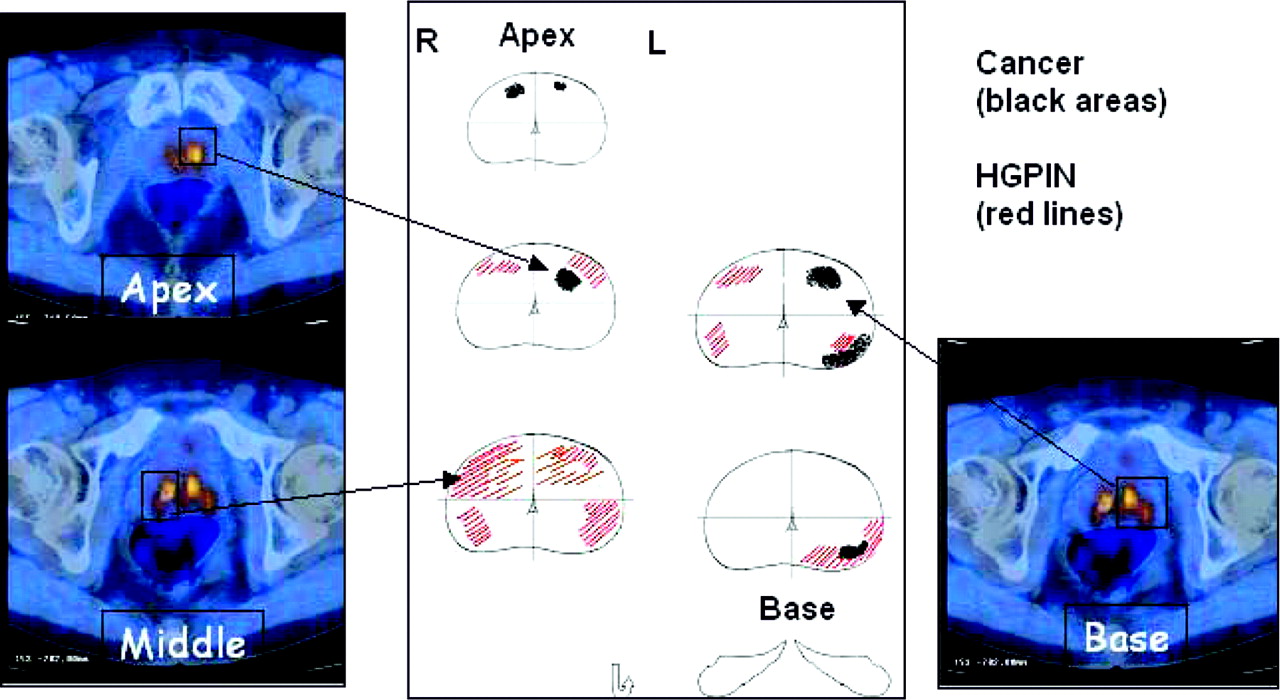
- FIGURE 1.
PET/CT results and histologic matching.
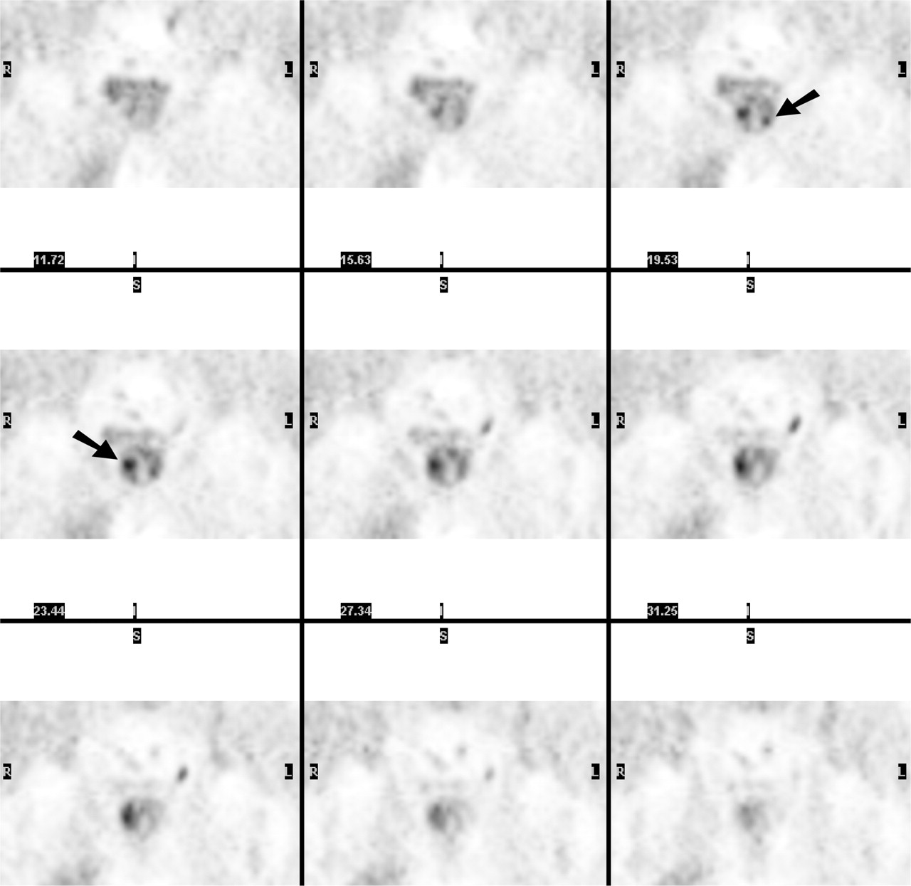
- FIGURE 2.
Patient 6: Coronal PET images show 2 foci of pathologic uptake in left and right mid regions (arrows), confirmed to be the only foci of cancer on histologic examination.
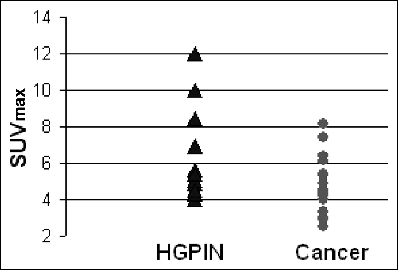
- FIGURE 3.
Correlation between SUVmax and lesion type.
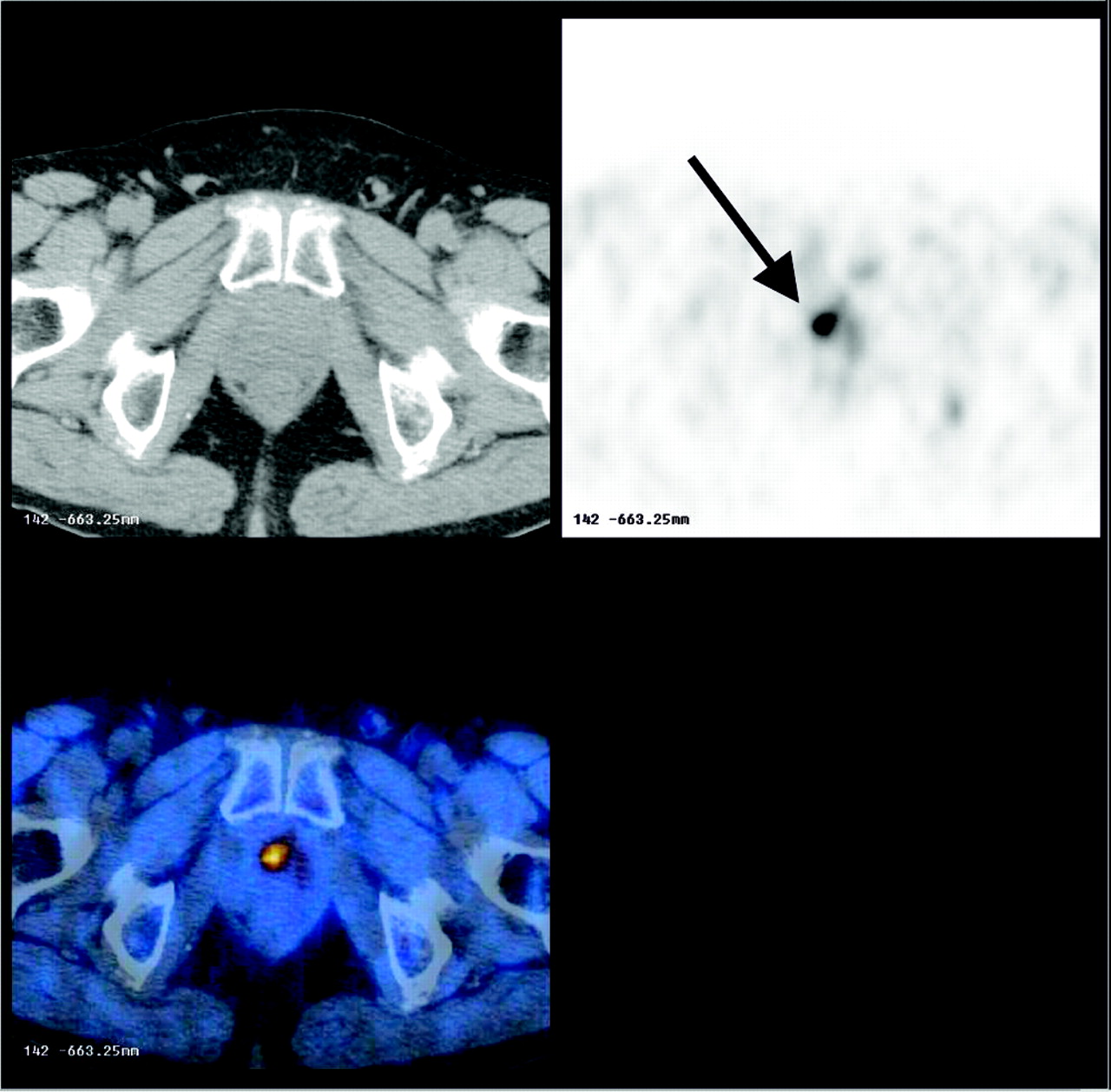
- FIGURE 4.
Patient 36: Transaxial PET/CT images show 1 focus of pathologic uptake in anterior transitional zone (arrow). First biopsy failed to detect cancer foci, whereas repeated biopsy using PET/CT images detected sites of cancer.

Tables
Patient no. Age (y) PSA (ng/mL) Free-to-total PSA DRE Hypoechogenic lesion on TRUS Prostate volume (cm3) No. of biopsy cores Treatment TNM Gleason score 1 65 9 11 − + 70 14 RRP T2c N0 Gs4+3 2 60 3 15 + + 30 12 RRP T3a N0 Gs3+4 3 56 6.4 12 − − 25 13 VLRP T3a N0 Gs3+3 4 60 9 13 − − 75 10 VLRP T2c N0 Gs3+4 5 65 15 9 − − 45 12 RRP T2c N0 Gs3+3 6 63 5 20 − − 50 12 VLRP T2c N0 Gs1+1 7 61 5 28 − + 75 12 RRP T3a N0 Gs4+3 8 57 13 11 − − 60 12 VLRP T3a N0 Gs3+3 9 64 2 7 − − 25 12 VLRP T2a N0 Gs2+1 10 69 9 2 + + 35 13 RRP T3a N0 Gs3+3 11 68 9 7 − − 50 12 RRP T2c N0 Gs3+3 12 70 28 13 − − 50 18 RRP T2c N0 Gs3+3 13 57 8 12 − − 50 12 VLRP T2c N0 Gs2+3 14 62 7 8 − − 35 15 RRP T2c N0 Gs3+1 15 51 10 11 + + 40 12 VLRP T3a N0 Gs3+3 16 75 8 7 + + 40 12 RRP T2c N1 Gs3+3 17 72 10 20 + + 55 12 RRP T2c N0 Gs2+3 18 64 21 16 − + 35 12 VLRP T3a N0 Gs3+4 19 58 5 9 − + 50 12 RRP T3a N0 Gs3+3 20 72 10 8 + + 30 12 VLRP T3a N0 Gs4+3 21 68 20 13 + + 20 10 RRP T3a N0 Gs3+3 22 60 8 9 − − 30 12 RRP T3a N0 Gs3+2 23 63 5 19 + + 40 15 VLRP T2b N0 Gs3+3 24 72 70 18 + + 50 10 RRP T3b N1 Gs4+3 25 63 7 13 + + 30 10 RRP T3b N1 Gs4+3 26 59 12 7 − − 50 12 VLRP T2c N0 Gs3+3 27 70 59 15 − + 70 14 RRP T2c N0 Gs3+2 28 65 5 7 − − 50 13 RRP T3a N0 Gs3+3 29 60 5 6 − − 30 12 RRP T2c N0 Gs3+3 30 68 7 9 − − 30 12 RRP T2c N0 Gs3+3 31 58 5 7 + + 25 12 RRP T3b N0 Gs4+3 32 51 12 11 + + 50 12 RRP T3b N1 Gs4+3 33 69 6.2 10 − − 50 12 RRP T2c N0 Gs2+2 34 71 17 13 − − 30 15 RRP T3a N0 Gs3+3 35 65 7 12 + + 40 12 RRP T3a N0 Gs3+3 36 52 5.2 8 − − 40 12 RRP T2c N0 Gs3+3 RRP = radical retropubic prostatectomy; VLRP = videolaparoscopic prostatectomy.
- TABLE 2
Clinical Characteristics of Control Group with Infiltrating Transitional Bladder Carcinoma
Patient no. Age (y) PSA (ng/mL) Free-to-total PSA DRE TRUS Prostate volume (cm3) TNM (bladder) Tumor grade 1 65 2.1 18 − − 30 T1 N0 2–3 2 70 2.8 24 − − 40 T3b N0 3 3 65 2.0 20 − − 30 T1 N0 3 4 64 1.3 30 − − 45 T1 N0 3 5 71 1.6 25 − − 40 T3b N0 3 Patient no. Biopsy PET SUVmax Background SUV Carcinoma HGPIN 1 La, Lm Rm, 4.9; La, 4 2.5 Ra, Rm, La, Lm 2 Ra, Rm, Rb, La, Lm, Lb Ra, 2.9; Rm, 2.9; La, 2.5 1.5 Ra, Rm, Rb, La, Lm, Lb 3 Lb Rm, 8.4; Lm, 6.1; Lb, 6.1 4.0 Lm, Lb Rm, Rb 4 Rm Ra, 3.3; Rm, 3 2.0 Ra, Rm, La, Lb Rm, Rb, Lm, Lb 5 Ra, Rb Ra, 5.4; Rm, 8.1; Rb, 8.1; Lm, 4.3; Lb, 4.5 2.9 Ra, Rm, Rb, La, Lm, Lb Lb 6 Lm Rm, 7.4; Lm, 6.4 3.0 Rm, Lm Rm, Lm 7 Lm, Lb Rm, 4.9; Rb, 5.7 2.0 Lm, Lb Rm, Rb, Lm, Lb 8 Rb, Rm, Lm, Lb Rm, 4.3; Rb, 5.3; La, 4.4; Lm, 4.2; Lb, 4.5 2.5 Rm, Rb, La, Lm, Lb Rm, Rb, Lb 9 Rm Rm, 4.6; Rb, 4.6 2.4 Rm, Rb Rb, Lb 10 Rb Rm, 6.4; Rb, 5.5; Lm, 5 2.7 Rm, Rb Lm, Lb 11 Rm Rm, 2.9; Lm, 2.8 1.8 Rm, Lm 12 Rm,* Lm* Ra, 8.4; Rm, 8.4; Rb, 8.4; Lm, 7.8; Lb, 7.8 2.5 Ra, Rm, Rb, Lm, Lb 13 Rb Rm, 5.5; Rb, 5.5 3.0 Rm, Rb, Lb Rm, Rb, Lm, Lb 14 La, Lm Rm, 4; Lm, 4.7 2.8 Ra, La, Lm, Lb Rm, Rb, Lm, Lb 15 Ra, Rm Ra, 4.3; La, 4.3 2.5 Ra, Rm, La Rm 16 Lm, Lb Rm, 3; Lm, 3; Lb, 3.2 2.0 Rm, Lm, Lb Rm, Rb, Lm, Lb 17 La, Lb Rm, 4.9; Rb, 4.9; Lm, 4.9; Lb, 4.9 2.5 Rm, Rb, Lm, Lb Rm, Rb, Lm, Lb 18 Rm Rm, 4.1; Rb, 4.4 2.3 Ra, Rm, Rb Rm, Lb 19 La, Lm Rm, 2.8; La, 3; Lm, 3; Lb, 3.1 1.9 Rm, La, Lm, Lb Ra, Rm, Rb, La, Lm, Lb 20 Ra, Rm, Rb, Lb Ra, 2.7; Rm, 2.7; Rb, 2.8 1.4 Ra, Rm, Rb, Lb Rm, Rb, Lm, Lb 21 Rm, Rb, Lb Ra, 4.9; Lm, 7; Lb, 7 3.0 Ra, Rm, Rb, La, Lm, Lb Rm, Rb, Lm, Lb 22 La, Lm, Lb Rb, 4.3; Lm, 5; Lb, 5 2.3 La, Lm, Lb Rm, Rb 23 Lm, Lb Rm, 4.5 2.2 La, Lm, Lb Rm, Lm, Lb 24 Ra, Rm, Rb, La, Lm, Lb Rm, 7; Rb, 7; Lm, 5.9 3.3 Ra, Rm, Rb, La, Lm, Lb 25 Rm, Rb, La, Lm, Lb Lm, 4.1; Lb, 3.2 2.0 Ra, Rm, Rb, La, Lm, Lb 26 La Ra, 4.6; La, 5.7; Lm, 5 2.4 Ra, Rm, La, Lm, Lb Lb 27 Rm, Lm Rm, 7.8; Rb, 7.8; La, 5.7; Lm, 5.7 3.0 Ra, Rm, Rb, La, Lm, Lb Ra, La, Lb 28 Lm, Lb Ra, 12; Rm, 12; Rb, 12; La, 10; Lm, 10; Lb, 10 5.0 Lm, Lb Rm, Rb, La, Lm, Lb 29 Ra, Rb, La, Lm, Lb Rb, 2.2; Lb, 2.2 1.6 Ra, Rb, Lm, Lb Rm, La, Lm 30 Rm, Lm, Lb Lm, 2.2 1.1 Rm, Lm, Lb Rm, Rb, Lm, Lb 31 Ra, Rm, La, Lm Rm, 5.8; Lm, 8.5; Lb, 8.5 2.8 Ra, Rm, La, Lm, Lb 32 Ra, Rm, Rb, La, Lm, Lb Rb, 3.3; Lm, 2.9 2.0 Ra, Rm, Rb, La, Lm, Lb Rb 33 Lm, Lb Ra, 5.3; Rm, 5.3; La, 5.3; Lm, 5.3; Lb, 5.3 3.0 Ra, Rm, Rb, Lm, Lb Rb, Lb 34 Ra,* Rm,* La,* Lm* Ra, 4; Rm, 6.6; La, 6.6; Lm, 4 2.0 Ra, Rm, La, Lm, Lb 35 Rm, Lm, Lb Rm, 5.6; Rb, 5.4; Lm, 6.9; Lb, 6.2 2.8 Rm, La, Lb Ra, Rm, Rb, La, Lm, Lb 36 Rb,* La,* Lm,* Lb* Rb, 4.6; La, 3.2; Lm, 4; Lb, 4.6 2.2 Rb, La, Lm, Lb Ra, Rm, Rb, La, Lm, Lb ↵* Positive on repeated biopsy.
La = left apex; Lb = left base; Lm = left middle; Ra = right apex; Rb = right base; Rm = right middle.
In this issue


Jump to section
Related Articles
Cited By...
- Clinical Evaluation of (4S)-4-(3-[18F]Fluoropropyl)-L-glutamate (18F-FSPG) for PET/CT Imaging in Patients with Newly Diagnosed and Recurrent Prostate Cancer
- PSMA Ligand PET/MRI for Primary Prostate Cancer: Staging Performance and Clinical Impact
- Update on 18F-Fluciclovine PET for Prostate Cancer Imaging
- Evaluation of Prostate Cancer with 11C- and 18F-Choline PET/CT: Diagnosis and Initial Staging
- 18F-Choline PET/MRI: The Additional Value of PET for MRI-Guided Transrectal Prostate Biopsies
- 18F-DCFBC PET/CT for PSMA-Based Detection and Characterization of Primary Prostate Cancer
- PET/MR in Oncology: Non-18F-FDG Tracers for Routine Applications
- Introducing Parametric Fusion PET/MRI of Primary Prostate Cancer
- Use of [11C]Choline PET-CT as a Noninvasive Method for Detecting Pelvic Lymph Node Status from Prostate Cancer and Relationship with Choline Kinase Expression
- How Many PET Tracers Do We Need?
- The Sensitivity of [11C]Choline PET/CT to Localize Prostate Cancer Depends on the Tumor Configuration
- Prostate Cancer: PET with 18F-FDG, 18F- or 11C-Acetate, and 18F- or 11C-Choline
- Detection of Aggressive Primary Prostate Cancer with 11C-Choline PET/CT Using Multimodality Fusion Techniques
- Tumor Cell Metabolism Imaging
- Initial Experience with the Radiotracer Anti-1-Amino-3-18F-Fluorocyclobutane-1-Carboxylic Acid with PET/CT in Prostate Carcinoma
- Imaging Prostate Cancer with 11C-Choline PET/CT