
Main menu

  • Home
  • Content
    • Current
    • Ahead of print
    • Past Issues
    • JNM Supplement
    • SNMMI Annual Meeting Abstracts
    • Continuing Education
    • JNM Podcasts
  • Subscriptions
    • Subscribers
    • Institutional and Non-member
    • Rates
    • Journal Claims
    • Corporate & Special Sales
  • Authors
    • Submit to JNM
    • Information for Authors
    • Assignment of Copyright
    • AQARA requirements
  • Info
    • Reviewers
    • Permissions
    • Advertisers
  • About
    • About Us
    • Editorial Board
    • Contact Information
  • More
    • Alerts
    • Feedback
    • Help
    • SNMMI Journals
  • SNMMI
    • JNM
    • JNMT
    • SNMMI Journals
    • SNMMI

User menu

  • Subscribe
  • My alerts
  • Log in
  • Log out
  • My Cart

Search

  • Advanced search
Journal of Nuclear Medicine
  • SNMMI
    • JNM
    • JNMT
    • SNMMI Journals
    • SNMMI
  • Subscribe
  • My alerts
  • Log in
  • Log out
  • My Cart
Journal of Nuclear Medicine

Advanced Search

  • Home
  • Content
    • Current
    • Ahead of print
    • Past Issues
    • JNM Supplement
    • SNMMI Annual Meeting Abstracts
    • Continuing Education
    • JNM Podcasts
  • Subscriptions
    • Subscribers
    • Institutional and Non-member
    • Rates
    • Journal Claims
    • Corporate & Special Sales
  • Authors
    • Submit to JNM
    • Information for Authors
    • Assignment of Copyright
    • AQARA requirements
  • Info
    • Reviewers
    • Permissions
    • Advertisers
  • About
    • About Us
    • Editorial Board
    • Contact Information
  • More
    • Alerts
    • Feedback
    • Help
    • SNMMI Journals
  • View or Listen to JNM Podcast
  • Visit JNM on Facebook
  • Join JNM on LinkedIn
  • Follow JNM on Twitter
  • Subscribe to our RSS feeds
OtherClinical Investigations

18F-FDG PET for Detecting Myocardial Viability: Validation of 3D Data Acquisition

Claudia Brogsitter, Thomas Grüning, Reiner Weise, Peter Wielepp, Oliver Lindner, Reiner Körfer and Wolfgang Burchert
Journal of Nuclear Medicine January 2005, 46 (1) 19-24;
Claudia Brogsitter
  • Find this author on Google Scholar
  • Find this author on PubMed
  • Search for this author on this site
Thomas Grüning
  • Find this author on Google Scholar
  • Find this author on PubMed
  • Search for this author on this site
Reiner Weise
  • Find this author on Google Scholar
  • Find this author on PubMed
  • Search for this author on this site
Peter Wielepp
  • Find this author on Google Scholar
  • Find this author on PubMed
  • Search for this author on this site
Oliver Lindner
  • Find this author on Google Scholar
  • Find this author on PubMed
  • Search for this author on this site
Reiner Körfer
  • Find this author on Google Scholar
  • Find this author on PubMed
  • Search for this author on this site
Wolfgang Burchert
  • Find this author on Google Scholar
  • Find this author on PubMed
  • Search for this author on this site
  • Article
  • Figures & Data
  • Info & Metrics
  • PDF
Loading

Article Figures & Data

Figures

  • Tables
  • FIGURE 1.
    • Download figure
    • Open in new tab
    • Download powerpoint
    FIGURE 1.

    Absolute activity concentration (Bq/mL) measured in each segment with 2D-G (A), 3D-10 (B), and 3D-5 (C) protocols, compared with 2D-NG acquisition. Each dot represents 1 segment, and a different color represents each of the 21 patients. Corresponding residual plots are in left upper corners. Values for slope of curve and Pearson correlation coefficient were, respectively, 1.04 and 0.98 for 2D-G, 0.97 and 0.97 for 3D-10, and 0.97 and 0.96 for 3D-5.
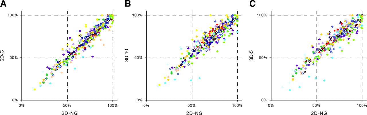
  • FIGURE 2.
    • Download figure
    • Open in new tab
    • Download powerpoint
    FIGURE 2.

    Relative activity concentration in each segment with 2D-G (A), 3D-10 (B), and 3D-5 (C) protocols, compared with 2D-NG acquisition. Segment with maximum absolute activity concentration in each patient equals 100%. Each dot represents 1 segment, and a different color represents each of the 20 patients. One patient with hibernating myocardium confirmed by 13NH3 PET was excluded.
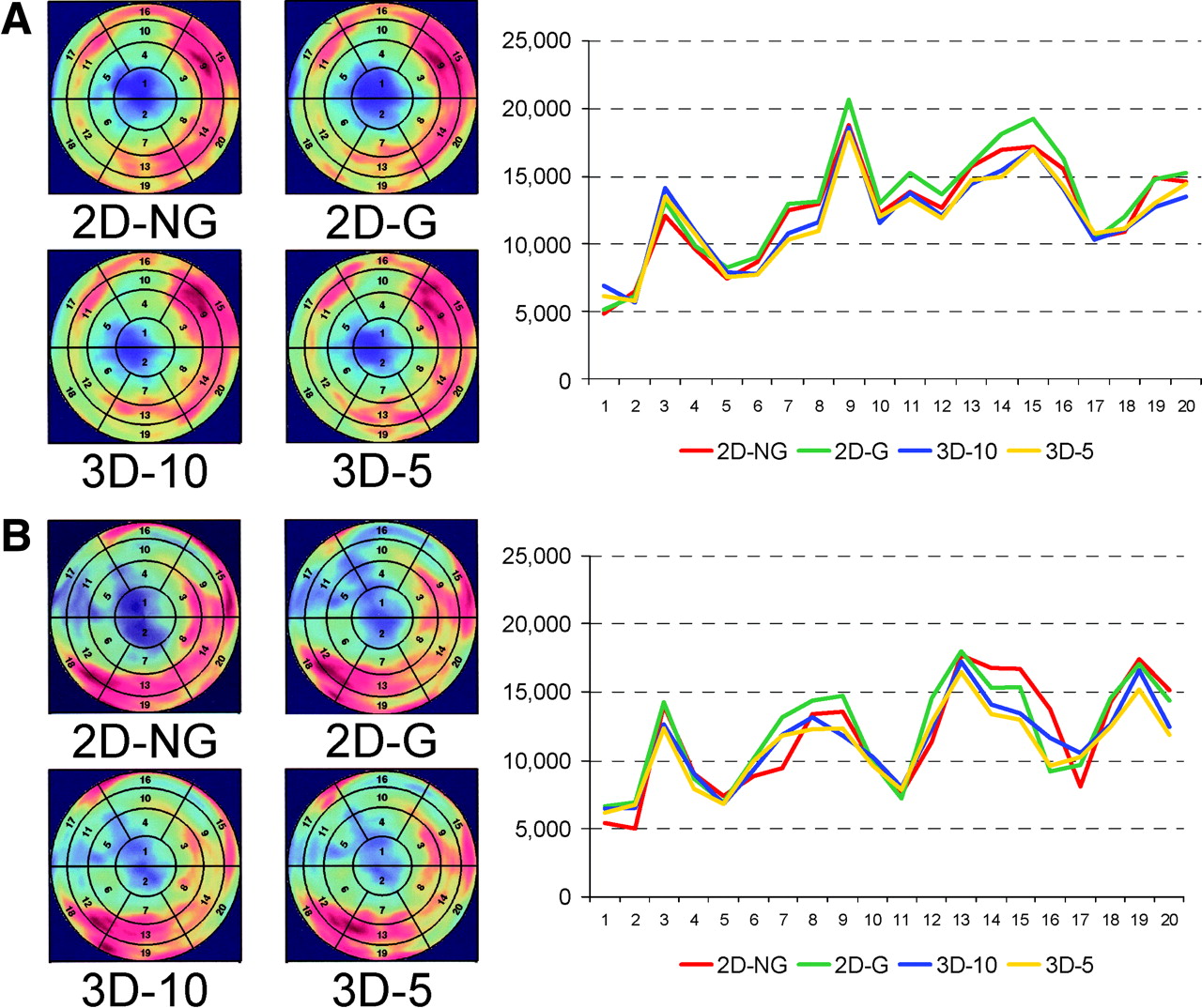
  • FIGURE 3.
    • Download figure
    • Open in new tab
    • Download powerpoint
    FIGURE 3.

    Regional variation of absolute activity concentration in each of 20 segments measured with 2D-NG, 2D-G, 3D-10, and 3D-5 protocols. Polar maps are shown for each protocol, and absolute activity concentrations (Bq/mL) have been plotted for each segment. Two representative patients (A and B) are shown.
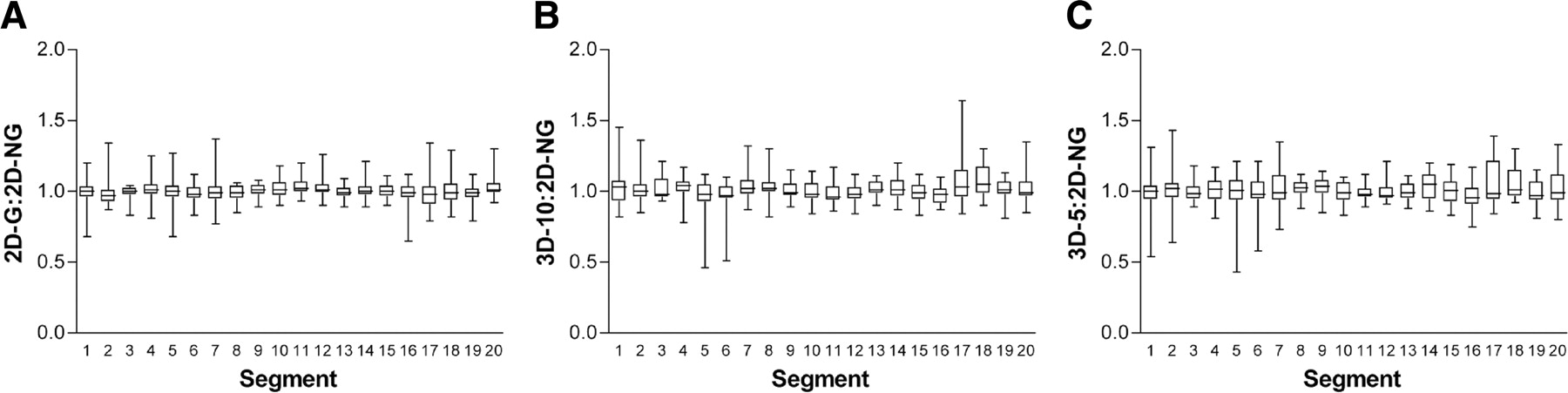
  • FIGURE 4.
    • Download figure
    • Open in new tab
    • Download powerpoint
    FIGURE 4.

    Mean regional activity distribution in all 20 segments measured with 2D-G (A), 3D-10 (B), and 3D-5 (C) protocols, expressed as a ratio to 2D-NG values.
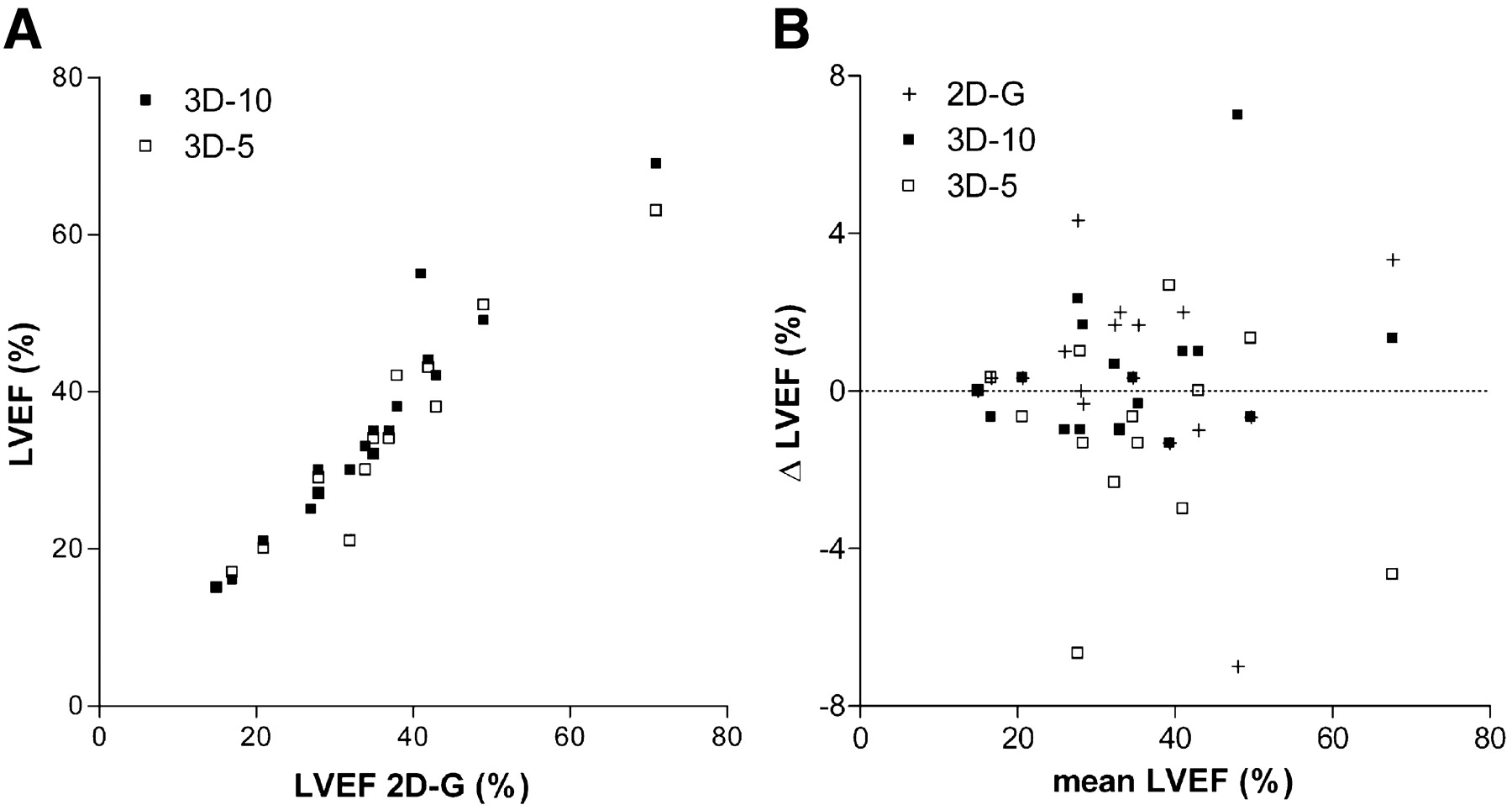
  • FIGURE 5.
    • Download figure
    • Open in new tab
    • Download powerpoint
    FIGURE 5.

    (A) Comparison of LVEF measured with 2D-G, 3D-10, and 3D-5 protocols. (B) Corresponding Bland–Altman plot. Slope of curve, {2D,3D} intercept, and Pearson correlation coefficient were 1.02, {0.4,-0.4}, and 0.96, respectively, for 3D-10 and 0.91, {-1.3,1.2}, and 0.96, respectively, for 3D-5.

Tables

  • Figures
    • View popup
    TABLE 1

    Count Statistics for 2D and 3D Acquisitions

    Parameter2D acquisition3D acquisition
    True coincidences(19.6 ± 4.98) × 103 s−1(164 ± 34.9) × 103 s−1
    Random coincidences(6.36 ± 2.65) × 103 s−1(124 ± 48.8) × 103 s−1
    True-to-random ratio3.261.40
    Single events(2.08 ± 0.57) × 106 s−1(6.33 ± 1.07) × 106 s−1
    Dead time5%15%
    • View popup
    TABLE 2

    Number of Segments Showing Viable Myocardium, Nontransmural Scarring, and Transmural Scarring, as Measured with 2D-NG, 2D-G, 3D-10, and 3D-5

    Acquisition typeViable myocardiumNontransmural scarringTransmural scarring
    2D-NG16114554
    2D-G14915556
    3D-1016514550
    3D-514416452
PreviousNext
Back to top

In this issue

Journal of Nuclear Medicine: 46 (1)
Journal of Nuclear Medicine
Vol. 46, Issue 1
January 1, 2005
  • Table of Contents
  • About the Cover
  • Index by author
  • Supplement "Clinical Practice of Molecular Radiotherapy"
Print
Download PDF
Article Alerts
Sign In to Email Alerts with your Email Address
Email Article

Thank you for your interest in spreading the word on Journal of Nuclear Medicine.

NOTE: We only request your email address so that the person you are recommending the page to knows that you wanted them to see it, and that it is not junk mail. We do not capture any email address.

Enter multiple addresses on separate lines or separate them with commas.
18F-FDG PET for Detecting Myocardial Viability: Validation of 3D Data Acquisition
(Your Name) has sent you a message from Journal of Nuclear Medicine
(Your Name) thought you would like to see the Journal of Nuclear Medicine web site.
Citation Tools
18F-FDG PET for Detecting Myocardial Viability: Validation of 3D Data Acquisition
Claudia Brogsitter, Thomas Grüning, Reiner Weise, Peter Wielepp, Oliver Lindner, Reiner Körfer, Wolfgang Burchert
Journal of Nuclear Medicine Jan 2005, 46 (1) 19-24;

Citation Manager Formats

  • BibTeX
  • Bookends
  • EasyBib
  • EndNote (tagged)
  • EndNote 8 (xml)
  • Medlars
  • Mendeley
  • Papers
  • RefWorks Tagged
  • Ref Manager
  • RIS
  • Zotero
Share
18F-FDG PET for Detecting Myocardial Viability: Validation of 3D Data Acquisition
Claudia Brogsitter, Thomas Grüning, Reiner Weise, Peter Wielepp, Oliver Lindner, Reiner Körfer, Wolfgang Burchert
Journal of Nuclear Medicine Jan 2005, 46 (1) 19-24;
Twitter logo Facebook logo LinkedIn logo Mendeley logo
  • Tweet Widget
  • Facebook Like
  • Google Plus One
Bookmark this article

Jump to section

  • Article
    • Abstract
    • MATERIALS AND METHODS
    • RESULTS
    • DISCUSSION
    • CONCLUSION
    • Footnotes
    • REFERENCES
  • Figures & Data
  • Info & Metrics
  • PDF

Related Articles

  • THIS MONTH IN JNM
  • PubMed
  • Google Scholar

Cited By...

  • Artifacts from Misaligned CT in Cardiac Perfusion PET/CT Studies: Frequency, Effects, and Potential Solutions
  • PET/CT: Challenge for Nuclear Cardiology
  • Google Scholar

More in this TOC Section

  • Feasibility of Ultra-Low-Activity 18F-FDG PET/CT Imaging Using a Long–Axial-Field-of-View PET/CT System
  • Cardiac Presynaptic Sympathetic Nervous Function Evaluated by Cardiac PET in Patients with Chronotropic Incompetence Without Heart Failure
  • Validation and Evaluation of a Vendor-Provided Head Motion Correction Algorithm on the uMI Panorama PET/CT System
Show more Clinical Investigations

Similar Articles

SNMMI

© 2025 SNMMI

Powered by HighWire