


Article Figures & Data
Figures
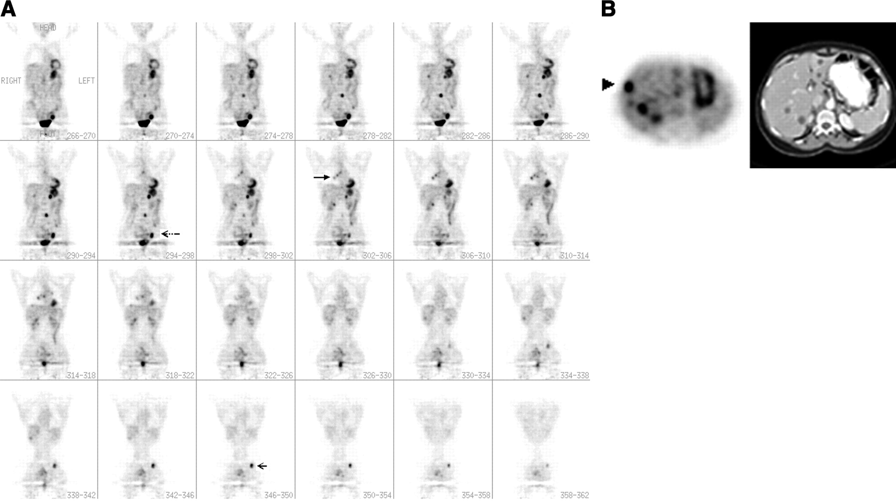
- FIGURE 1.
(A) Whole-body PET scan of a patient with a recently removed stage II melanoma of the right arm shows several lesions in the right hilum (large arrow), the liver, the left groin (dotted arrow), the lumbar spine, and the left ischium (small arrow). (B) CT of the chest, abdomen, and pelvis was able to identify only the liver metastases (right panel); PET showed the same lesions in the liver but detected an additional focus in the anterior right liver (arrowhead, left panel). The diagnosis of metastatic spread was further established by follow-up of the patient.
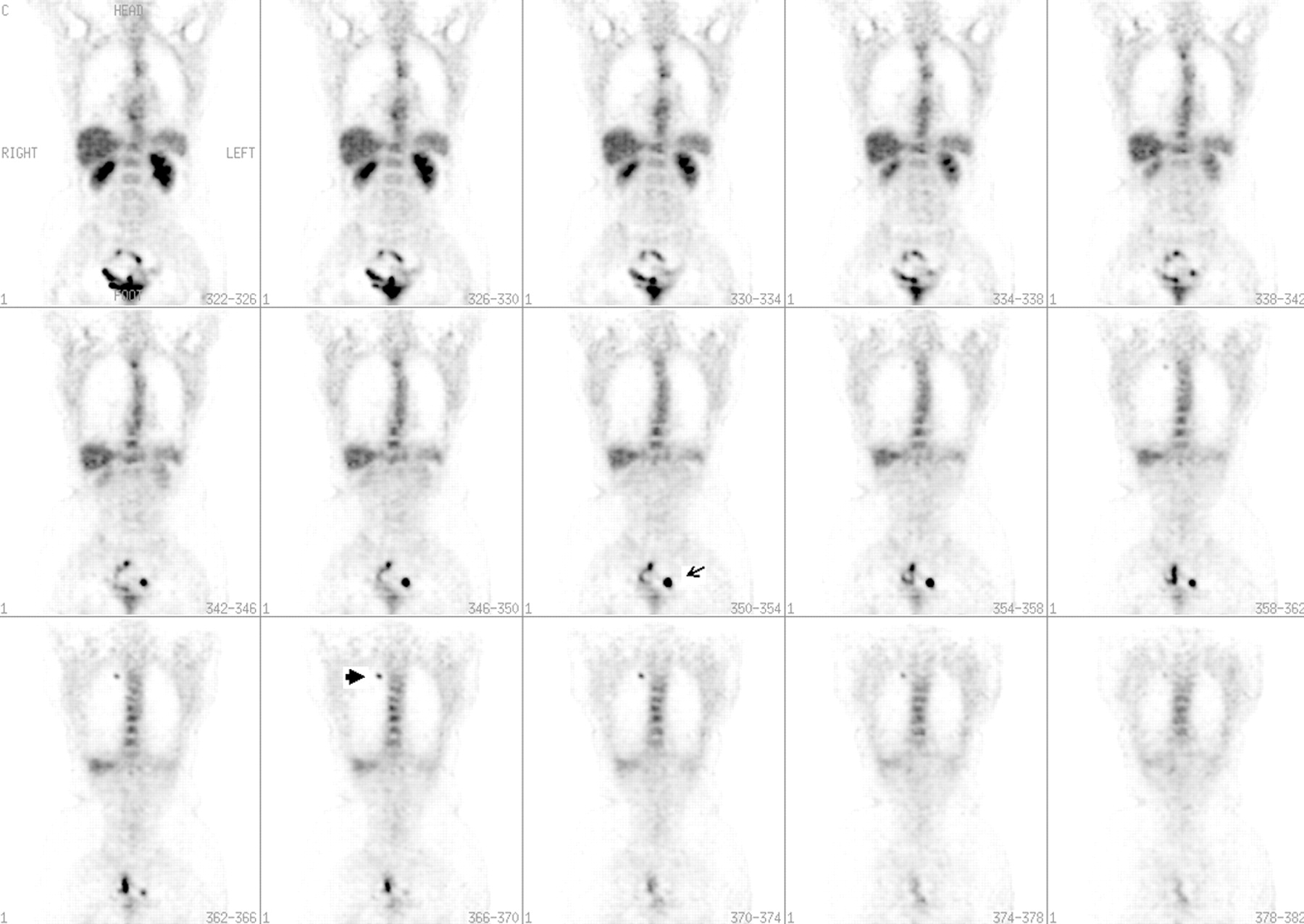
- FIGURE 2.
Whole-body PET scan shows focal uptake in the apex of the right lung, in the same location as a 2-cm lesion observed on a prior chest CT scan (large arrow). PET also detected several lesions that extended to the midline in the left pelvis and were not found by other CPs (small arrow). These lesions were histologically confirmed to be lymph node metastases.
Tables
- TABLE 1
A Comparison of PET and Body CT Results in the Detection of Active Melanoma in 115 Patients
Site PET CT TN TP FN FP SE (%) SP (%) Acc (%) TN TP FN FP SE (%) SP (%) Acc (%) Lung 68 16 12 5 57 92 83 51 26 2 22 93 70 77 Liver 84 11 2 — 85 100 98 67 10 3 17 77 80 79 Bone 88 8 — 1 100 99 99 86 1 7 3 12 96 89 Lymph node 60 30 4 3 88 95 93 51 19 15 12 56 81 72 Abdomen 83 6 2 2 75 97 96 71 5 3 14 62 84 82 TN = true negative; TP = true positive; FN = false negative; FP = false positive; SE = sensitivity; SP = specificity; Acc = accuracy.


Jump to section
Related Articles
Cited By...
- Nuclear Medicine and Molecular Imaging in Nodal Staging and Surveillance of Ocular Melanoma: Case Reports and Review of the Literature
- Prospective Comparison of [18F]Fluorodeoxyglucose Positron Emission Tomography and Computed Tomography in Patients With Melanoma With Palpable Lymph Node Metastases: Diagnostic Accuracy and Impact on Treatment
- Role of Modern Imaging Techniques for Diagnosis of Infection in the Era of 18F-Fluorodeoxyglucose Positron Emission Tomography
- Role of Nuclear Medicine in the Management of Cutaneous Malignant Melanoma
- Diagnostic Performance of Whole Body Dual Modality 18F-FDG PET/CT Imaging for N- and M-Staging of Malignant Melanoma: Experience With 250 Consecutive Patients