
Main menu

  • Home
  • Content
    • Current
    • Ahead of print
    • Past Issues
    • JNM Supplement
    • SNMMI Annual Meeting Abstracts
    • Continuing Education
    • JNM Podcasts
  • Subscriptions
    • Subscribers
    • Institutional and Non-member
    • Rates
    • Journal Claims
    • Corporate & Special Sales
  • Authors
    • Submit to JNM
    • Information for Authors
    • Assignment of Copyright
    • AQARA requirements
  • Info
    • Reviewers
    • Permissions
    • Advertisers
  • About
    • About Us
    • Editorial Board
    • Contact Information
  • More
    • Alerts
    • Feedback
    • Help
    • SNMMI Journals
  • SNMMI
    • JNM
    • JNMT
    • SNMMI Journals
    • SNMMI

User menu

  • Subscribe
  • My alerts
  • Log in
  • Log out
  • My Cart

Search

  • Advanced search
Journal of Nuclear Medicine
  • SNMMI
    • JNM
    • JNMT
    • SNMMI Journals
    • SNMMI
  • Subscribe
  • My alerts
  • Log in
  • Log out
  • My Cart
Journal of Nuclear Medicine

Advanced Search

  • Home
  • Content
    • Current
    • Ahead of print
    • Past Issues
    • JNM Supplement
    • SNMMI Annual Meeting Abstracts
    • Continuing Education
    • JNM Podcasts
  • Subscriptions
    • Subscribers
    • Institutional and Non-member
    • Rates
    • Journal Claims
    • Corporate & Special Sales
  • Authors
    • Submit to JNM
    • Information for Authors
    • Assignment of Copyright
    • AQARA requirements
  • Info
    • Reviewers
    • Permissions
    • Advertisers
  • About
    • About Us
    • Editorial Board
    • Contact Information
  • More
    • Alerts
    • Feedback
    • Help
    • SNMMI Journals
  • View or Listen to JNM Podcast
  • Visit JNM on Facebook
  • Join JNM on LinkedIn
  • Follow JNM on Twitter
  • Subscribe to our RSS feeds
OtherClinical Investigations

Overexpression of Glut1 in Lymphoid Follicles Correlates with False-Positive 18F-FDG PET Results in Lung Cancer Staging

Jin-Haeng Chung, Kyung-Ja Cho, Seung-Sook Lee, Hee Jong Baek, Jong-Ho Park, Gi Jeong Cheon, Chang-Woon Choi and Sang Moo Lim
Journal of Nuclear Medicine June 2004, 45 (6) 999-1003;
Jin-Haeng Chung
  • Find this author on Google Scholar
  • Find this author on PubMed
  • Search for this author on this site
Kyung-Ja Cho
  • Find this author on Google Scholar
  • Find this author on PubMed
  • Search for this author on this site
Seung-Sook Lee
  • Find this author on Google Scholar
  • Find this author on PubMed
  • Search for this author on this site
Hee Jong Baek
  • Find this author on Google Scholar
  • Find this author on PubMed
  • Search for this author on this site
Jong-Ho Park
  • Find this author on Google Scholar
  • Find this author on PubMed
  • Search for this author on this site
Gi Jeong Cheon
  • Find this author on Google Scholar
  • Find this author on PubMed
  • Search for this author on this site
Chang-Woon Choi
  • Find this author on Google Scholar
  • Find this author on PubMed
  • Search for this author on this site
Sang Moo Lim
  • Find this author on Google Scholar
  • Find this author on PubMed
  • Search for this author on this site
  • Article
  • Figures & Data
  • Info & Metrics
  • PDF
Loading

Article Figures & Data

Figures

  • Tables
  • FIGURE 1.
    • Download figure
    • Open in new tab
    • Download powerpoint
    FIGURE 1.

    18F-FDG PET of mediastinal lymph nodes. (A) 18F-FDG PET image of TP lymph node in 64-y-old man. 18F-FDG PET scan shows hypermetabolic lung mass in right middle lobe with standardized uptake value (SUV) of 8.4 and small hypermetabolic lymph node in prevascular space with SUV of 6.3. Lymph node had high activity of 18F-FDG with focal appearance on 18F-FDG PET, so lesion was classified preoperatively as a metastatic lymph node. After surgery, adenocarcinoma and lymph node metastasis were proven on pathologic examination of surgical specimen. (B) 18F-FDG PET image of FP lymph node in 64-y-old man. 18F-FDG PET scan shows hypermetabolic lung mass in right upper lobe with SUV of 12.5 and small hypermetabolic lesion in right paratracheal lymph node with SUV of 4.3. Lymph node had high activity of 18F-FDG with focal appearance on 18F-FDG PET, so lesion was classified preoperatively as a metastatic lymph node. After surgery, squamous cell carcinoma was proven in the main mass, but there was no metastasis in lymph node.

  • FIGURE 2.
    • Download figure
    • Open in new tab
    • Download powerpoint
    FIGURE 2.

    Glut1 immunostaining in metastatic non-small cell carcinomas. (A) Squamous cell carcinoma with strong membranous Glut1 immunostaining (STAIN, ×200). (B) Adenocarcinoma with focal cytoplasmic Glut1 immunostaining (immunostain, ×200).
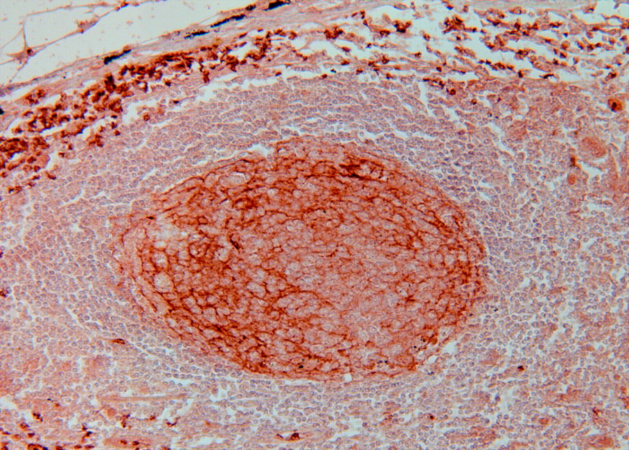
  • FIGURE 3.
    • Download figure
    • Open in new tab
    • Download powerpoint
    FIGURE 3.

    Glut1 immunostaining in lymphoid follicles. Note intense linear membranous staining in lymphoid follicular cells and erythrocytes as an internal positive control (immunostain, ×200).

Tables

  • Figures
    • View popup
    TABLE 1

    Number of Studied Lymph Nodes by Histologic Subtype and Results of 18F-FDG PET

    18F-FDG PETSQCCADCOthersTotal
    FN011112
    FP129627
    TN1419841
    TP1712231
    Total435117111
    • SQCC = squamous cell carcinoma; ADC = adenocarcinoma.

    • View popup
    TABLE 2

    Features of Histologically Proven Negative Lymph Nodes

    PET resultsFP (n = 27)TN (n = 41)P value
    Size (mm)11.0 ± 2.99.7 ± 2.80.071
    Follicular hyperplasia2.7 ± 1.11.6 ± 0.7<0.001
    Glut1 score3.0 ± 0.81.5 ± 0.9<0.001
    • View popup
    TABLE 3

    Features of Histologically Proven Positive Lymph Nodes

    PET resultsFN (n = 12)TP (n = 31)P value
    Size (mm)11.2 ± 2.713.3 ± 5.00.584
    Tumor volume (%)52.5 ± 31.267.9 ± 35.00.095
    Glut1 score1.8 ± 0.73.1 ± 0.9<0.001
PreviousNext
Back to top

In this issue

Journal of Nuclear Medicine: 45 (6)
Journal of Nuclear Medicine
Vol. 45, Issue 6
June 1, 2004
  • Table of Contents
  • About the Cover
  • Index by author
Print
Download PDF
Article Alerts
Sign In to Email Alerts with your Email Address
Email Article

Thank you for your interest in spreading the word on Journal of Nuclear Medicine.

NOTE: We only request your email address so that the person you are recommending the page to knows that you wanted them to see it, and that it is not junk mail. We do not capture any email address.

Enter multiple addresses on separate lines or separate them with commas.
Overexpression of Glut1 in Lymphoid Follicles Correlates with False-Positive 18F-FDG PET Results in Lung Cancer Staging
(Your Name) has sent you a message from Journal of Nuclear Medicine
(Your Name) thought you would like to see the Journal of Nuclear Medicine web site.
Citation Tools
Overexpression of Glut1 in Lymphoid Follicles Correlates with False-Positive 18F-FDG PET Results in Lung Cancer Staging
Jin-Haeng Chung, Kyung-Ja Cho, Seung-Sook Lee, Hee Jong Baek, Jong-Ho Park, Gi Jeong Cheon, Chang-Woon Choi, Sang Moo Lim
Journal of Nuclear Medicine Jun 2004, 45 (6) 999-1003;

Citation Manager Formats

  • BibTeX
  • Bookends
  • EasyBib
  • EndNote (tagged)
  • EndNote 8 (xml)
  • Medlars
  • Mendeley
  • Papers
  • RefWorks Tagged
  • Ref Manager
  • RIS
  • Zotero
Share
Overexpression of Glut1 in Lymphoid Follicles Correlates with False-Positive 18F-FDG PET Results in Lung Cancer Staging
Jin-Haeng Chung, Kyung-Ja Cho, Seung-Sook Lee, Hee Jong Baek, Jong-Ho Park, Gi Jeong Cheon, Chang-Woon Choi, Sang Moo Lim
Journal of Nuclear Medicine Jun 2004, 45 (6) 999-1003;
Twitter logo Facebook logo LinkedIn logo Mendeley logo
  • Tweet Widget
  • Facebook Like
  • Google Plus One
Bookmark this article

Jump to section

  • Article
    • Abstract
    • MATERIALS AND METHODS
    • RESULTS
    • DISCUSSION
    • CONCLUSION
    • Acknowledgments
    • Footnotes
    • REFERENCES
  • Figures & Data
  • Info & Metrics
  • PDF

Related Articles

  • No related articles found.
  • PubMed
  • Google Scholar

Cited By...

  • Biological Correlation of 18F-FDG Uptake on PET in Pulmonary Neuroendocrine Tumors
  • EBUS-TBNA in the differential diagnosis of pulmonary artery sarcoma and thromboembolism
  • Biologic Correlation of 2-[18F]-Fluoro-2-Deoxy-D-Glucose Uptake on Positron Emission Tomography in Thymic Epithelial Tumors
  • 18F-FDG Uptake in Lung, Breast, and Colon Cancers: Molecular Biology Correlates and Disease Characterization
  • 18F-FDG and 18F-FLT Do Not Discriminate Between Reactive and Metastatic Lymph Nodes in Oral Cancer
  • 18F-FDG Uptake in Reactive Neck Lymph Nodes of Oral Cancer: Relationship to Lymphoid Follicles
  • Fluorine-18-{alpha}-Methyltyrosine Positron Emission Tomography for Diagnosis and Staging of Lung Cancer: A Clinicopathologic Study
  • In Vitro Studies on the Signal Transduction of Thyroidal Uptake of 18F-FDG and 131I-Iodide
  • GLUT1 Expression in Tissue and 18F-FDG Uptake
  • Google Scholar

More in this TOC Section

  • Feasibility of Ultra-Low-Activity 18F-FDG PET/CT Imaging Using a Long–Axial-Field-of-View PET/CT System
  • Cardiac Presynaptic Sympathetic Nervous Function Evaluated by Cardiac PET in Patients with Chronotropic Incompetence Without Heart Failure
  • Validation and Evaluation of a Vendor-Provided Head Motion Correction Algorithm on the uMI Panorama PET/CT System
Show more Clinical Investigations

Similar Articles

SNMMI

© 2025 SNMMI

Powered by HighWire