


Article Figures & Data
Figures
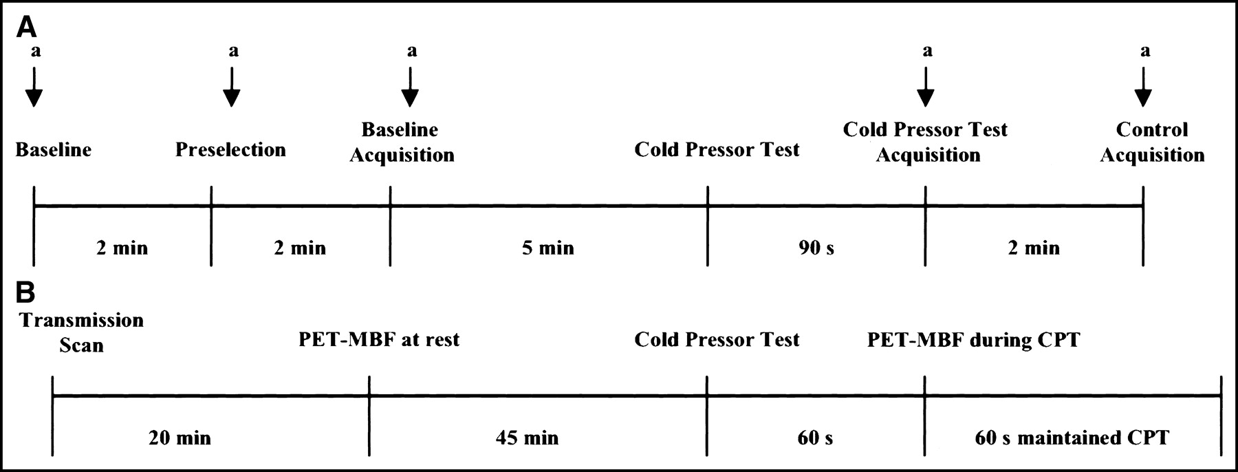
- FIGURE 1.
Study protocol. (A) Study inclusion at coronary angiography and quantitative assessment of epicardial vasoreactivity during CPT. a = angiogram. (B) Within 20 d of study inclusion MBF assessment with PET at rest and during CPT.
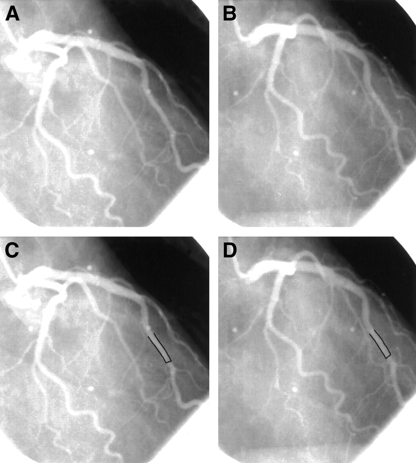
- FIGURE 2.
(A) Normal coronary angiogram of the left coronary tree in the RAO view of a control patient. (B) Corresponding coronary angiogram during sympathetic activation by CPT. (C and D) Quantitative coronary angiographic evaluation of the proximal-mid LAD segment at rest (mean diameter, 2.0 mm) (C) and during CPT (mean diameter, 2.5 mm) (D).
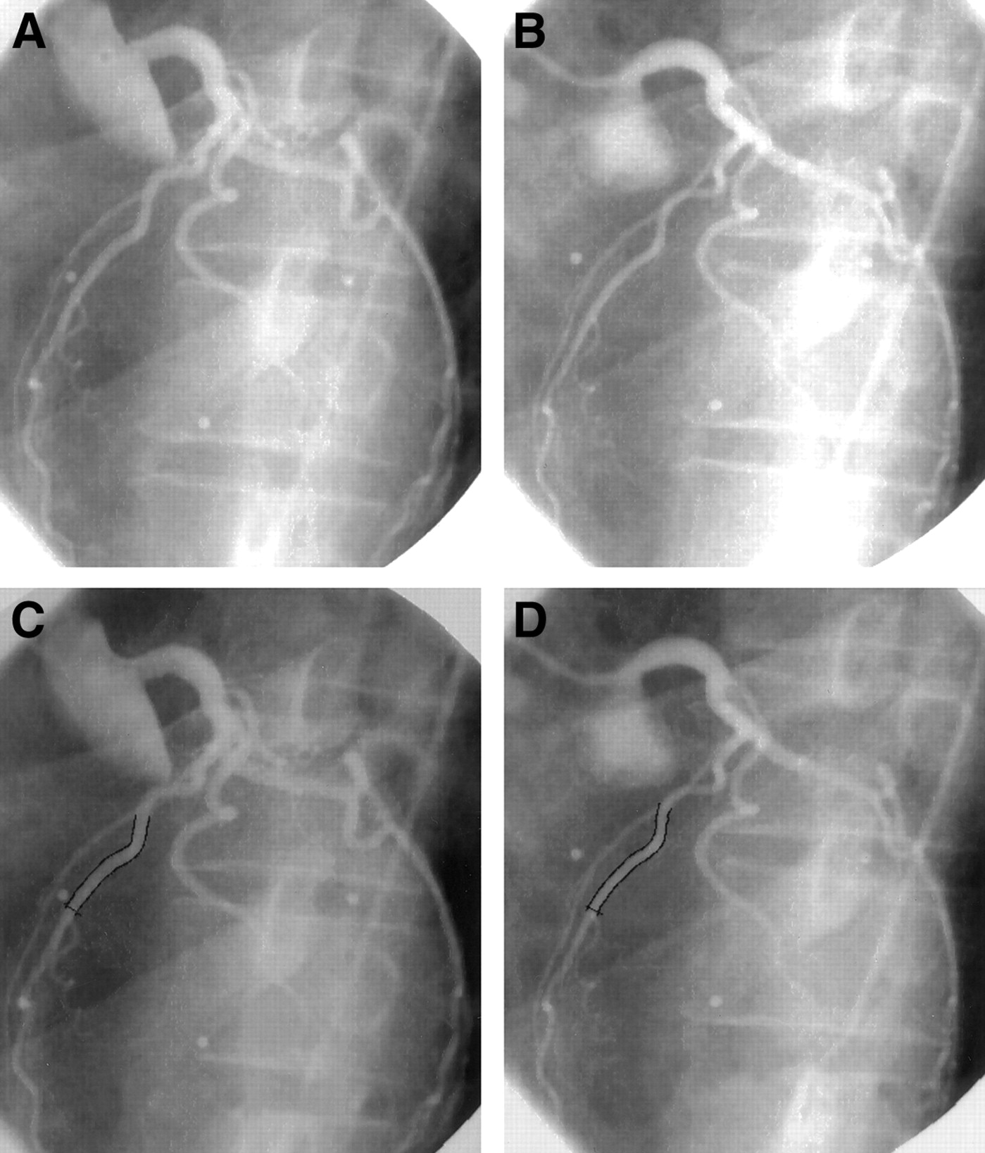
- FIGURE 3.
(A) Normal coronary angiogram of the left coronary artery tree in the LAO view in a chronic smoker at baseline. (B) Corresponding coronary angiogram during sympathetic activation by CPT. (C and D) Quantitative coronary angiographic evaluation of the proximal-mid LAD segment at rest (mean diameter, 1.89 mm) (C) and during CPT (mean diameter, 1.57 mm) (D).
- FIGURE 4.
Correlation of CPT-induced change in RPP and epicardial LA in normal control patients (○) and in the group at risk for CAD (•), demonstrating a significant correlation in normal patients but no relation for patients at risk for CAD.
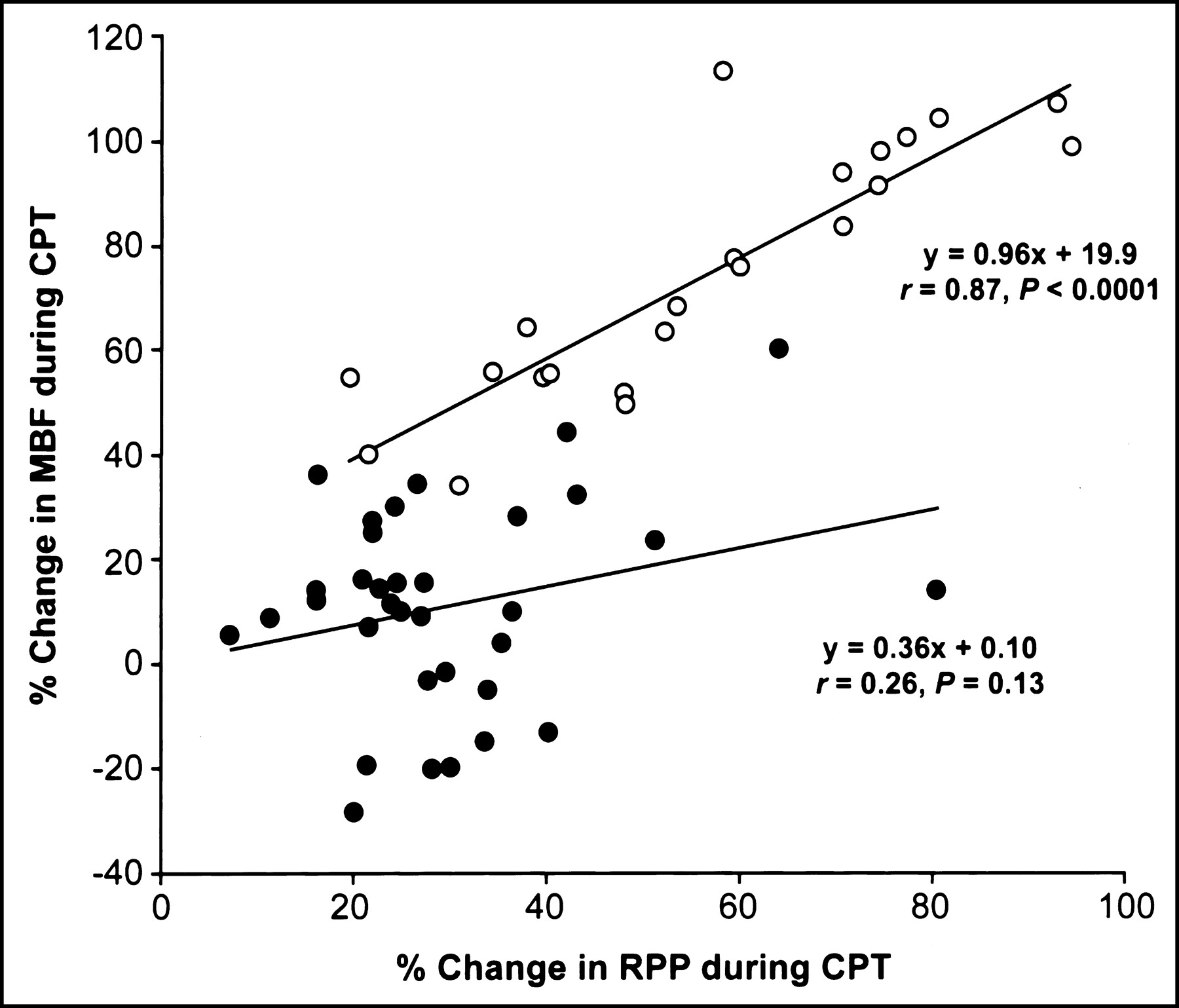
- FIGURE 5.
Correlation of CPT-induced change in RPP and regional MBF in normal control patients (○) and in the group at risk for CAD (•), revealing a significant correlation in normal patients but no relation for patients at risk for CAD.
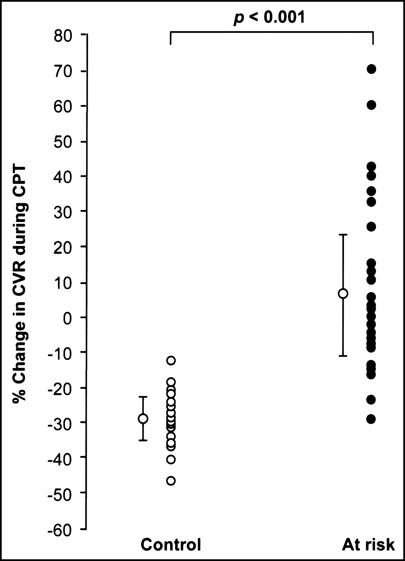
- FIGURE 6.
Percent change of CVR in the control group and in patients at risk for CAD.
- FIGURE 7.
Percent change of epicardial LA and MBF to CPT of the study population.


Tables
Characteristic Control group (n = 22) At-risk group (n = 34) Age (y) 57 ± 6 56 ± 6 Sex (F/M) 14/8 19/15 Hypertension 0 16 Hypercholesterolemia 0 10 Smoking 0 8 Lipid status Serum cholesterol level (mg/dL) 186 ± 50 218 ± 40* Serum LDL level (mg/dL) 114 ± 40 144 ± 24* Serum HDL level (mg/dL) 53 ± 12 47 ± 12 Triglyceride level 125 ± 38 140 ± 52 Glucose level (mg/dL) 88 ± 7 95 ± 15 Epicardial LA (mm2) Before CPT 5.01 ± 1.07 5.13 ± 1.49 After CPT 5.88 ± 0.89 4.24 ± 1.12 % Change 19 ± 9 −16 ± 12* MBF (mL · g−1 · min−1) Before CPT 0.77 ± 0.16 0.76 ± 0.20 After CPT 1.34 ± 0.34 0.83 ± 0.25 % Change 74 ± 23 11 ± 19* Coronary vascular resistance (mm Hg/mL · g−1 · min−1) Before CPT 114 ± 23 122 ± 27 After CPT 80 ± 14 128 ± 32 % Change −43 ± 13 6 ± 17* ↵* P < 0.001 vs. normal controls.
LDL = low-density lipoprotein; HDL = high-density lipoprotein.
Values are mean ± SD.
Test QCA PET Rest CPT Rest CPT HR (bpm) 62 ± 8 67 ± 7* 60 ± 9 66 ± 10† SBP (mm Hg) 108 ± 13 136 ± 17* 111 ± 11 139 ± 17† DBP (mm Hg) 75 ± 12 81 ± 9* 73 ± 8 81 ± 8† RPP 6,674 ± 995 9,146 ± 1,431* 6,636 ± 1,221 9,199 ± 1,634† %ΔRPP 37 ± 17* 40 ± 21† MAP (mm Hg) 87 ± 6 100 ± 7* 86 ± 5 101 ± 7† ↵* P ≤ 0.0001, CPT vs. rest for each corresponding hemodynamic parameter in QCA study evaluation.
↵† P ≤ 0.0001, CPT vs. rest for each corresponding hemodynamic parameter in PET study evaluation.
HR = heart rate; SBP = systolic blood pressure; DBP = diastolic blood pressure; RPP = systolic blood pressure × heart rate; %ΔRPP = percent change of RPP from rest during CPT; MAP = mean arterial blood pressure.
Values are mean ± SD.
In this issue


Jump to section
Related Articles
Cited By...
- Total-Body Perfusion Imaging with [11C]-Butanol
- Coronary Microvascular Dysfunction: Clinical Considerations and Noninvasive Diagnosis
- Myocardial Blood Flow and Coronary Flow Reserve During 3 Years Following Bioresorbable Vascular Scaffold Versus Metallic Drug-Eluting Stent Implantation: The VANISH Trial
- Clinical Quantification of Myocardial Blood Flow Using PET: Joint Position Paper of the SNMMI Cardiovascular Council and the ASNC
- Atherosclerotic Plaque Characteristics by CT Angiography Identify Coronary Lesions That Cause Ischemia: A Direct Comparison to Fractional Flow Reserve
- Structural Abnormalities of the Coronary Arterial Wall--in Addition to Luminal Narrowing--Affect Myocardial Blood Flow Reserve
- Cardiac PET Imaging for the Detection and Monitoring of Coronary Artery Disease and Microvascular Health
- Coronary Circulatory Function Abnormalities in Insulin Resistance: Insights From Positron Emission Tomography
- Improvement in coronary vascular dysfunction produced with euglycaemic control in patients with type 2 diabetes
- Smoking Cessation Normalizes Coronary Endothelial Vasomotor Response Assessed with 15O-Water and PET in Healthy Young Smokers
- Repeatability of Cold Pressor Test-Induced Flow Increase Assessed with H215O and PET
- Relationship Between Increasing Body Weight, Insulin Resistance, Inflammation, Adipocytokine Leptin, and Coronary Circulatory Function
- Coronary Circulatory Dysfunction in Insulin Resistance, Impaired Glucose Tolerance, and Type 2 Diabetes Mellitus
- Positron Emission Tomography-Measured Abnormal Responses of Myocardial Blood Flow to Sympathetic Stimulation Are Associated With the Risk of Developing Cardiovascular Events