


Article Figures & Data
Figures
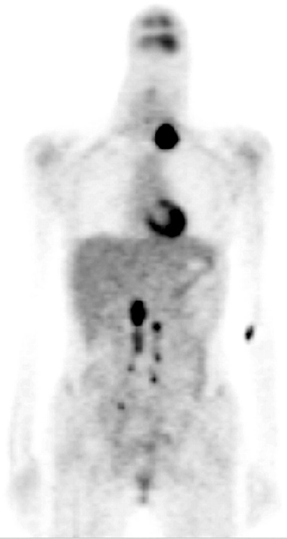
- FIGURE 1.
A 25-y-old woman with poorly differentiated squamous cell carcinoma of uterine cervix, FIGO stage IIIb, underwent concurrent chemoradiation therapy. Three months later, a left neck mass was palpated. Abdominopelvic MRI and chest CT showed no definite abnormal findings except an enlarged supraclavicular LN. Balancing between salvage RT and palliation treatment, PET was performed and suggested nodal metastases at the left supraclavicular, the bilateral upper and lower paraaortic, and the bilateral pelvic regions. After the left supraclavicular and paraaortic nodal metastases were confirmed histopathologically, she received palliation treatment.
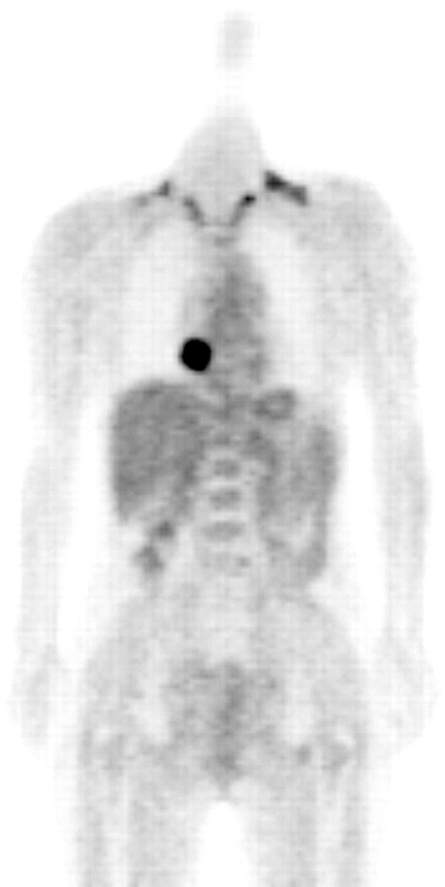
- FIGURE 2.
A 52-y-old woman with poorly differentiated squamous cell carcinoma of uterine cervix, FIGO stage IIa, underwent concurrent chemoradiation therapy. Four months after complement of treatment, an elevated serum SCC-Ag of 2.23 ng/mL was noted. Abdominopelvic MRI and chest RT showed negative findings. Two months later, her serum SCC-Ag level was 7.36 ng/mL. Conventional images still showed negative findings. A PET scan was then obtained and disclosed a metastatic lesion in the right lower lung. She subsequently received pneumonectomy and was well for 1 y.
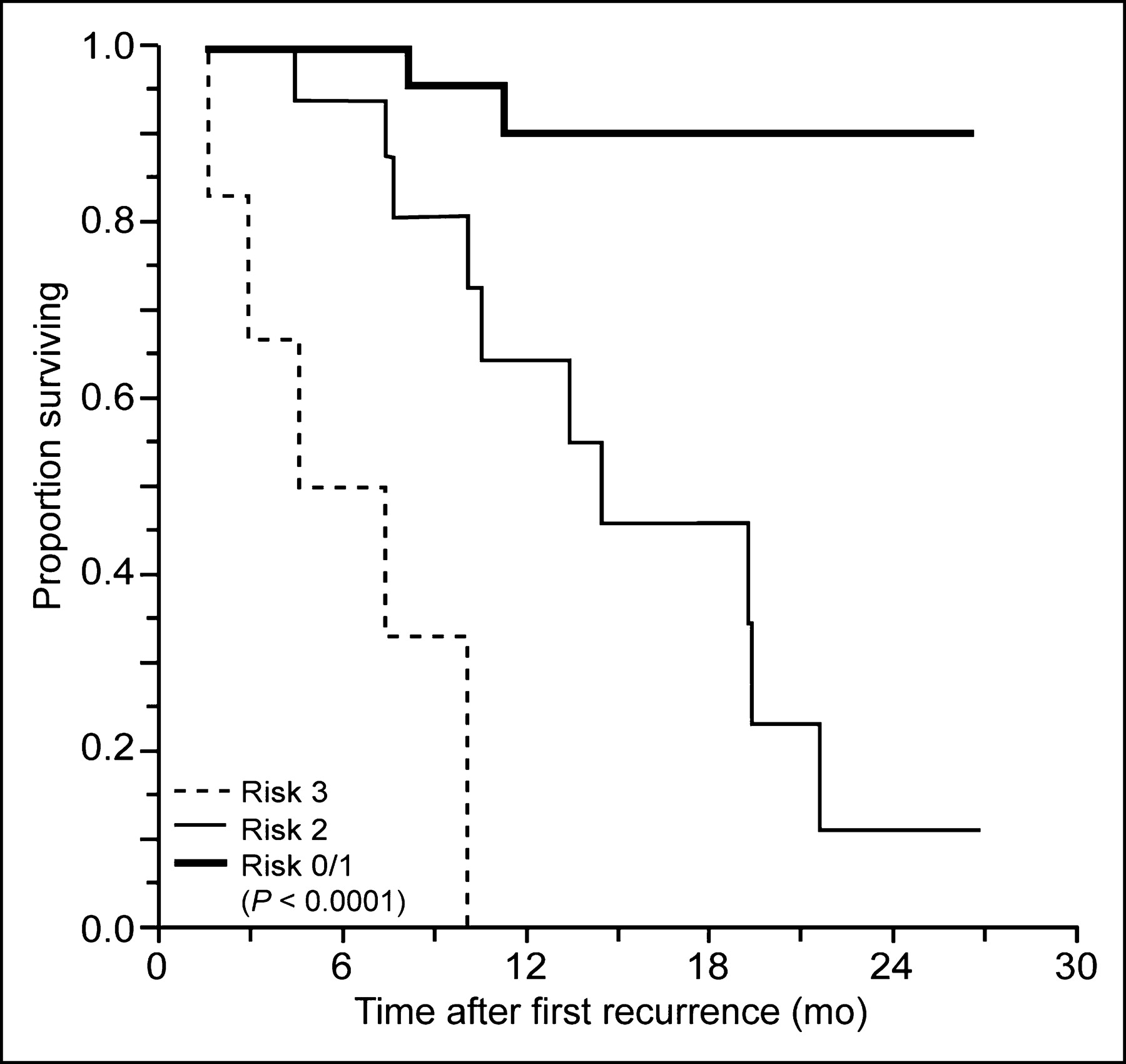
- FIGURE 3.
Kaplan–Meier curves for 2-y OS rates in patients with recurrent cervical cancer. Patients are categorized by risk score of ≤1 (bold solid line), 2 (thin solid line), and 3 (dashed line) (P < 0.0001).


Tables
- TABLE 1
Results of 18F-FDG PET (40 Minutes and 3 Hours) and MRI in Recurrent Cervical Cancer Patients (n = 55) and Lesions (n = 550)
Site TP TN FP FN Sensitivity (%) [95% CI] Specificity (%) [95% CI] PPV (%) [95% CI] NPV (%) [95% CI] Accuracy (%) [95% CI] Peritoneum PET 7 45 2 1 87.5 [47.3–99.7] 95.7 [85.5–99.5] 77.8 [40.0–97.2] 97.8 [88.5–99.9] 94.5 [84.9–98.9] CT/MRI 0 47 0 8 0 [—] 100 [—] N/A 85.5 [73.3–93.5] 85.5 [73.3–93.5] Bone PET 0 54 1 0 N/A 98.2 [90.3–100] 0 [—] 100 [—] 98.2 [90.3–100] CT/MRI 0 52 3 0 N/A 94.5 [84.9–98.9] 0 [—] 100 [—] 94.5 [84.9–98.9] Liver/spleen PET 2 52 1 0 100 [—] 98.1 [89.9–100] 66.7 [9.4–99.2] 100 [—] 98.2 [90.3–100] CT/MRI 0 52 1 2 0 [—] 98.1 [89.9–100] 0 [—] 96.3 [87.3–99.5] 94.5 [84.9–98.9] Lung PET 7 46 0 2 77.8 [40.0–97.2] 100 [—] 100 [—] 95.8 [85.7–99.5] 96.4 [87.5–99.6] CT/MRI 4 46 0 5 44.4 [13.7–78.8] 100 [—] 100 [—] 90.2 [78.6–96.7] 90.9 [80.0–97.0] Mediastinum PET 10 44 1 0 100 [—] 97.8 [88.2–99.9] 90.9 [58.7–99.8] 100 [—] 98.2 [90.3–100] CT/MRI 2 43 2 8 20.0 [2.5–55.6] 95.6 [84.9–99.5] 50.0 [6.8–93.2] 84.3 [71.4–93.0] 81.8 [69.1–90.9] Supraclavicular LN PET 11 41 1 2 84.6 [54.6–98.1] 97.6 [87.4–99.9] 91.7 [61.5–99.8] 95.3 [84.2–99.4] 94.5 [84.9–98.9] CT/MRI 9 42 0 4 69.2 [38.6–90.9] 100 [—] 100 [—] 91.3 [79.2–97.6] 92.7 [82.4–98.0] Paraaortic LN PET 15 38 0 2 88.2 [63.6–98.5] 100 [—] 100 [—] 95.0 [83.1–99.4] 96.4 [87.5–99.6] CT/MRI 9 37 1 8 52.9 [27.8–77.0] 97.4 [86.2–99.9] 90.0 [55.5–99.7] 82.2 [67.9–92.0] 83.6 [71.2–92.2] Pelvic LN PET 10 43 1 1 90.9 [58.7–99.8] 97.7 [88.0–99.9] 90.9 [58.7–99.8] 97.7 [88.0–99.9] 96.4 [87.5–99.6] CT/MRI 3 44 0 8 27.3 [6.0–61.0] 100 [—] 100 [—] 84.6 [71.9–93.1] 85.5 [73.3–93.5] Inguinal LN PET 4 51 0 0 100 [—] 100 [—] 100 [—] 100 [—] 100 [—] CT/MRI 2 51 0 2 50.0 [6.8–93.2] 100 [—] 100 [—] 96.2 [87.0–1.0] 96.4 [87.5–99.6] Metastatic lesions PET 66 414 7 8 89.2 [79.8–95.2] 98.3 [96.6–99.3] 90.4 [81.2–96.1] 98.1 [96.3–99.2] 97.0 [95.1–98.3] CT/MRI 29 414 7 45 39.2 [28.0–51.2] 98.3 [96.6–99.3] 80.6 [64.0–91.8] 90.2 [87.1–92.8] 89.5 [86.5–92.1] Central or pelvic recurrent or persistent tumors PET 18 34 1 2 90.0 [68.3–98.8] 97.1 [85.1–99.9] 94.7 [74.0–99.9] 94.4 [81.3–99.3] 94.5 [84.9–98.9] CT/MRI 16 34 1 4 80.0 [56.3–94.3] 97.1 [85.1–99.9] 94.1 [71.3–99.9] 89.5 [75.2–97.1] 90.9 [80.0–97.0] Total lesions PET 84 448 8 10 89.4 [81.3–94.8] 98.2 [96.6–99.2] 91.3 [83.6–96.2] 97.8 [96.0–98.9] 96.7 [94.9–98.0] CT/MRI 45 448 8 49 47.9 [37.5–58.4] 98.2 [96.6–99.2] 84.9 [72.4–93.3] 90.1 [87.2–92.6] 89.6 [86.8–92.1] PPV = positive predictive value; NPV = negative predictive value; N/A = not applicable.
- TABLE 2
Clinical Features and Univariate Analyses of Prognostic Covariates in Study Subjects (n = 55)
Feature No. of patients No. of deaths (%) OS (%) [95% CI] 6 mo 12 mo 24 mo P value* Initial FIGO stage IB–IIA 25 5 (20.0) 96.0 [88.3–100] 79.5 [61.2–97.8] 70.7 [47.6–93.7] 0.1184 IIB–IVA 30 13 (43.3) 88.5 [76.2–100] 62.1 [40.8–83.4] 13.6 [0–36.1] Histopathology SCC 44 17 (38.6) 90.2 [8.10–99.3] 63.2 [46.2–80.3] 28.3 [5.0–51.6] 0.1259 Adeno-adenosquamous 11 1 (9.1) 100 [—] 100 [—] 100 [—] Differentiation Well or moderate 34 12 (35.3) 90.9 [81.0–100] 90.9 [81.0–100] 33.0 [5.4–60.6] 0.8392 Poor 21 6 (28.6) 94.4 [83.9–100] 94.4 [83.9–100] 22.9 [0–60.5] Primary treatment Radical surgery 20 2 (10.0) 100 [—] 93.3 [80.7–100] 77.8 [48.0–100] 0.0081 RT 35 16 (45.7) 87.6 [76.2–99.0] 56.3 [36.2–76.4] 12.3 [0–32.8] Pelvic node metastasis before primary treatment Negative 23 5 (21.7) 100 [—] 91.3 [79.8–100] 86.1 [71.4–100] 0.0585 Positive 32 13 (40.6) 100 [—] 93.3 [84.4–100] 51.4 [29.5–73.4] Symptoms Symptomatic 18 8 (44.4) 75.6 [54.8–96.5] 57.6 [30.6–84.7] 21.7 [0–55.6] 0.0536 Asymptomatic 37 10 (27.0) 100 [—] 76.5 [59.9–93.1] 31.1 [0–62.9] SCC-Ag† (ng/mL) ≤4 26 3 (11.5) 95.5 [86.6–100] 88.6 [73.4–100] 59.1 [10.8–100] 0.0041 >4 26 14 (53.8) 88.1 [75.5–100] 48.5 [25.4–71.6] 11.1 [0–30.5] Locations of recurrences Central or pelvic 11 4 (36.4) 100 [—] 74.1 [42.5–100] 37.0 [0–90.7] 0.6951 Distant 27 6 (22.2) 96.3 [89.2–100] 75.8 [54.2–97.3] 32.5 [0–79.4] Pelvic and distant 17 8 (47) 80.4 [60.4–100] 58.4 [32.8–84.1] 36.5 [7.3–65.8] Salvage treatment‡ Curative intent 28 6 (21.4) 96.4 [89.6–100] 81.3 [64.4–98.3] 48.8 [12.4–85.1] 0.0642 Surgery§ 13 5 (38.5) 91.7 [76.0–100] 60.2 [21.3–99.0] 0 [—] CCRT 15 1 (6.7) 100 [—] 90.0 [71.4–100] 90.0 [71.4–100] Curative: no change 19 6 (31.6) 94.7 [84.7–100] 74.8 [53.2–95.4] 37.4 [0–75.6] 0.372 Curative: modified 9 0 (0) 100 [—] 100 [—] 100 [—] Palliation 27 12 (44.4) 88.6 [76.4–100] 65.6 [45.9–85.3] 27.8 [0–56.9] SCC = squamous cell carcinoma; CCRT = concurrent chemoradiation therapy.
↵* P values were determined using log-rank test.
↵† SCC-Ag levels were the value of at time of recurrence. Three patients were unavailable due to elevation of CEA.
↵‡ Difference (P = 0.0249) was not significant with Bonferroni correction for multiple comparisons.
↵§ Surgery consisted of paraaortic, neck, or inguinal lymph excision, radical hysterectomy, or pelvic exenteration. Adjuvant therapy consisted of intraoperative RT chemoradiation or RT to previously unradiated sites, postoperative adjuvant chemotherapy, or postoperative chemoradiation or RT to previously unradiated sites.
- TABLE 4
HRs and Modification of Salvage Treatment Due to 18F-FDG PET According to Risk Score (n = 52)
Risk score Treatment planning No. of patients No. of deaths (%) HR for death (95% CI) P value 0–1 27 2 (7.4) 1 (reference) No change 10 1 (10.0) Change: curative intent 7 0 (0) Change: palliation 10 1 (10.0) 2 19 10 (52.6) 6.91 (1.49–32.14) 0.014 No change 5 3 (60.0) Change: curative intent 2 0 (0) Change: palliation 12 7 (58.3) 3 6 5 (83.3) 60.46 (9.68–378.09) 0 < 0.0001 No change 1 1 (100) Change: curative intent 0 0 (0) Change: palliation 5 4 (80.0) * P values were determined using Cox proportional hazards model.
In this issue


Jump to section
Related Articles
Cited By...
- Para-aortic lymph node recurrence after curative radiotherapy for cervical cancer
- NCCN Task Force: Clinical Utility of PET in a Variety of Tumor Types
- Clarifying the Diagnosis of Clinically Suspected Recurrence of Cervical Cancer: Impact of 18F-FDG PET
- PET in Cervical Cancer -- Implications for `Staging,' Treatment Planning, Assessment of Prognosis, and Prediction of Response
- Expanding Role of Positron Emission Tomography in Cancer of the Uterine Cervix
- Oncologic Imaging in Gynecologic Malignancies
- MR imaging in cervical cancer: seeing is believing: The 2004 Mackenzie Davidson Memorial Lecture
- 18F-FDG PET Imaging in Posttherapy Monitoring of Cervical Cancers: From Diagnosis to Prognosis