
Main menu

  • Home
  • Content
    • Current
    • Ahead of print
    • Past Issues
    • JNM Supplement
    • SNMMI Annual Meeting Abstracts
    • Continuing Education
    • JNM Podcasts
  • Subscriptions
    • Subscribers
    • Institutional and Non-member
    • Rates
    • Journal Claims
    • Corporate & Special Sales
  • Authors
    • Submit to JNM
    • Information for Authors
    • Assignment of Copyright
    • AQARA requirements
  • Info
    • Reviewers
    • Permissions
    • Advertisers
  • About
    • About Us
    • Editorial Board
    • Contact Information
  • More
    • Alerts
    • Feedback
    • Help
    • SNMMI Journals
  • SNMMI
    • JNM
    • JNMT
    • SNMMI Journals
    • SNMMI

User menu

  • Subscribe
  • My alerts
  • Log in
  • My Cart

Search

  • Advanced search
Journal of Nuclear Medicine
  • SNMMI
    • JNM
    • JNMT
    • SNMMI Journals
    • SNMMI
  • Subscribe
  • My alerts
  • Log in
  • My Cart
Journal of Nuclear Medicine

Advanced Search

  • Home
  • Content
    • Current
    • Ahead of print
    • Past Issues
    • JNM Supplement
    • SNMMI Annual Meeting Abstracts
    • Continuing Education
    • JNM Podcasts
  • Subscriptions
    • Subscribers
    • Institutional and Non-member
    • Rates
    • Journal Claims
    • Corporate & Special Sales
  • Authors
    • Submit to JNM
    • Information for Authors
    • Assignment of Copyright
    • AQARA requirements
  • Info
    • Reviewers
    • Permissions
    • Advertisers
  • About
    • About Us
    • Editorial Board
    • Contact Information
  • More
    • Alerts
    • Feedback
    • Help
    • SNMMI Journals
  • View or Listen to JNM Podcast
  • Visit JNM on Facebook
  • Join JNM on LinkedIn
  • Follow JNM on Twitter
  • Subscribe to our RSS feeds
OtherContinuing Education

Preoperative Localization and Radioguided Parathyroid Surgery

Giuliano Mariani, Seza A. Gulec, Domenico Rubello, Giuseppe Boni, Marco Puccini, Maria Rosa Pelizzo, Gianpiero Manca, Dario Casara, Guido Sotti, Paola Erba, Duccio Volterrani and Armando E. Giuliano
Journal of Nuclear Medicine September 2003, 44 (9) 1443-1458;
Giuliano Mariani
  • Find this author on Google Scholar
  • Find this author on PubMed
  • Search for this author on this site
Seza A. Gulec
  • Find this author on Google Scholar
  • Find this author on PubMed
  • Search for this author on this site
Domenico Rubello
  • Find this author on Google Scholar
  • Find this author on PubMed
  • Search for this author on this site
Giuseppe Boni
  • Find this author on Google Scholar
  • Find this author on PubMed
  • Search for this author on this site
Marco Puccini
  • Find this author on Google Scholar
  • Find this author on PubMed
  • Search for this author on this site
Maria Rosa Pelizzo
  • Find this author on Google Scholar
  • Find this author on PubMed
  • Search for this author on this site
Gianpiero Manca
  • Find this author on Google Scholar
  • Find this author on PubMed
  • Search for this author on this site
Dario Casara
  • Find this author on Google Scholar
  • Find this author on PubMed
  • Search for this author on this site
Guido Sotti
  • Find this author on Google Scholar
  • Find this author on PubMed
  • Search for this author on this site
Paola Erba
  • Find this author on Google Scholar
  • Find this author on PubMed
  • Search for this author on this site
Duccio Volterrani
  • Find this author on Google Scholar
  • Find this author on PubMed
  • Search for this author on this site
Armando E. Giuliano
  • Find this author on Google Scholar
  • Find this author on PubMed
  • Search for this author on this site
  • Article
  • Figures & Data
  • Info & Metrics
  • PDF
Loading

Article Figures & Data

Figures

  • FIGURE 1.
    • Download figure
    • Open in new tab
    • Download powerpoint
    FIGURE 1.

    Diagrammatic representation of routes of descent of thymus, parathyroid glands, and “last branchial body” during fetal life. Parathyroid glands originating more cranially in third branchial pouch migrate in close association with thymus to reach final position more caudally with respect to parathyroid glands originating in fourth branchial pouch. Final location of last branchial body is intimately embedded in thyroid gland, to constitute parafollicular C-cells producing calcitonin. Thyroid gland originates at “blind foramen” along midline and migrates down to first tracheal ring (modified from (115)).
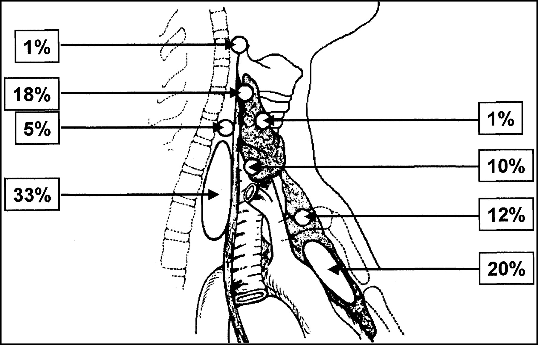
  • FIGURE 2.
    • Download figure
    • Open in new tab
    • Download powerpoint
    FIGURE 2.

    Anatomic locations of abnormal parathyroid glands found at reoperation by single group. Most common ectopic sites mirror routes of descent of upper parathyroid glands (short migration path) and of lower parathyroid glands (longer migration path in association with thymus) (modified from (116)).
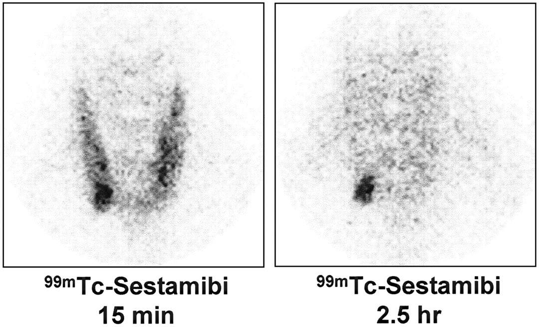
  • FIGURE 3.
    • Download figure
    • Open in new tab
    • Download powerpoint
    FIGURE 3.

    Classical single-tracer, double-phase parathyroid scintigraphy with 99mTc-sestamibi. (Left) Image at 15 min shows physiologic early uptake in thyroid gland, with clear focus of moderately increased accumulation at lower pole of right thyroid lobe. (Right) Late scan shows almost complete washout of 99mTc-sestamibi from thyroid gland, with obvious focal retention of radioactivity at lower pole of right thyroid lobe. Minimally invasive radioguided surgery confirmed presence of parathyroid adenoma.
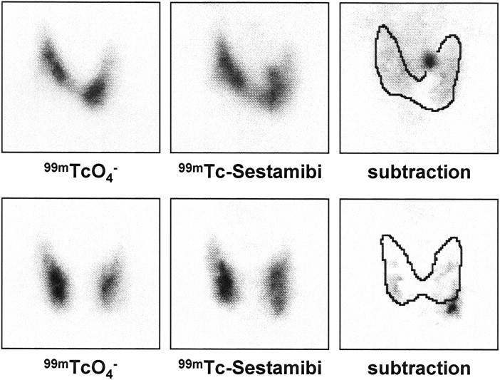
  • FIGURE 4.
    • Download figure
    • Open in new tab
    • Download powerpoint
    FIGURE 4.

    Parathyroid scintigraphy according to dual-tracer protocol (99mTc-pertechnetate and 99mTc-sestamibi), with administration of KClO4− at start of 99mTc-pertechnetate imaging. (Top left) Virtually normal thyroid gland. (Top center) Five-minute scan recorded within 35 min of 99mTc-sestamibi injection shows summation scan suggesting presence of adenoma of upper left parathyroid gland, better outlined when subtracting 99mTc-pertechnetate scan from summation scan (top right). Minimally invasive radioguided surgery confirmed presence of parathyroid adenoma. (Bottom left) Virtually normal thyroid gland. (Bottom center) Five-minute scan recorded within 35 min of 99mTc-sestamibi injection shows summation scan suggesting presence of adenoma of lower left parathyroid gland, better outlined when subtracting 99mTc-pertechnetate scan from summation scan (bottom right). Minimally invasive radioguided surgery confirmed presence of parathyroid adenoma.
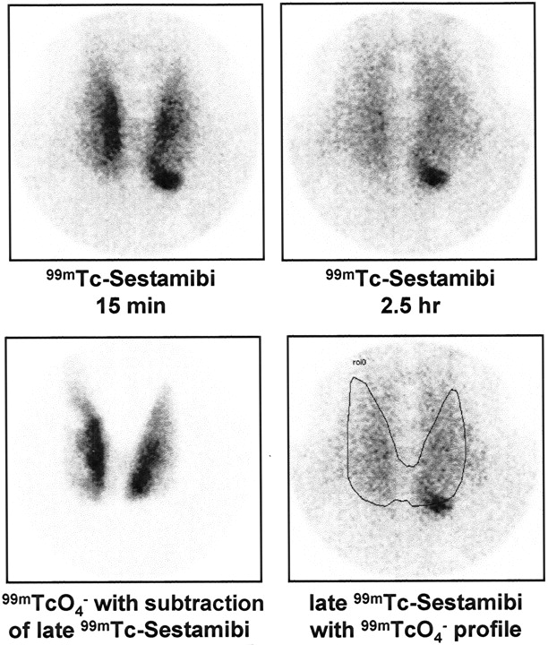
  • FIGURE 5.
    • Download figure
    • Open in new tab
    • Download powerpoint
    FIGURE 5.

    Double-phase parathyroid scintigraphy with 99mTc-sestamibi in patient with multinodular goiter. (Top left) Image at 15 min shows physiologic early uptake in thyroid gland, with clear focus of increased accumulation at lower pole of left thyroid lobe. (Top right) Late 99mTc-sestamibi scan shows some residual activity in thyroid gland, with obvious focal 99mTc-sestamibi retention at lower pole of left thyroid lobe. Second tracer (99mTc-pertechnetate) was administered after recording delayed 99mTc-sestamibi scan. (Bottom left) Image obtained by subtracting delayed 99mTc-sestamibi scan from summation scan recorded after 99mTc-pertechnetate administration was used to draw profile of thyroid gland, which was then superimposed on delayed 99mTc-sestamibi scan (bottom right) for better anatomic localization of parathyroid adenoma (confirmed by minimally invasive radioguided surgery).
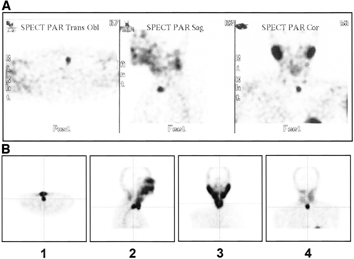
  • FIGURE 6.
    • Download figure
    • Open in new tab
    • Download powerpoint
    FIGURE 6.

    Anatomic localization of ectopic parathyroid adenomas by 99mTc-sestamibi SPECT. (A) Patient with persistent hyperparathyroidism after total thyroidectomy and bilateral neck exploration performed because of concomitant multinodular goiter and PHPT. Tomographic slices in transverse (left), sagittal (center), and coronal (right) planes define anatomic location of single focus of intense 99mTc-sestamibi uptake in anterior planes of upper mediastinum. Minimally invasive reoperation under radioguidance enabled surgeon to remove parathyroid adenoma located within thymus. (B) Tomographic slices in transverse (1), sagittal (2), and coronal (3 and 4) planes define anatomic location of single focus of intense 99mTc-sestamibi uptake behind thyroid gland. Surgery revealed parathyroid adenoma located in retroesophageal space.
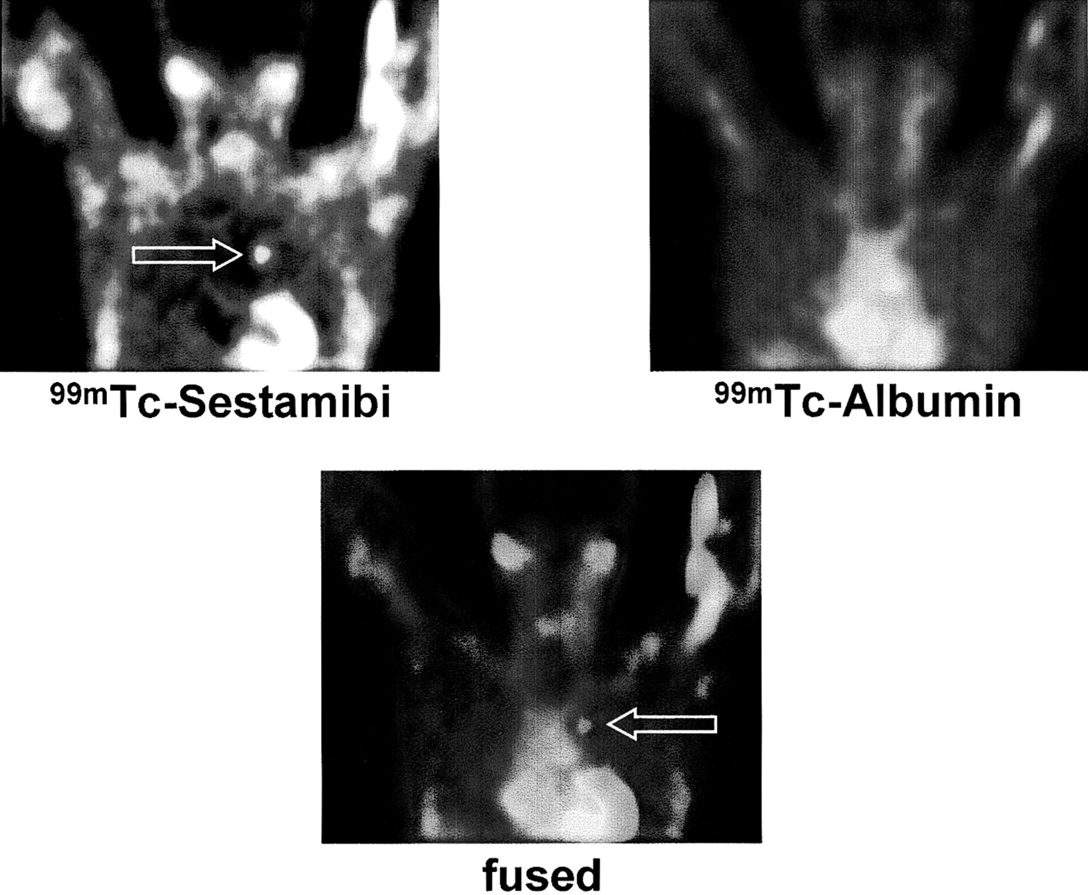
  • FIGURE 7.
    • Download figure
    • Open in new tab
    • Download powerpoint
    FIGURE 7.

    Anatomic localization of ectopic parathyroid adenoma by combined 99mTc-sestamibi and 99mTc-albumin SPECT. (Top left) Tomographic coronal slice obtained after 99mTc-sestamibi administration demonstrates focal area of abnormal tracer uptake in mediastinum, whose exact anatomic location with respect to other mediastinal structures remains uncertain. (Top right) Dose of 99mTc-albumin was then injected (without moving patient) as indicator of intravascular space, and another SPECT image was acquired under same conditions as 99mTc-sestamibi SPECT. (Bottom) Image fusion of corresponding coronal slices demonstrates that lesion with focal uptake of 99mTc-sestamibi is located in aorto-pulmonary window. Subsequent radioguided surgery confirmed presence of parathyroid adenoma in anatomic location identified by fusion analysis.

PreviousNext
Back to top

In this issue

Journal of Nuclear Medicine
Vol. 44, Issue 9
September 1, 2003
  • Table of Contents
  • Index by author
Print
Download PDF
Article Alerts
Sign In to Email Alerts with your Email Address
Email Article

Thank you for your interest in spreading the word on Journal of Nuclear Medicine.

NOTE: We only request your email address so that the person you are recommending the page to knows that you wanted them to see it, and that it is not junk mail. We do not capture any email address.

Enter multiple addresses on separate lines or separate them with commas.
Preoperative Localization and Radioguided Parathyroid Surgery
(Your Name) has sent you a message from Journal of Nuclear Medicine
(Your Name) thought you would like to see the Journal of Nuclear Medicine web site.
Citation Tools
Preoperative Localization and Radioguided Parathyroid Surgery
Giuliano Mariani, Seza A. Gulec, Domenico Rubello, Giuseppe Boni, Marco Puccini, Maria Rosa Pelizzo, Gianpiero Manca, Dario Casara, Guido Sotti, Paola Erba, Duccio Volterrani, Armando E. Giuliano
Journal of Nuclear Medicine Sep 2003, 44 (9) 1443-1458;

Citation Manager Formats

  • BibTeX
  • Bookends
  • EasyBib
  • EndNote (tagged)
  • EndNote 8 (xml)
  • Medlars
  • Mendeley
  • Papers
  • RefWorks Tagged
  • Ref Manager
  • RIS
  • Zotero
Share
Preoperative Localization and Radioguided Parathyroid Surgery
Giuliano Mariani, Seza A. Gulec, Domenico Rubello, Giuseppe Boni, Marco Puccini, Maria Rosa Pelizzo, Gianpiero Manca, Dario Casara, Guido Sotti, Paola Erba, Duccio Volterrani, Armando E. Giuliano
Journal of Nuclear Medicine Sep 2003, 44 (9) 1443-1458;
Twitter logo Facebook logo LinkedIn logo Mendeley logo
  • Tweet Widget
  • Facebook Like
  • Google Plus One
Bookmark this article

Jump to section

  • Article
    • Abstract
    • ANATOMY, PHYSIOLOGY, AND PATHOPHYSIOLOGY
    • SURGICAL TREATMENT
    • PREOPERATIVE PARATHYROID LOCALIZATION
    • INTRAOPERATIVE PARATHYROID LOCALIZATION
    • GUIDELINES AND CONTROVERSIES IN RADIOGUIDED MIP
    • CONCLUSION
    • Footnotes
    • REFERENCES
  • Figures & Data
  • Info & Metrics
  • PDF

Related Articles

  • No related articles found.
  • PubMed
  • Google Scholar

Cited By...

  • Imaging of the Thyroid and Parathyroid Using a Cardiac Cadmium-Zinc-Telluride Camera: Phantom Studies
  • Radioguided parathyroidectomy in forearm graft for recurrent hyperparathyroidism
  • Parathyroid Imaging and Localization Using SPECT/CT: Initial Results
  • Preoperative 123I/99mTc-Sestamibi Subtraction SPECT and SPECT/CT in Primary Hyperparathyroidism
  • Pinhole Versus Parallel-Hole Collimators for Parathyroid Imaging: An Intraindividual Comparison
  • Differences in Accuracy of 99mTc-Sestamibi Scanning Between Severe and Mild Forms of Primary Hyperparathyroidism
  • Subtraction SPECT for Parathyroid Scintigraphy Based on Maximization of Mutual Information
  • Is the "Ideal" {gamma}-Probe for Intraoperative Radioguided Surgery Conceivable?
  • Radioguided Surgery of Primary Hyperparathyroidism Using the Low-Dose 99mTc-Sestamibi Protocol: Multiinstitutional Experience from the Italian Study Group on Radioguided Surgery and Immunoscintigraphy (GISCRIS)
  • The Value of 99mTc-Sestamibi SPECT/CT over Conventional SPECT in the Evaluation of Parathyroid Adenomas or Hyperplasia
  • Google Scholar

More in this TOC Section

  • Approaches to Imaging Immune Activation Using PET
  • Large Language Models and Large Multimodal Models in Medical Imaging: A Primer for Physicians
  • Precision Oncology in Melanoma: Changing Practices
Show more Continuing Education

Similar Articles

SNMMI

© 2025 SNMMI

Powered by HighWire