


Abstract
99mTc-TRODAT-1 ([2-[[2-[[[3-(4-chlorophenyl)-8-methyl-8-azabicyclo[3,2,1]oct-2-yl]methyl](2-mercaptoethyl)amino]ethyl]amino]ethanethiolato(3-)-N2,N2′,S2,S2′]oxo-[1R-(exo-exo)]) is a potential agent for dopamine transporter (DAT) SPECT, whereas 6-18F-fluoro-l-dopa (18F-FDOPA) PET has been used for the quantitative assessment of presynaptic nigrostriatal dopaminergic function. The current study investigated the relationship between the 2 imaging modalities in evaluating patients with Parkinson’s disease (PD). Methods: Twenty patients in whom PD was diagnosed by generally accepted criteria were recruited. In addition to visual inspection, specific uptake ratios (SURs) of 99mTc-TRODAT-1 in the striatum and putamen were measured bilaterally. For PET, all patients received 100 mg of carbidopa 90 min before 18F-FDOPA (300 MBq) injection. Images were acquired between 120 and 150 min after injection, using a whole-body PET scanner with settings identical to those for the SPECT studies. The SURs for PET were calculated similarly to those for SPECT. Individual SURs of the striatum or putamen from SPECT were correlated with the corresponding PET values using linear regression. Results: A consistent image pattern between SPECT and PET was achieved by visual inspection except for 3 cases. In 1 case (a patient with Hoehn and Yahr Scale I PD), the SPECT images were more compatible with the patient’s clinical findings whereas PET showed nearly normal uptake. In the other 2 cases (both patients with Hoehn and Yahr Scale II PD), PET correlated better with the clinical findings. The caudate and putamen nuclei were more discernable on PET. An acceptable correlation of SUR, however, was found between SPECT and PET in both the striatum and the putamen (P < 0.01 for both). Conclusion: The comparability of 99mTc-TRODAT-1 SPECT and 18F-FDOPA PET suggests that 99mTc-TRODAT-1 SPECT may provide a reliable alternative to 18F-FDOPA PET in the evaluation of clinical PD patients.
Parkinson’s disease (PD) is a common neurodegenerative disorder characterized by the progressive degeneration of dopaminergic neurons in the substantia nigra and loss of its nerve terminal in the basal ganglia structures, especially in the striatum (1). An accurate diagnosis of PD is important for patient management and epidemiologic studies. The diagnosis of PD still depends mainly on clinical criteria (1,2). Although loss of approximately 80% of dopamine innervations is needed before symptoms manifest (3), the insidious onset and varied presentation of PD still often obscure clinical diagnosis. In clinicopathologic studies, up to 25% of cases with an antemortem clinical diagnosis of PD were found to have other diseases at postmortem examination (1–3). Improvement of accuracy in diagnosing PD is therefore needed (1,4).
The dopamine transporter (DAT), a protein in the presynaptic membrane on the terminal of dopaminergic projections, plays a critical role in the regulation of the extracellular dopamine concentration (5) and has been considered a marker of dopamine terminal innervations (6). Degeneration of dopaminergic projections from the substantia nigra to the striatum results in loss of DAT (5). Because the DAT is located only on dopaminergic nerve terminals (7), a close relationship between DAT concentration and striatal dopamine levels (8,9) and the presence of PD has been observed in postmortem studies (10,11). Both PET and SPECT have been used as in vivo methods to evaluate neuronal loss in PD (4,12,13). Although PET provides higher resolution and better quantitative capacity than SPECT, SPECT is more practical as a routine procedure. Several 123I-labeled DAT SPECT imaging agents based on cocaine or the closely related tropane derivatives have been reported (12,14,15). These studies indicated that this technique might be useful in identifying individuals in the preclinical and asymptomatic phase of the disease. However, because of the limited availability and relatively high cost of 123I, few 123I-labeled DAT ligands have been routinely applied for DAT imaging (10).
99mTc has a suitable energy and half-life for imaging, is relatively inexpensive, and is readily obtainable. Imaging with 99mTc-labeled ligands would therefore be more suitable for routine use. Several 99mTc-labeled tropanes have been reported (10,16,17), among which, a 99mTc-labeled tropane derivative, [2-[[2-[[[3-(4-chlorophenyl)-8-methyl-8-azabicyclo[3,2,1]oct-2-yl]methyl](2-mercaptoethyl)amino]ethyl]amino]ethanethiolato(3-)-N2,N2′,S2,S2′]oxo-[1R-(exo-exo)] (TRODAT-1), has been intensively studied and has already shown promise in humans and other primates (10,17–19). In this study, we investigated the imaging correlation between 99mTc-TRODAT-1 SPECT and 6-18F-fluoro-l-dopa (18F-FDOPA) PET, a commonly used technique for evaluating central dopaminergic metabolism, in patients with PD.
MATERIALS AND METHODS
Subjects
Twenty patients with various severities of PD were studied. PD was diagnosed according to generally accepted criteria (1,20). Patients received neurologic examinations by 2 experienced neurologists. A clinical diagnosis of idiopathic PD required display of at least 2 of the following symptoms: resting tremor, akinesia, and rigidity, with a favorable response to l-dopa therapy. All PD patients were scored with the Hoehn and Yahr Scale (HYS), which ranges from HYS I to HYS IV (21). Four patients were HYS I (2 men and 2 women; mean age, 61 y; age range, 49–78 y), 6 were HYS II (5 men and 1 woman; mean age, 65 y; age range, 48–76 y), 7 were HYS III (2 men and 5 women; mean age, 64 y; age range, 48–77 y), and 3 were HYS IV (2 men and 1 woman; mean age, 63 y; age range, 59–66 y). All subjects consumed a low-protein diet for 24 h before the examinations. Antiparkinsonian medication (l-dopa) was discontinued for at least 12 h before commencement and until completion of the SPECT or PET studies. Four age-matched healthy volunteers (2 men and 2 woman; mean age, 63 y; age range, 51–68 y) served as controls. Each subject underwent 99mTc-TRODAT-1 SPECT and 18F-FDOPA PET within a month of each other and gave written informed consent. This study was approved by the institution research boards of our hospitals and the Department of Health of our country.
Radiopharmaceuticals
The method of preparing 99mTc-TRODAT-1 was a modification of a previously described method (19). Briefly, 99mTc-TRODAT-1 was prepared from a freeze-dried kit by adding 1,110 MBq of freshly eluted 99mTc-pertechnetate to 5 mL of saline preparation (20). The 99mTc-TRODAT-1 was obtained in a neutral solution (pH 7.0–7.5) with greater than 90% radiochemical purity over 6 h, as determined by high-performance liquid chromatography. The shelf life of the lyophilized kit was more than 2 mo when it was stored at room temperature. 18F-FDOPA was prepared as previously described (22).
Imaging and Data Analysis
99mTc-TRODAT-1 SPECT.
The subjects were placed supine, and the position of their head was fixed with a holder. After injection of 740 MBq of 99mTc-TRODAT-1, the brain SPECT studies commenced 3 h later, using a dual-head camera equipped with ultra-high-resolution fanbeam collimators (Helix SPX; Elscint). Data were acquired in a 128 × 128 matrix with a 1.4 zoom through a 360° rotation (180° for each head) at 3° intervals, for 30 s per angle step. Images were reconstructed using the backprojection method with a Metz filter. Attenuation correction was performed by the first-order method of Chang. The SPECT images were analyzed along the level of the canthomeatal line. Regions of interest were marked for the striatum and putamen of each hemisphere, in reference to the corresponding MR image, on composite images of the 3 slices showing the highest basal ganglia activity. The occipital cortices (OC) were also drawn in the same way and served as background areas. The numbers of pixels (2.96 mm2 per pixel) in regions of interest were around 170 for the striatum, 115 for the putamen, and 210 for background areas. Specific uptake ratios (SURs) in the striatum and putamen were calculated by subtracting the mean counts per pixel in the OC from the mean counts per pixel in the whole striatum or putamen region and dividing the result by the mean counts per pixel in the background: (striatum − OC)/OC or (putamen − OC)/OC (19).
18F-FDOPA PET.
An 8-ring whole-body PET scanner (PC4096-15WB; Scanditronix) was used to yield 15 simultaneous planes with an axial resolution of 6.5 mm in full width at half maximum and an in-plane resolution of 8 mm at the center of the field of view. A special head holder was applied for each study to minimize head motion during acquisition. Correction for tissue attenuation of 511-keV γ-radiation was measured with an external 68Ge pin source. Scanning was done during wakefulness, after pretreatment with an oral administration of 100 mg of carbidopa (l-aromatic amino acid decarboxylase inhibitor to block metabolism of dopa by the non–central nervous system tissues) 90 min before tracer injection. The subjects were positioned and immobilized in the scanner with the canthomeatal line parallel to the detector rings. Static 18F-FDOPA PET was performed starting 120 min after intravenous injection of 185 MBq (mean specific activity, 20–40 MBq/μmol) of 18F-dopa (in 10 mL of normal saline solution, over 30 s). The data were collected for 30 min. The axial field of view was 9.75 cm starting from the canthomeatal line caudally. Image reconstruction used filtered backprojection. A Hann filter was used with a filter width of 4.2 mm, a pixel size of 2 mm × 2 mm, a matrix size of 128 × 128, and an interslice distance of 6.5 mm. The location and size of determined regions of interest of the striatum and its subnuclei were analogous to those used in the SPECT analyses (23).
Statistical Analysis
Individual SURs of the striatum or putamen from SPECT were correlated with the corresponding PET values using linear regression. The correlation between averaged striatum and putamen SURs of subjects and their score on the HYS was measured by the Spearman rank correction coefficient. Significance was defined as P < 0.05.
RESULTS
For both 99mTc-TRODAT-1 SPECT and 18F-FDOPA PET, greater loss of uptake was found in the putamen than in the caudate and in the contralateral side to the more affected limbs, as interpreted by both visual and quantitative analysis (Table 1).
Demographic Characteristics of PD Patients and Their Imaging Findings
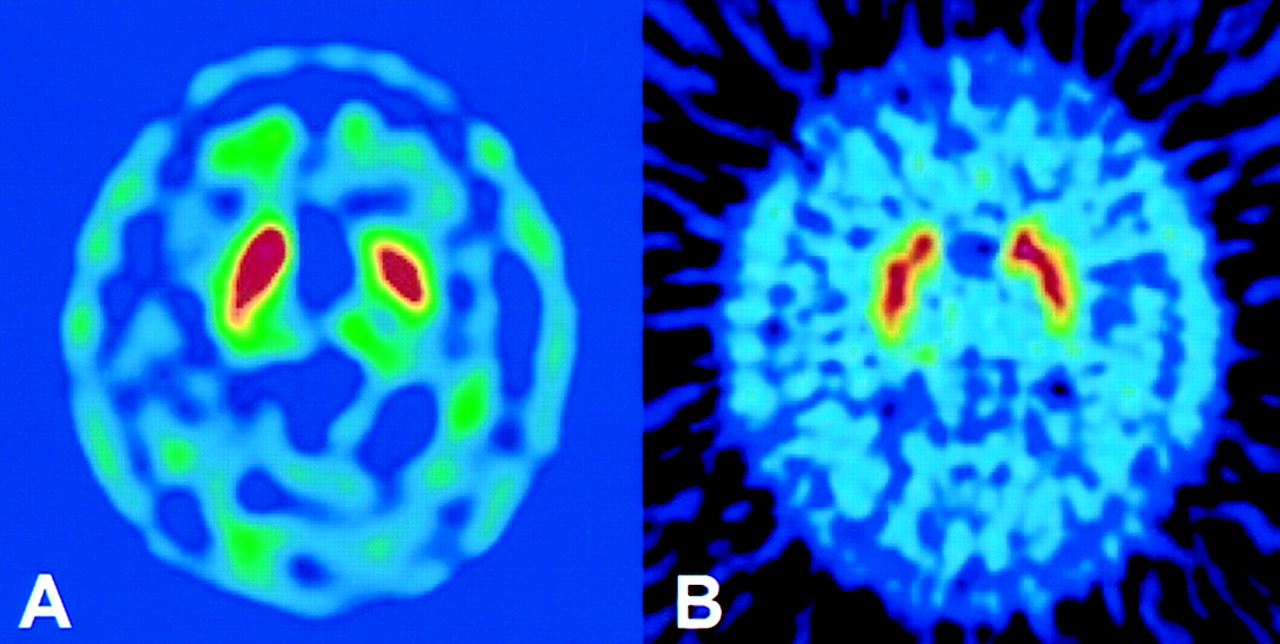
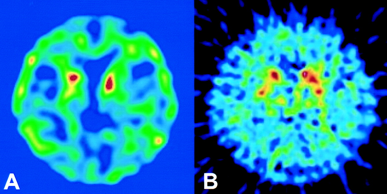
A consistent image pattern between SPECT and PET was achieved by visual inspection for all but 3 cases. Representative images for consistent control and disease cases are shown in Figures 1 and 2. In 1 of the 3 inconsistent cases (HYS I), the SPECT images were more compatible with the patient’s clinical presentation whereas the PET images showed bilateral, symmetric uptake in the striatum (Fig. 3). In the other 2 inconsistent cases (both HYS II), the PET images correlated better with the clinical findings (Fig. 4) whereas the SPECT images indicated more advanced disease. Two cases (9 and 10) had profoundly reduced SPECT SURs in the putamen (0.02 and 0.16). Although SPECT produced images that were comparable with the PET images, as shown in Figure 1, the caudate and putamen nuclei were more discernable on the PET images than on the SPECT images. A statistically significant correlation of SUR, however, was found between SPECT and PET for both the striatum (n = 40, r = 0.42, P < 0.01) and the putamen (n = 40, r = 0.47, P < 0.01) (Figs. 5 and 6). Notably, the ranges of the SURs for both the striatum and the putamen appeared wider in the SPECT images than in the PET images (slopes of PET vs. SPECT = 0.32 for the striatum and 0.45 for the putamen). Individual SURs for the putamen and striatum for different HYS scores are also shown in Figures 5 and 6. The average SURs for the putamen and striatum negatively correlated with the HYS score of patients (ρ = −0.74 and −0.77 for PET; −0.69 and −0.63 for SPECT, P < 0.01).

Representative transverse 99mTc-TRODAT-1 SPECT images (A) and concordant 18F-FDOPA PET images (B) of healthy volunteer. Bilateral, symmetrically normal uptake in striatum is seen on both SPECT and PET.

Representative transverse 99mTc-TRODAT-1 SPECT images (A) and concordant 18F-FDOPA PET images (B) of patient with HYS II PD (patient 16). Markedly decreased uptake in right putamen, with less apparent decrease in left putamen, was found on both SPECT and PET.
Transverse 99mTc-TRODAT-1 SPECT images (A) and discordant 18F-FDOPA PET images (B) of patient with HYS I PD (patient 1). SPECT images showed greater loss of uptake in left putamen and appeared more correlated with clinical manifestations than did corresponding 18F-FDOPA PET images.

Transverse 99mTc-TRODAT-1 SPECT images (A) and discordant 18F-FDOPA PET images (B) of patient with HYS II PD (patient 15). PET images correlated better with clinical staging than did corresponding 99mTc-TRODAT-1 SPECT images, which appeared to overestimate disease severity.
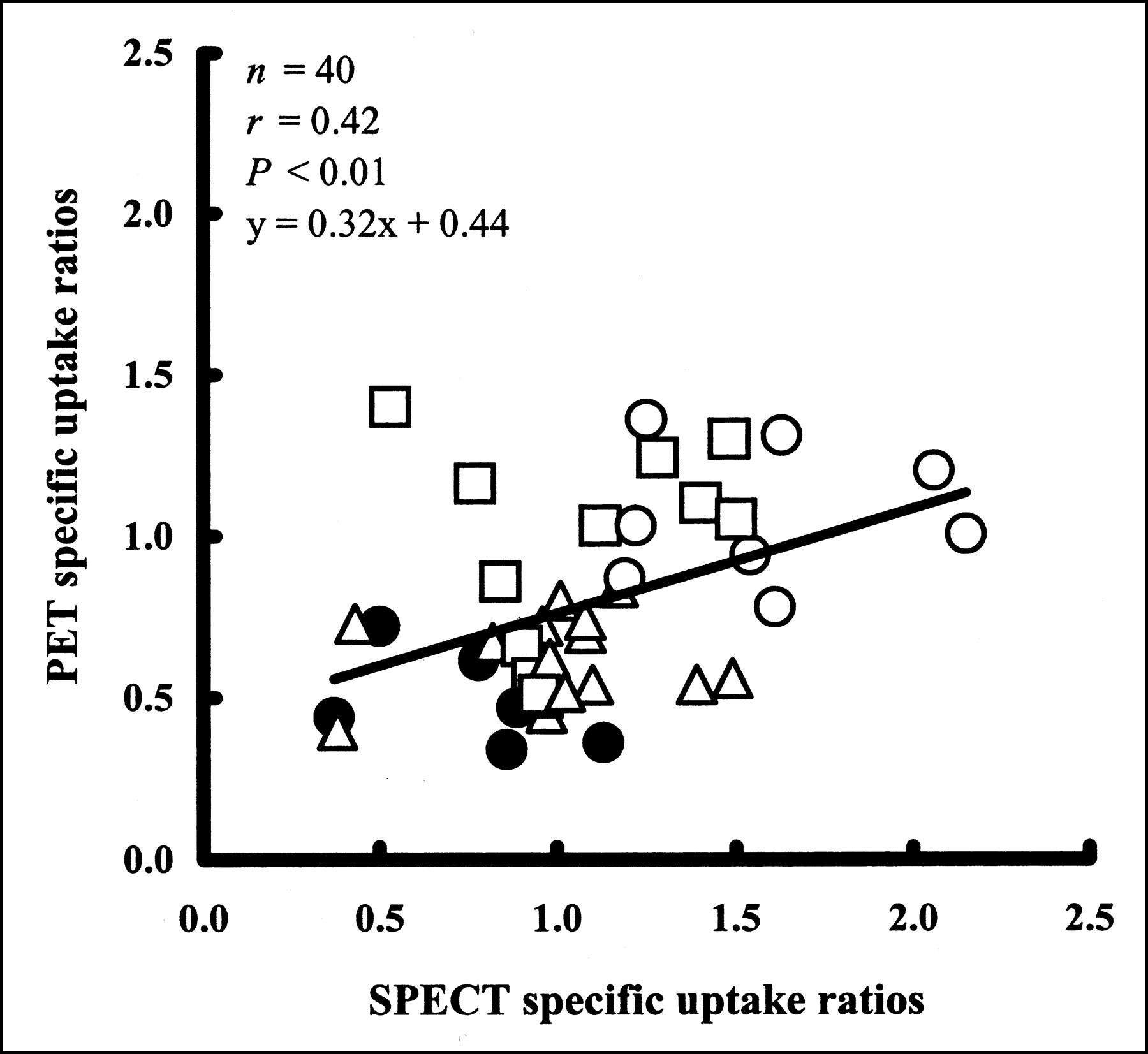
Correlation of 99mTc-TRODAT-1 SPECT and 18F-FDOPA PET in striatum as measured by SURs. Straight line is linear regression line. Each point represents an individual striatum. ○ = HYS I; □ = HYS II; ▵ = HYS III; • = HYS IV.
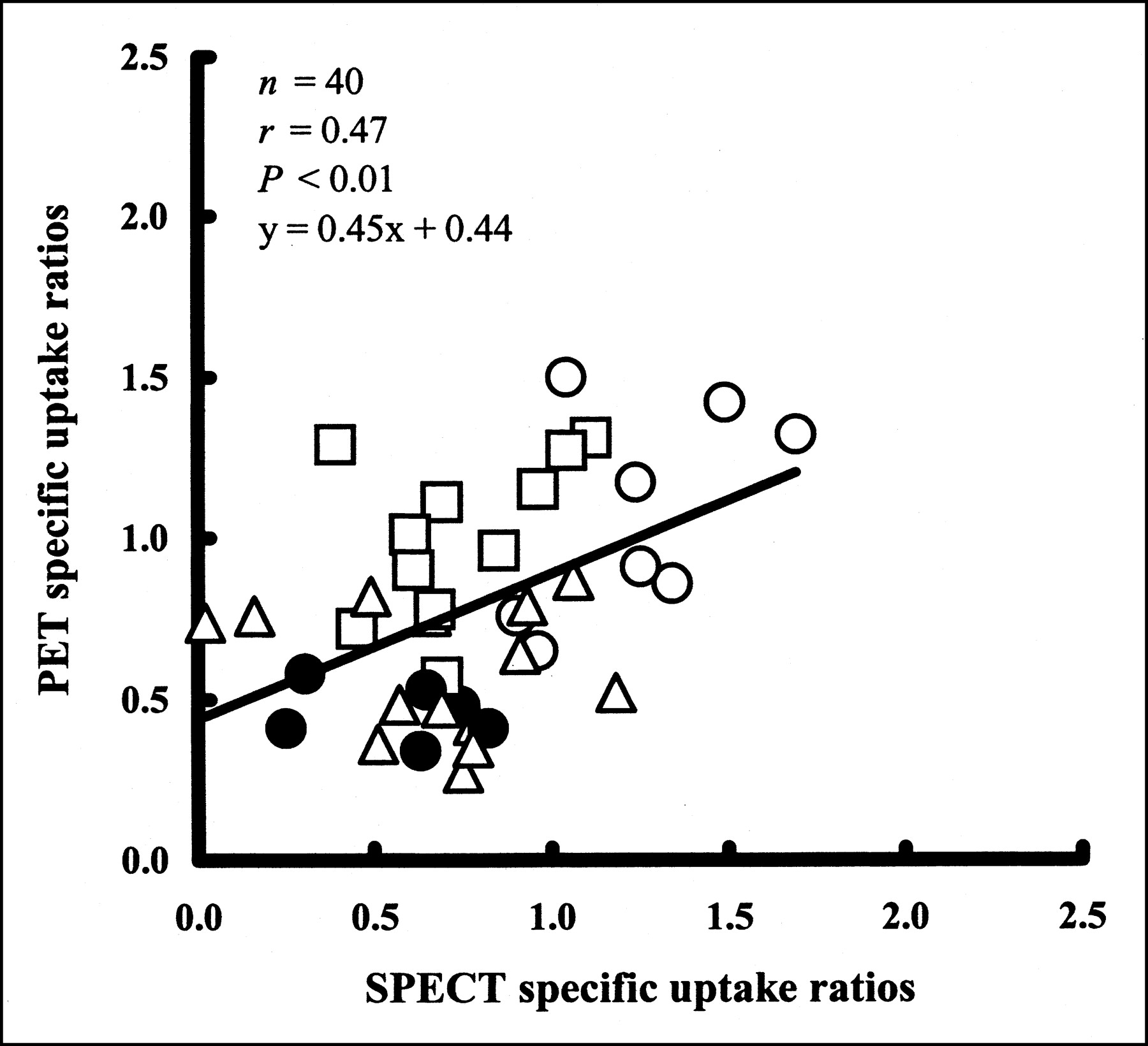
Correlation of 99mTc-TRODAT-1 SPECT and 18F-FDOPA PET in putamen as measured by SURs. Straight line is linear regression line. Each point represents an individual putamen. ○ = HYS I; □ = HYS II; ▵ = HYS III; • = HYS IV.
DISCUSSION
In this study, significant comparability was found between 99mTc-TRODAT-1 SPECT and 18F-FDOPA PET as interpreted by visual inspection and SUR measurements. The significant correlation between 99mTc-TRODAT-1 SPECT and 18F-FDOPA PET implies that an acceptable striatum imaging quality and semiquantitation were achievable by 99mTc-TRODAT-1 SPECT using conventional dual-head nuclear medicine facilities at 3 h after injection. The results are consistent with those of 18F-FDOPA and 11C-WIN 35,428 DAT PET (24,25), suggesting that kit-based 99mTc-TRODAT-1 may serve as a sensitive and objective in vivo marker to reflect the onset and severity of PD.
With the progress of nuclear neuroimaging, 18F-FDOPA PET has become a standard procedure for evaluating dopaminergic metabolism of the central nervous system (25). The good imaging quality and quantitative capacity of PET allow discernable detection of caudate and putamen nuclei. However, more hospitals are equipped with SPECT scanners; therefore, SPECT is more practical as a routine procedure. The easy preparation of 99mTc-TRODAT-1 from lyophilized kits and the availability of SPECT scanners equipped with dual-head cameras, the most commonly used nuclear medicine facilities worldwide, could be an ideal combination for daily clinical application (15).
The implications of using 99mTc-TRODAT-1 SPECT for monitoring central dopamine-related disorders have recently been intensively investigated. Comparison of 99mTc-TRODAT-1 SPECT with 18F-FDOPA PET in patients with PD, however, has not been reported. Although prior studies using ligands for PET have demonstrated its utility for measuring the structural and biochemical integrity of dopaminergic neurons in vivo (12,26–28), DAT-targeting radiopharmaceuticals are increasingly being recognized as more effective markers (10,14,15,20,29). In PD studies, Kish et al. found a good correlation between loss of DATs and loss of dopamine neurons (5). In our study, the greater reduction of striatal uptake in the putamen contralateral to the more affected side in PD patients agrees with this theory. Such changes were also consistent with previous SPECT data using other tracers for DAT imaging that correlated with 18F-FDOPA PET–measured disease severity (13,14,25). Disease progression of PD, especially in the early stages, is asymmetric (5,10), which may cause overlap of individual SURs among patients with different HYS scores. However, when SURs of the bilateral putamen or striatum were averaged, significant negative correlations were found between the SURs determined from both PET and SPECT and the HYS scores. A larger study is required to confirm this finding. To reduce the effects of age-related differences in DAT density (10,19), we selected patients who were more than 48 y old and matched the ages among the groups. Patients with clinical HYS V were excluded because of poor study compliance.
Postmortem studies have demonstrated a close relationship between DAT concentrations and striatal dopamine levels (25,30). Curiously, in our study, we found a case (HYS I) in which the SPECT images were more compatible with the patient’s clinical findings whereas the PET images showed less reduction of striatum uptake. Postmortem investigations suggest that the levels of dopamine neuronal markers vary considerably in PD (9). A previous study using 11C-WIN 35,428 for DAT imaging found that in patients with mild PD, 18F-FDOPA uptake was reduced less than 11C-WIN 35,428 uptake (24). Retention of the tracer not only reflects nerve terminal loss but also critically depends on aromatic l-amino acid decarboxylase (AADC) activity (31,32). Frost et al. reported that 18F-FDOPA reflects primarily AADC activity, which might be increased in residual dopaminergic nerve terminals (24) and therefore underestimate presynaptic terminal loss (33). Torstenson et al. also found that, apart from the structural integrity, the intrinsic activity of the dopamine autoreceptors might also govern the influx ratio of radiolabeled l-dopa (34). Differences in the mechanism of measurement of dopaminergic nerve function between DAT and 18F-FDOPA imaging may at least in part explain discrepancies between these imaging techniques in some patients.
In patients with PD, there was a marked reduction of striatal dopamine concentrations and a corresponding loss of DATs (25,28). Ligands, such as TRODAT-1, that bind specifically to the presynaptically located DATs may more directly reflect the loss of dopaminergic nerve terminals in the striatum (7,31–33). Previous studies have also shown a correlation between semiquantitative SPECT and the clinical severity of PD (13–15,29), suggesting that this method can be used to evaluate the presence and progression of PD. In our study, 2 patients with clinical HYS II each showed better correlations between PET and the clinical findings, whereas TRODAT-1 DAT SPECT indicated a greater loss of striatum uptake. There were also 2 patients (9 and 10) with HYS III who showed profound loss of SPECT SURs in the putamen, possibly due in part to disease progression per se (5) or to relatively high background counts. The results appeared to reflect the fact that PET provides more integrated information for dopamine metabolism than does SPECT in patients with PD (31,32). From this point of view, DAT SPECT, especially in early-stage PD patients, might tend to overestimate disease progression (31). Nevertheless, an acceptable linear correlation of SURs between SPECT and PET in our patient group suggested that 99mTc-TRODAT-1 DAT SPECT might provide a suitable alternative to 18F-FDOPA PET in the clinical evaluation of patients with PD. The PET images for our study were obtained using an old-generation machine in 2-dimensional mode, which might be the reason for the noisy images presented here. Using current high-sensitivity scanners in 3-dimensional mode would further improve imaging quality.
Although PET has advantages over SPECT in resolution and quantification, PET is more expensive, less widely available, more time consuming, and more manpower intensive (31). In contrast, 99mTc is produced in generators, is easy to label, is inexpensive, produces suitable energy, and has an appropriate half-life for imaging. 99mTc-TRODAT-1 is therefore a suitable DAT imaging agent for routine evaluation of PD patients. The different dosage of 99mTc-TRODAT-1 and 123I-2β-carbomethoxy-3β-(4-iodophenyl)tropane (740 vs. 185–333 MBq) may also contribute to the relatively good quality of the SPECT images in our study, compared with the images from a previous study (25). In our SPECT study, the caudate and the anterior part of the putamen could not be clearly separated, possibly because of the limited camera resolution. However, measurement of uptake activity in the striatum and its subnuclei might not be a problem when an appropriate imaging technique together with an anatomically driven region-of-interest strategy is applied.
In view of the acceptable image quality for visual interpretation and the wide range of measured SURs for different stages of PD, as found in our and other studies (10,18,19), we speculate that 99mTc-TRODAT-1 SPECT may provide a useful approach for the diagnosis of PD and the assessment of its severity and perhaps for discriminating the etiologies of Parkinson-like disorders. Clinical applications of 99mTc-TRODAT-1 SPECT in neuropsychiatry are still under investigation. Although an acceptable correlation between 99mTc-TRODAT-1 DAT SPECT and 18F-FDOPA PET using either visual interpretation or SURs was found in our study, a study with a large number of subjects and a longer follow-up period is needed to further validate this issue.
CONCLUSION
The present study showed that changes of 99mTc-TRODAT-1 uptake in the striatum and its subregions in patients with PD were concordant with those of 18F-FDOPA PET, clinical presentations, and previous reports. Our observations suggest that 99mTc-TRODAT-1 DAT SPECT using a conventional nuclear medicine camera system may provide a suitable alternative to 18F-FDOPA PET in evaluating clinical PD patients.
Acknowledgments
The authors thank Chia-Jung Chang and Miriam Wang for able technical help; Prof. Cheng-Yu Chen, a senior neuroradiologist, for assistance with image integration; and Prof. Hank F. Kung at the University of Pennsylvania for help in preparing this manuscript. This study was supported by the National Science Council, the Atomic Energy Council, and the Ministry of Economic Affairs of Taiwan under grants NSC 90-2314-B-016-100, NSC 91-NU-7-016-002, and EC-2A-17-0304.
Footnotes
Received Nov. 12, 2002; revision accepted Mar. 5, 2003.
For correspondence or reprints contact: Wen-Sheng Huang, MD, Department of Nuclear Medicine, Tri-Service General Hospital, 325, Section 2, Cheng-Kung Rd., Taipei, Taiwan 114.
E-mail: wshuang{at}ms22.url.com.tw


