


Article Figures & Data
Figures
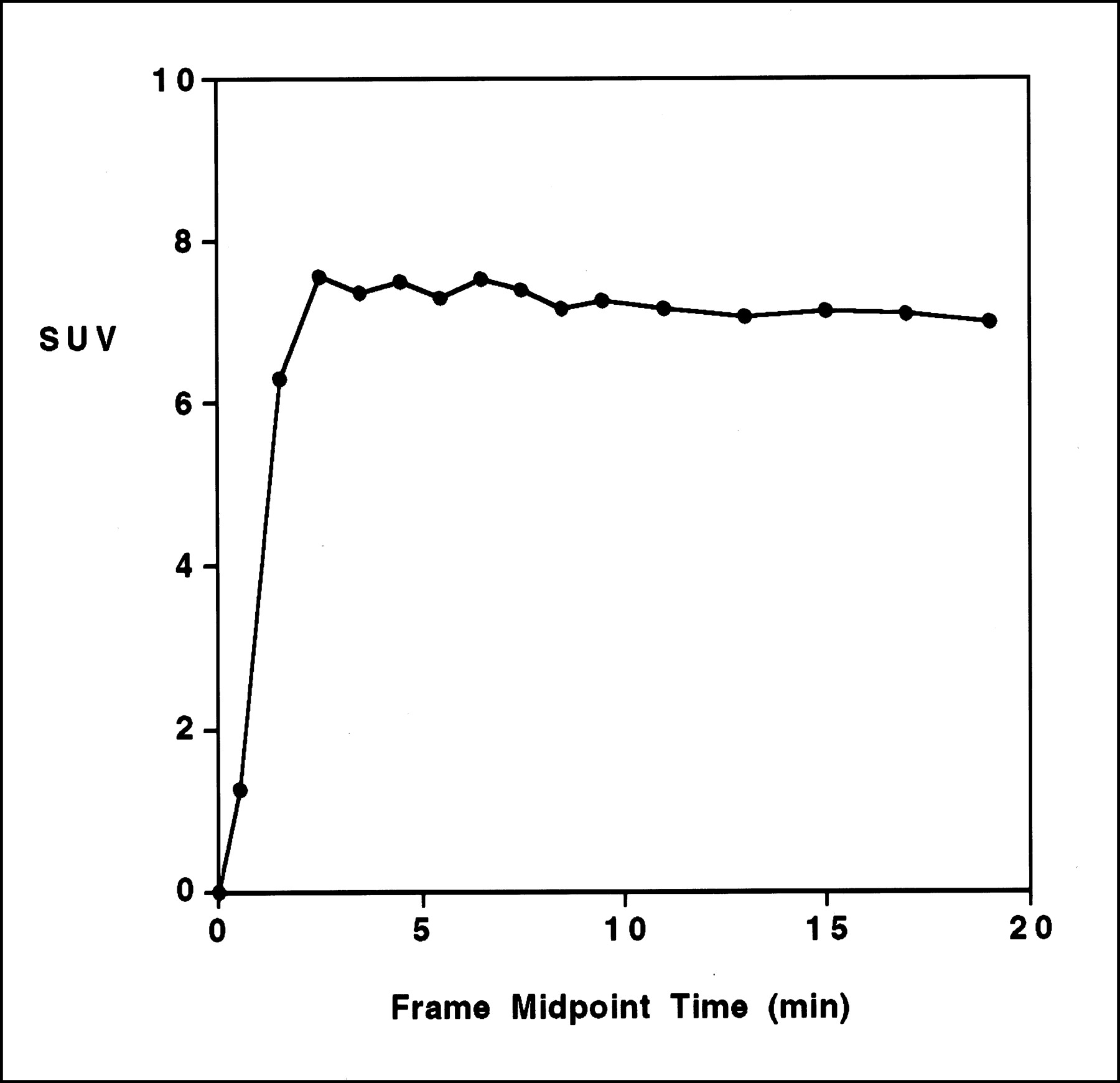
- FIGURE 1.
Time–activity curve of 11C-choline PET in patient with uterine cervical cancer (patient 12) shows rapid initial uptake in tumor tissue with almost constant level afterward.
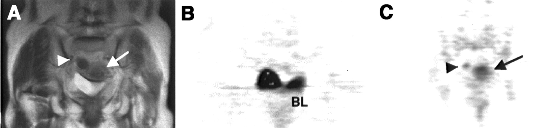
- FIGURE 2.
Coronal T2-weighted MR image (A) shows enlarged isointense area of endometrium (arrow), indicating presence of tumor. Arrowhead points to intestine. This patient was diabetic. Blood sugar levels were 177 mg/dL (9.8 mmol/L) and 188 mg/dL (10.4 mmol/L) at time of 18F-FDG PET and 11C-choline PET, respectively. In 18F-FDG PET image (B), tumor uptake is negative in presence of hyperglycemia. 11C-Choline PET image (C) demonstrates increased tracer uptake (arrow), corresponding to endometrial tumor. 11C-Choline accumulation in bladder was only minimal, but small hot spot was seen in intestine (arrowhead). Surgical histologic examination revealed clear cell carcinoma (stage IB). BL = bladder activity.
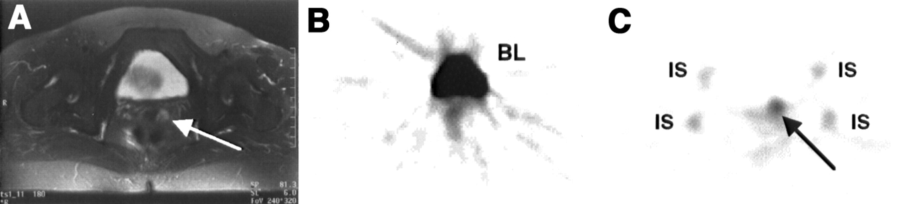
- FIGURE 3.
Transaxial T2-weighted MR image (A) shows small hyperintense lesion in uterine cervix (arrow). In 18F-FDG PET image (B), tumor visualization is obscured by high bladder activity. 11C-Choline PET image (C) clearly shows intense tumor uptake (arrow), corresponding to cervical cancer. No bladder activity is seen in 11C-choline PET image. Surgical histologic examination revealed spindle cell carcinoma (9 × 6 mm, stage IB). BL = bladder activity; IS = physiologic uptake of 11C-choline in bilateral ischia.
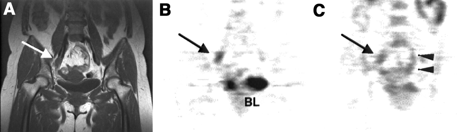
- FIGURE 4.
Coronal T1-weighted MR image (A) demonstrates right parailiac lymphadenopathy (arrow) in patient with uterine cervical cancer. 18F-FDG PET image (B) clearly shows intense uptake in right parailiac region (arrow), indicating lymph node metastasis. Although 11C-choline PET image (C) detects abnormal activity (arrow), physiologic bowel uptake (arrowheads) mimics parailiac lymph node metastases. Lymphadenopathy was not seen on follow-up MR images after transarterial infusion of chemotherapy. BL = bladder activity.
- FIGURE 5.
Whole-body 18F-FDG PET (A) and 11C-choline PET (B) images show hot spot in para-aortic lymph node metastasis (arrows) in patient with recurrent ovarian cancer. High physiologic uptake of 11C-choline in liver, pancreas, and intestine hampers interpretation of abdomen and pelvis.


Tables
Patient no. Age (y) Histology of primary tumor Tumor Tumor SUV Stage Size (cm) 18F-FDG 11C-CHO Untreated primary tumor (n = 18) Uterine corpus cancer 1 49 Endometrial adenocarcinoma IIIA 4 16.81 5.77 2 66 Endometrial adenocarcinoma IB 1 3.79 2.83 3 72 Endometrial adenocarcinoma IB 2.5 12.55 5.52 4 42 Endometrial adenocarcinoma IB 2.5 9.09 6.33 5 62 Endometrial adenocarcinoma IB 2.2 6.13 4.93 6 40 Endometrial adenocarcinoma IA 2.5 6.83 3.06 7 63 Endometrial adenocarcinoma IA 2 6.54 6.79 8 77 Carcinosarcoma IIIA 4 7.43 3.45 9 72 Carcinosarcoma IC 3 7.29 2.13 10 74 Clear cell carcinoma IB 1.7 FN 4.81 11 68 Atypical hyperplasia 0 NA FN FN Uterine corpus cancer 12 68 Squamous cell carcinoma IIB 4 12.53 7.07 13 36 Squamous cell carcinoma IIB 3 9.49 4.43 14 47 Squamous cell carcinoma IIB 7.5 14.57 5.27 15 53 Squamous cell carcinoma IIB 3.7 10.95 5.53 16 54 Spindle cell carcinoma IB 0.9 FN 3.86 17 75 Ovarian mucinous cystadenocarcinoma IA 12 9.31 2.42 18 51 Pelvic inflammatory disease NA 5 8.13 6.02 (FP) (FP) Recurrent tumor (n = 3) 19 66 Ovarian serous adenocarcinoma NA 1.5 3.75 3.63 20 69 Ovarian serous adenocarcinoma NA 2 FN FN 21 47 Ovarian serous adenocarcinoma NA NA FN FN FN = false-negative; NA = not applicable; FP = false-positive.
Tumor size is longest diameter.
In this issue


Jump to section
Related Articles
Cited By...
- Diagnostic Accuracy of FEC-PET/CT, FDG-PET/CT, and Diffusion-Weighted MRI in Detection of Nodal Metastases in Surgically Treated Endometrial and Cervical Carcinoma
- Activation of Phosphatidylcholine Cycle Enzymes in Human Epithelial Ovarian Cancer Cells
- Gene Expression Patterns and Tumor Uptake of 18F-FDG, 18F-FLT, and 18F-FEC in PET/MRI of an Orthotopic Mouse Xenotransplantation Model of Pancreatic Cancer
- Detection of Hepatocellular Carcinoma Using 11C-Choline PET: Comparison with 18F-FDG PET
- Expanding Role of Positron Emission Tomography in Cancer of the Uterine Cervix
- Comparison of Sigma-Ligands and Metabolic PET Tracers for Differentiating Tumor from Inflammation
- Oncologic Imaging in Gynecologic Malignancies
- Alterations of Choline Phospholipid Metabolism in Ovarian Tumor Progression