


Article Figures & Data
Figures
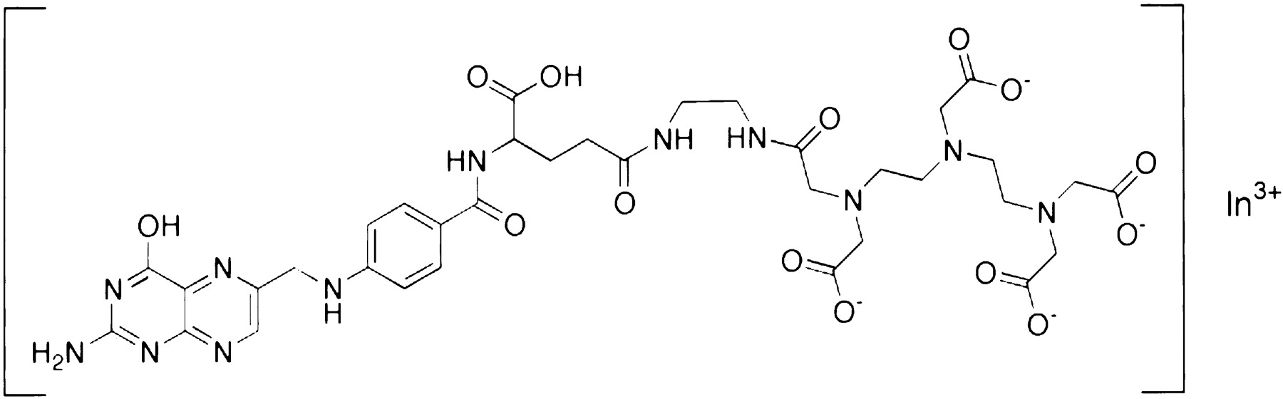
- FIGURE 1.
Structural formula of 111In-DTPA-folate.
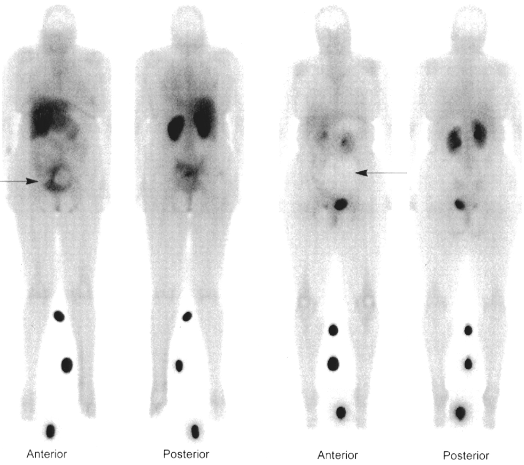
- FIGURE 2.
Anterior and posterior planar scintigrams obtained 4 h after administration of 111In-DTPA-folate. The 2 leftmost images, of patient 15, demonstrate increased tracer uptake (arrow) corresponding to solid tumor component in rim of poorly differentiated adenocarcinoma of left ovary. The 2 rightmost images, of patient 9, show region of focally decreased activity (arrow) corresponding to simple serous benign cyst. (Foci of increased activity between legs of each patient are reference sources used for dosimetry calculations.)

Tables
Classification n % n % Benign 18 54.5 Borderline malignancy 1 3.0 New ovarian malignancy 7 21.2 Stage I 1 14 Stage II 2 29 Stage III 4 57 Stage IV 0 0 Recurrent malignancy 7 21.2 Ovarian 5 71 Endometrial 2 29 Total 33 100 Pathology Patient no. Serum folate (ng/mL) Serum CA-125 (U/mL) Age (y) Folic acid dose (mg) Image scoring* Tumor type Masked Unmasked A B A B Malignant 4 19.3 36 76 0 ++ ++ ++ ++ Mixed endometrioid and serous carcinoma 7 10.0 1,221 49 5.0 ++ ++ ++ ++ Serous ovarian carcinoma 8 9.6 69 61 5.0 ++ ++ ++ ++ Endometrioid adenocarcinoma 11 16.4 1,632 76 0 ++ ++ ++ ++ Serous carcinoma 13 12.2 410 52 0 ++ ++ ++ ++ Endometrial adenocarcinoma 15 6.0 46 1.0 ++ ++ ++ ++ Ovarian adenocarcinoma 37 5.2 2,123 55 2.0 ++ ++ ++ ++ Ovarian/mesodermal, carcinosarcoma Borderline malignant 6 42 61 0 + 0 + nr Serous tumor (borderline malignancy) Benign 5 6.4 67 54 0 0 0 0 0 Mature teratoma 9 9.8 53 5.0 0 0 0 0 Simple serous cyst 10 16.2 11 50 1.0 0 0 0 0 Serous cystadenofibroma 14 >32.0 46 10.0 0 0 0 0 Ovarian endometrial cyst 16 7.7 64 34 1.0 0 0 0 0 Endometriosis 20 23.6 18 47 1.0 0 0 0 0 Myometrial leiomyomata 21 8.2 38 1.0 0 0 0 0 Fibrovascular adhesions 24 3.4 10 45 2.0 0 0 0 0 Brenner tumor 25 12.4 229 47 1.0 0 0 0 0 Endometriosis 35 15.4 <10 44 2.0 0 0 0 0 Serous cystadenoma 36 16.1 <10 67 2.0 0 0 0 0 Hydrosalpinx 27 36.0 30 39 1.0 0 nr nr nr Mucinous cystadenoma 29 82 0 0 nr 0 nr Serous cystadenoma 12 17.2 78 0 ++ ++ ++ ++ Adenomatoid hilar Leydig cell hyperplasia 23 12.8 24 48 1.0 + + + + Endometriosis 17 6.6 <10 52 0 ++ ++ 0 0 Mature cystic teratoma 22 52.9 141 37 2.0 ++ + ++ 0 Endometriosis 34 11.1 68 2.0 0 0 0 + Serous cystadenoma ↵* ++ = increased uptake of 111In-DTPA-folate; + = faint uptake of 111In-DTPA-folate; 0 = no uptake of 111In-DTPA-folate; nr = images not read.
- TABLE 4
Summary of 111In-DTPA-Folate Imaging Results from Patients Presenting with Newly Detected Abdominal Masses
Parameter Masked Unmasked Reader A Reader B Reader A Reader B M B M B* M B* M B* Scoring Increased 7 3 7 2 7 2 7 1 Faint 0 1 0 2 0 1 0 2 None 0 14 0 12 0 14 0 13 Sensitivity 7/7 (100%) 7/7 (100%) 7/7 (100%) 7/7 (100%) Specificity 14/18 (78%) 12/16 (75%) 14/17 (82%) 13/16 (81%) ↵* Two subjects with benign masses did not have complete masked and unmasked scoring by both readers; thus the variation in totals.
M = malignant; B = benign.
Data from the 1 borderline-malignant tumor are not included in this table.
Patient no. Serum folate (ng/mL) Serum CA-125 (U/mL) Age (y) Folic acid dose (mg) Image scoring* Pathology of previous cancer Tumor type Masked Unmasked A B A B 18 23.4 139 70 0 ++ ++ ++ ++ Ovarian High-grade solid carcinoma with transitional cell features 19† 9.7 36 75 0 ++ ++ ++ ++ Ovarian Adenocarcinoma with clear cell features 28 11.9 60 0 0 0 + + Ovarian Adenocarcinoma, poorly differentiated, splenic flexure tumor 31 15.1 107 31 0 0 0 ++ ++ Ovarian Metastatic papillary serous carcinosarcoma 33‡ 35.8 95 61 0 0 0 0 0 Ovarian Adenocarcinoma, poorly differentiated, pelvic ovarian tumor 30 15.7 22 60 0 0 + ++ ++ Endometrial Metastatic adenocarcinoma, primary endometrial, left iliac lymph node 38 10.9 421 55 2.0 0 nr + nr Endometrial Metastatic papillary serous and clear cell carcinosarcoma, poorly differentiated - TABLE 6
Summary of Results from Patients with Recurrent Ovarian and Endometrial Malignancies
Tracer uptake Masked reader Unmasked reader A B A B Increased 2 2 4 4 Faint 0 1 2 1 None 5 3 1 1 Sensitivity 2/7 (29%) 3/6 (50%) 6/7 (86%) 5/6 (83%)
In this issue


Jump to section
Related Articles
Cited By...
- Identification of a PET Radiotracer for Imaging of the Folate Receptor-{alpha}: A Potential Tool to Select Patients for Targeted Tumor Therapy
- Molecular Imaging of Ovarian Cancer
- Development and Pre-clinical Evaluation of New 68Ga-NOTA-folate Conjugates for PET Imaging of Folate Receptor-positive Tumors
- Imaging the Folate Receptor on Cancer Cells with 99mTc-Etarfolatide: Properties, Clinical Use, and Future Potential of Folate Receptor Imaging
- A Folate Receptor-{alpha}-Specific Ligand That Targets Cancer Tissue and Not Sites of Inflammation
- Folic Acid Conjugates for Nuclear Imaging of Folate Receptor-Positive Cancer
- In Vivo Assay of Folate Receptors in Nonfunctional Pituitary Adenomas with 99mTc-Folate SPECT/CT
- A New 18F-Labeled Folic Acid Derivative with Improved Properties for the PET Imaging of Folate Receptor-Positive Tumors
- Pemetrexed Improves Tumor Selectivity of 111In-DTPA-Folate in Mice with Folate Receptor-Positive Ovarian Cancer
- SPECT Study of Folate Receptor-Positive Malignant and Normal Tissues in Mice Using a Novel 99mTc-Radiofolate
- In vivo quantitation of rare circulating tumor cells by multiphoton intravital flow cytometry
- Effects of Antifolate Drugs on the Cellular Uptake of Radiofolates In Vitro and In Vivo
- Synthesis and Preclinical Evaluation of a Folic Acid Derivative Labeled with 18F for PET Imaging of Folate Receptor-Positive Tumors