
Main menu

  • Home
  • Content
    • Current
    • Ahead of print
    • Past Issues
    • JNM Supplement
    • SNMMI Annual Meeting Abstracts
    • Continuing Education
    • JNM Podcasts
  • Subscriptions
    • Subscribers
    • Institutional and Non-member
    • Rates
    • Journal Claims
    • Corporate & Special Sales
  • Authors
    • Submit to JNM
    • Information for Authors
    • Assignment of Copyright
    • AQARA requirements
  • Info
    • Reviewers
    • Permissions
    • Advertisers
  • About
    • About Us
    • Editorial Board
    • Contact Information
  • More
    • Alerts
    • Feedback
    • Help
    • SNMMI Journals
  • SNMMI
    • JNM
    • JNMT
    • SNMMI Journals
    • SNMMI

User menu

  • Subscribe
  • My alerts
  • Log in
  • My Cart

Search

  • Advanced search
Journal of Nuclear Medicine
  • SNMMI
    • JNM
    • JNMT
    • SNMMI Journals
    • SNMMI
  • Subscribe
  • My alerts
  • Log in
  • My Cart
Journal of Nuclear Medicine

Advanced Search

  • Home
  • Content
    • Current
    • Ahead of print
    • Past Issues
    • JNM Supplement
    • SNMMI Annual Meeting Abstracts
    • Continuing Education
    • JNM Podcasts
  • Subscriptions
    • Subscribers
    • Institutional and Non-member
    • Rates
    • Journal Claims
    • Corporate & Special Sales
  • Authors
    • Submit to JNM
    • Information for Authors
    • Assignment of Copyright
    • AQARA requirements
  • Info
    • Reviewers
    • Permissions
    • Advertisers
  • About
    • About Us
    • Editorial Board
    • Contact Information
  • More
    • Alerts
    • Feedback
    • Help
    • SNMMI Journals
  • View or Listen to JNM Podcast
  • Visit JNM on Facebook
  • Join JNM on LinkedIn
  • Follow JNM on Twitter
  • Subscribe to our RSS feeds
Research ArticleFocus on Molecular Imaging

Imaging the Folate Receptor on Cancer Cells with 99mTc-Etarfolatide: Properties, Clinical Use, and Future Potential of Folate Receptor Imaging

Alan H. Maurer, Philip Elsinga, Stefano Fanti, Binh Nguyen, Wim J.G. Oyen and Wolfgang A. Weber
Journal of Nuclear Medicine May 2014, 55 (5) 701-704; DOI: https://doi.org/10.2967/jnumed.113.133074
Alan H. Maurer
1Department of Radiology, Temple University Hospital and School of Medicine, Philadelphia, Pennsylvania
  • Find this author on Google Scholar
  • Find this author on PubMed
  • Search for this author on this site
Philip Elsinga
2Department of Nuclear Medicine and Molecular Imaging, University Medical Center Groningen, University of Groningen, Groningen, The Netherlands
  • Find this author on Google Scholar
  • Find this author on PubMed
  • Search for this author on this site
Stefano Fanti
3Department of Nuclear Medicine, Azienda Ospedaliero-Universitaria di Bologna, Policlinico S. Orsola-Malpighi, Bologna, Italy
  • Find this author on Google Scholar
  • Find this author on PubMed
  • Search for this author on this site
Binh Nguyen
4Clinical Development, Endocyte Inc., West Lafayette, Indiana
  • Find this author on Google Scholar
  • Find this author on PubMed
  • Search for this author on this site
Wim J.G. Oyen
5Department of Nuclear Medicine, Radboud University Nijmegen Medical Centre, Nijmegen, The Netherlands; and
  • Find this author on Google Scholar
  • Find this author on PubMed
  • Search for this author on this site
Wolfgang A. Weber
6Molecular Imaging and Therapy Service, Memorial Sloan-Kettering Cancer Center, New York, New York
  • Find this author on Google Scholar
  • Find this author on PubMed
  • Search for this author on this site
  • Article
  • Figures & Data
  • Info & Metrics
  • PDF
Loading

Abstract

Folate receptor (FR) can be used as a therapeutic target because of its expression on different epithelial cancers, such as ovarian, non–small cell lung, endometrial, and breast cancer. Assessing FR expression in tumors may help to identify patients who can benefit from FR-targeted therapeutics, such as vintafolide and farletuzumab. Different methods exist to detect FR expression. Tissue sampling has limited clinical utility, mainly because it requires an invasive procedure. 99mTc-etarfolatide, a 99mTc-labeled folate conjugate, is in late-phase trials in Europe and the United States. It allows noninvasive, whole-body imaging of the FR. This review focuses on this FR-imaging agent and how it may be used to direct FR-targeted therapy.

  • folate receptor
  • imaging
  • 99mTc-etarfolatide
  • SPECT
  • SPECT/CT

FOLATE RECEPTOR AND CANCER

Folate is a water-soluble B vitamin that is essential for cellular processes such as nucleotide biosynthesis (1). Cellular folate transport can be mediated by folate receptors (FRs), of which the α and β subtypes are the most studied (2).

FR expression has been shown in varying levels in many epithelial tumors (3). Few nonmalignant tissues, such as epithelial cells in the kidney and placenta, show FR expression, as most cells use the reduced folate carrier for folate transport (4). For some tumors, such as ovarian cancer, FR expression is associated with tumor stage and grade and is a negative prognostic factor (5,6). FRs are involved in tumor progression in various ways. First, the fact that FRs transport folate into cells appears to support proliferation of malignant cells (7). Second, FR expression was found to be associated with chemotherapy resistance in ovarian cancer, potentially by mediating apoptosis resistance (6). Third, FR expression seems to support cell motility, as its expression in ovarian cancer is associated with downregulated E-cadherin expression (7).

Targeting of FR is an attractive anticancer treatment with limited toxicity. Different treatments targeting the FR have been developed, such as farletuzumab, vintafolide, EC1456 (a folate-tubulysin conjugate in phase 1 trials), and IMGN853 (an anti-FR antibody-maytansinoid conjugate in phase 1 trials) (8). Farletuzumab and vintafolide have shown clinical promise in phase 2 and 3 trials (3,9). Farletuzumab is an FRα-specific monoclonal antibody that can induce cell death through complement-dependent cytotoxicity and antibody-dependent cell-mediated cytotoxicity (9). Vintafolide can deliver chemotherapy to FR-expressing cells, as it is a conjugate of folate and the chemotherapeutic agent desacetylvinblastine monohydrazide (3).

Because activated macrophages express FRβ, targeting FR may also be effective in treatment of inflammatory diseases, such as rheumatoid arthritis and osteoarthritis (8,10).

FR EXPRESSION ASSESSMENT

Assessing tumor FR expression can be both a prognostic and a diagnostic tool. Several methods have been used for FR detection, such as immunohistochemistry, polymerase chain reaction, quantitation polymerase chain reaction, or in situ hybridization (5,6). Although these methods can have high specificity and sensitivity, their clinical use requires a biopsy, typically taken only once of a single lesion. This gives an incomplete picture, as FR expression can be heterogeneous (11,12).

This cellular heterogeneity can occur between the primary lesion and distant metastases and during tumor evolution due to therapeutic resistance (12), thus creating a challenge to providing an accurate assessment of the patient’s FR status. Whole-body imaging using FR-radiolabeled conjugates can address this challenge by allowing noninvasive assessment of FR expression for multiple lesions and at different times (3,4,13). Additionally, FR imaging using radiolabeled conjugates leads to visualization of all functional FRs and not only FRα, a fact that is important because FR-targeted treatments need to be actively transported into the cell by functional FRs (of any isoform) (11).

Early studies of radiolabeled FRα-specific antibody MOv18 demonstrated ability to visualize FR expression in pelvic tumors (14). More recent studies have focused on the radiolabeled conjugates 111In-DTPA-folate (15) and 99mTc-etarfolatide (11).

99MTC-ETARFOLATIDE CHARACTERISTICS

99mTc-etarfolatide takes advantage of the more optimal SPECT imaging characteristics of technetium (half-life of 6 h and 140-keV photon) and has been evaluated in several clinical trials (1,3,11,13,16).

Preclinical studies showed that, compared with unmodified folate, 99mTc-etarfolatide has an affinity value for human FRs of 0.92 and a 3.2 nM dissociation constant, making 99mTc-etarfolatide suitable to visualize FR expression with high selectivity (17,18). The radiation dosimetry estimates of 99mTc-etarfolatide are similar to those of other 99mTc agents (average effective dose, 0.011 mSv/MBq), with the highest estimated doses being to the kidneys and urinary bladder wall (11). A 2-compartment pharmacokinetic model demonstrated rapid urinary and serum clearance, with relatively slow 99mTc-etarfolatide distribution from peripheral tissues into blood (11).

99MTC-ETARFOLATIDE IN CLINICAL TRIALS

99mTc-etarfolatide is being studied as a companion diagnostic imaging agent in multiple vintafolide clinical trials. To date, no safety concerns have been identified and the only 99mTc-etarfolatide–related adverse events were lower abdominal pain, nausea, and vomiting (occurring in <1% of patients) (11). Several phase 2 trials showed that 99mTc-etarfolatide imaging may be able to identify patients who are most likely to benefit from vintafolide (3,13). In a phase 2 trial (NCT00507741) of single-arm vintafolide treatment, heavily pretreated patients with ovarian cancer were categorized as FR(100%) if all evaluated lesions expressed FR (n = 14), FR(10%–90%) if at least one but not all lesions expressed FR (n = 22), and FR(0%) if no lesions expressed FR (n = 3) (13). The disease control rate (complete response + partial response + stable disease) was 57% in FR(100%) patients, compared with 36% and 33% in FR(10%–90%) and FR(0%) patients, respectively. Median overall survival also correlated with FR expression: 14.6 mo in FR(100%) patients, compared with 9.6 mo and 3.0 mo for FR(10%–90%) and FR(0%) patients, respectively (13).

Additionally, a randomized phase 2 trial (PRECEDENT, NCT00722592) confirmed that 99mTc-etarfolatide imaging can identify patients likely to benefit from vintafolide. This trial compared the efficacy of vintafolide combined with pegylated liposomal doxorubicin (PLD; combination arm) with that of PLD alone (control arm) in patients with recurrent platinum-resistant ovarian cancer (3). Median progression-free survival for FR(100)% patients (n = 23) was 5.5 mo in the combination arm and 1.5 mo in the control arm (P = 0.01), compared with 3.8 versus 5.4 mo (P = 0.468) for FR(0%) patients (n = 13) (3). The short progression-free survival of FR(100%) patients treated with PLD alone, compared with that of FR(0%) patients (1.5 mo vs. 5.4 mo, respectively), was suggestive of the potential tumor-promoting effects of FR. Furthermore, this phase 2 trial showed that FR(100%) patients significantly benefited from the addition of vintafolide to PLD treatment. In line with the mechanism of action of vintafolide, the progression-free survival of FR(0%) patients was not significantly altered by combination treatment.

Several studies exploring the potential of 99mTc-etarfolatide to assess FR2 expression are ongoing: a phase 3 trial (PROCEED, NCT01170650) in patients with 99mTc-etarfolatide–positive platinum-resistant ovarian cancer treated with vintafolide combined with PLD versus PLD alone; a randomized phase 2 trial (TARGET, NCT01577654) in patients with 99mTc-etarfolatide–positive lung cancer treated with vintafolide combined with docetaxel versus docetaxel alone versus vintafolide alone; and a phase 1 study (NCT01688791) in 99mTc-etarfolatide–positive patients with solid tumors for whom standard therapy has failed, assessing the maximum tolerated dose of vintafolide in combination with paclitaxel and carboplatin.

99MTC-ETARFOLATIDE IMAGING

Although early clinical trials have suggested that 99mTc-etarfolatide imaging can be used to select patients who are likely to respond to vintafolide therapy, there are both physiologic and technical factors that can affect imaging results and interpretation.

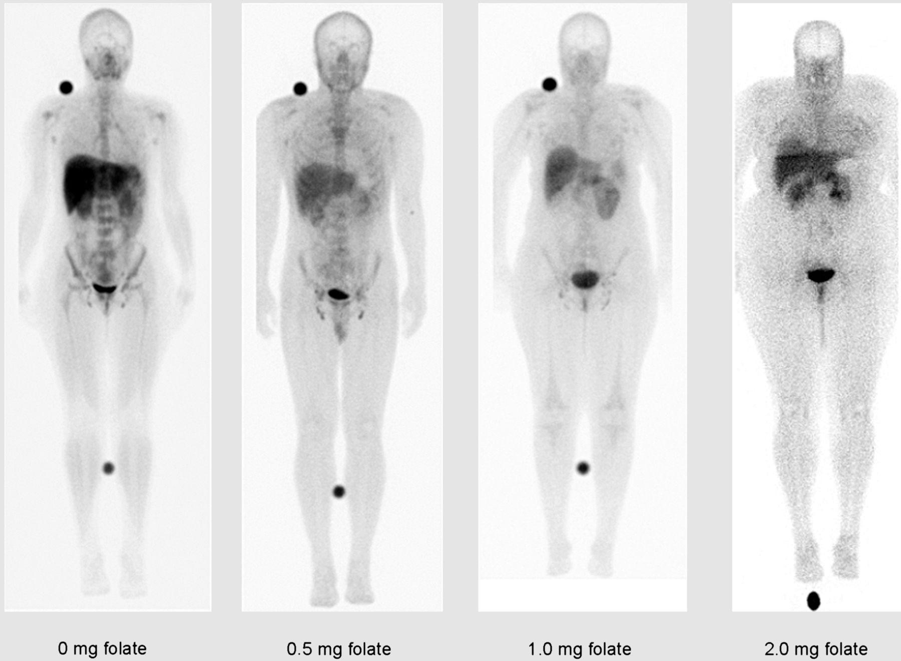
First, FR expression and 99mTc-etarfolatide physiologic uptake, which occurs in liver, kidneys, spleen, bladder, and, to a lesser extent, bone marrow, may complicate interpretation of FR expression in lesions close to these organs (11) (Fig. 1). To partially saturate FRs, and to reduce 99mTc-etarfolatide uptake in these organs, a small amount of folic acid is injected before 99mTc-etarfolatide administration.

FIGURE 1.
  • Download figure
  • Open in new tab
  • Download powerpoint
FIGURE 1.

99mTc-etarfolatide biodistribution pattern (1 h after injection) after folate preinjection in healthy volunteers. In absence of folate preinjection, 99mTc-etarfolatide uptake is seen in liver, kidney, spleen, bladder, and bone marrow. This background organ activity is decreased with preinjected folate.

Because activated macrophages express FRβ and show uptake of 99mTc-etarfolatide, areas of infection or inflammation may falsely appear as FR-positive malignant lesions (8,19) (Fig. 2). Another FR-imaging agent, a conjugate of a reduced and alkylated form of folic acid (N5, N10-dimethyltetrahydrofolic acid) and 99mTc (2,5-dimethoxytetrahydrofuran-99mTc), has been found to more specially bind to FRα, which is often expressed on malignant cells, and less to FRβ, often expressed by peripheral blood monocytes or inflammatory macrophages (20).

FIGURE 2.
  • Download figure
  • Open in new tab
  • Download powerpoint
FIGURE 2.

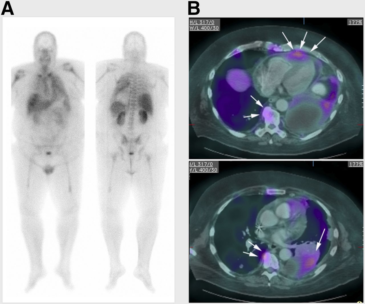
(A) Whole-body planar images obtained approximately 1 h after 99mTc-etarfolatide administration in lung cancer patient. (B) 99mTc-etarfolatide SPECT images were manually fused to patient’s prior CT images. SPECT/CT transaxial fusion images delineate tumor in left lower lobe (single arrow), with diffuse pleural spread of tumor and pericardial metastasis (triple arrows). 99mTc-etarfolatide uptake in inflammatory lesions such as benign osteophytes can be seen (double arrows).

The early imaging studies with 99mTc-etarfolatide used separate SPECT and CT images with only either visual side-by-side comparison or manual SPECT/CT fusion, because hybrid SPECT/CT cameras were not available. This technical limitation of using 2 noncontemporaneous studies may have limited these early studies. Integrated SPECT/CT cameras are becoming more prevalent. SPECT/CT fusion imaging should improve spatial localization and determination of FR-positive or -negative lesions (Figs. 2 and 3).

FIGURE 3.
  • Download figure
  • Open in new tab
  • Download powerpoint
FIGURE 3.

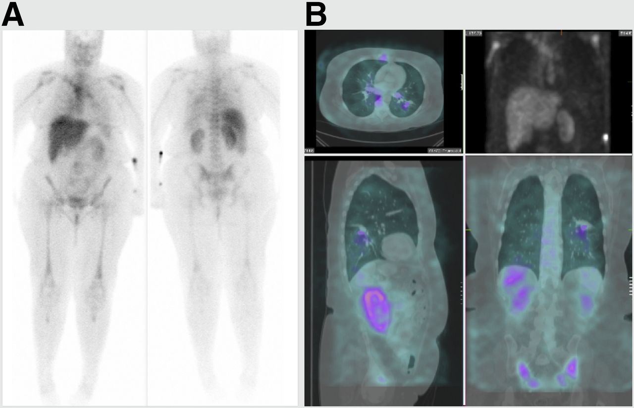
(A) Whole-body planar 99mTc-etarfolatide study for known pulmonary nodule obtained approximately 1 h after 99mTc-etarfolatide administration. As with other imaging agents, planar 2-dimensional imaging lacks image contrast and sensitivity for detecting 99mTc-etarfolatide binding to small pulmonary nodule in left lower lobe. (B) 99mTc-etarfolatide SPECT images with maximum intensity projection (upper right), transaxial (upper left), sagittal (lower left), and coronal (lower right) were manually fused to patient’s prior CT images, allowing for better anatomic localization and confirmation of FR binding to pulmonary nodule in this biopsy-proven non–small cell lung cancer.

At present, limited data exist on the correlation between FR expression as assessed by immunohistochemistry and 99mTc-etarfolatide uptake. One study showed a 61% agreement between methods (κ coefficient, 0.096; 95% confidence interval, –0.085 to 0.277) (11). The agreement was 72% for positive results and 38% for negative results (99mTc-etarfolatide–positive/–negative patients who are also FR immunohistochemistry–positive/–negative). These poor agreement scores could be related to tumor heterogeneity of FR expression, as immunohistochemistry analysis of primary tumors was compared with imaging of metastases (11). In addition, the immunohistochemistry did not identify FRβ, whereas 99mTc-etarfolatide imaging does.

PET FR IMAGING

Folate can be labeled with 18F (21–25). To date, 18F-fluorodeoxyglucose-folate and 3′-aza-2′-18F-fluorofolic acid seem the most promising PET FR imaging agents. Both have shown good tumor visualization, with limited accumulation in nonmalignant tissues (24,25). 68Ga-labeled radiofolates are also of interest because of their favorable biodistribution profile and 1-step radiolabeling (26,27). However, the short physical half-life of 68Ga (68 min) limits the time window for imaging compared with 18F (110 min) (21). Although there are promising preclinical results, clinical studies of PET folate imaging agents are needed for their further development.

POTENTIAL FUTURE APPLICATION OF 99MTC-ETARFOLATIDE IMAGING

99mTc-etarfolatide imaging may have other clinical applications. Because FR expression is prognostic for ovarian and lung cancer, 99mTc-etarfolatide imaging may be used as a prognostic tool (6,28). Intraoperative FR imaging with optical probes can improve staging and facilitates cytoreductive surgery (29). 99mTc-etarfolatide imaging may aid in selecting patients for intraoperative fluorescence FR imaging and help to identify deep-seated lesions that could be missed by intraoperative optical imaging because of limited signal penetration.

Preclinical studies have demonstrated that 99mTc-etarfolatide imaging can be used to visualize macrophage activation in atherosclerotic plaques and arthritis and to assess disease activity (30–32). One clinical study showed that 99mTc-etarfolatide imaging may be more sensitive than physical examination for assessing disease activity in rheumatoid arthritis patients (33). Other FR-imaging agents have potential to visualize macrophage activation in osteoarthritis and atherosclerosis (34,35).

CONCLUSION

99mTc-etarfolatide imaging has been shown to be valuable for selecting patients who may benefit from FR-targeted treatments. 99mTc-etarfolatide imaging may potentially also be useful for tumor staging or restaging, prognosis assessment, selection of patients for intraoperative fluorescence FR imaging, and assessment of inflammatory disease activity such as rheumatoid arthritis and osteoarthritis.

DISCLOSURE

Editorial and medical writing assistance (by Anna Hooijkaas, PhD, TRM Oncology, The Hague, The Netherlands) was funded by Endocyte Inc. Alan Maurer is a consultant and on the advisory board for Endocyte Inc. Philip Elsinga is a consultant for MSD. Stefano Fanti is on the advisory board for Endocyte Inc. Binh Nguyen is an employee of Endocyte Inc. Wim Oyen is a consultant for MSD and on the advisory board for Endocyte Inc. Wolfgang Weber is on the advisory boards for Endocyte Inc. and MSD. No other potential conflict of interest relevant to this article was reported.

Footnotes

  • Published online Apr. 14, 2014.

  • © 2014 by the Society of Nuclear Medicine and Molecular Imaging, Inc.

REFERENCES

  1. 1.↵
    1. Teng L,
    2. Xie J,
    3. Teng L,
    4. Lee RJ
    . Clinical translation of folate receptor-targeted therapeutics. Expert Opin Drug Deliv. 2012;9:901–908.
    OpenUrlCrossRefPubMed
  2. 2.↵
    1. Elnakat H,
    2. Ratnam M
    . Distribution, functionality and gene regulation of folate receptor isoforms: implications in targeted therapy. Adv Drug Deliv Rev. 2004;56:1067–1084.
    OpenUrlCrossRefPubMed
  3. 3.↵
    1. Naumann RW,
    2. Coleman RL,
    3. Burger RA,
    4. et al
    . PRECEDENT: a randomized phase II trial comparing vintafolide (EC145) and pegylated liposomal doxorubicin (PLD) in combination versus PLD alone in patients with platinum-resistant ovarian cancer. J Clin Oncol. 2013;31:4400–4406.
    OpenUrlAbstract/FREE Full Text
  4. 4.↵
    1. Jackman AL,
    2. Leamon CP
    1. Muller C,
    2. Schibli R
    . Folate-receptor targeted radionuclide imaging agents. In: Jackman AL, Leamon CP, eds. Targeted Drug Strategies for Cancer and Inflammation. New York, NY: Springer; 2011:65–92.
  5. 5.↵
    1. Kalli KR,
    2. Oberg AL,
    3. Keeney GL,
    4. et al
    . Folate receptor alpha as a tumor target in epithelial ovarian cancer. Gynecol Oncol. 2008;108:619–626.
    OpenUrlCrossRefPubMed
  6. 6.↵
    1. Chen YL,
    2. Chang MC,
    3. Huang CY,
    4. et al
    . Serous ovarian carcinoma patients with high alpha-folate receptor had reducing survival and cytotoxic chemo-response. Mol Oncol. 2012;6:360–369.
    OpenUrlCrossRefPubMed
  7. 7.↵
    1. Siu MK,
    2. Kong DS,
    3. Chan HY,
    4. et al
    . Paradoxical impact of two folate receptors, FRα and RFC, in ovarian cancer: effect on cell proliferation, invasion and clinical outcome. PLoS ONE. 2012;7:e47201.
    OpenUrlCrossRefPubMed
  8. 8.↵
    1. Low PS,
    2. Henne WA,
    3. Doorneweerd DD
    . Discovery and development of folic-acid-based receptor targeting for imaging and therapy of cancer and inflammatory diseases. Acc Chem Res. 2008;41:120–129.
    OpenUrlCrossRefPubMed
  9. 9.↵
    1. Armstrong DK,
    2. White AJ,
    3. Weil SC,
    4. Phillips M,
    5. Coleman RL
    . Farletuzumab (a monoclonal antibody against folate receptor alpha) in relapsed platinum-sensitive ovarian cancer. Gynecol Oncol. 2013;129:452–458.
    OpenUrlCrossRefPubMed
  10. 10.↵
    1. Xia W,
    2. Hilgenbrink AR,
    3. Matteson EL,
    4. Lockwood MB,
    5. Cheng JX,
    6. Low PS
    . A functional folate receptor is induced during macrophage activation and can be used to target drugs to activated macrophages. Blood. 2009;113:438–446.
    OpenUrlAbstract/FREE Full Text
  11. 11.↵
    1. Fisher RE,
    2. Siegel BA,
    3. Edell SL,
    4. et al
    . Exploratory study of 99mTc-EC20 imaging for identifying patients with folate receptor-positive solid tumors. J Nucl Med. 2008;49:899–906.
    OpenUrlAbstract/FREE Full Text
  12. 12.↵
    1. Almendro V,
    2. Marusyk A,
    3. Polyak K
    . Cellular heterogeneity and molecular evolution in cancer. Annu Rev Pathol. 2013;8:277–302.
    OpenUrlCrossRefPubMed
  13. 13.↵
    1. Morris RT,
    2. Joyrich RN,
    3. Naumann RW,
    4. et al
    . Phase 2 study of treatment of advanced ovarian cancer with folate-receptor–targeted therapeutic (vintafolide) and companion SPECT-based imaging agent (99mTc-etarfolatide). Ann Oncol. In press.
  14. 14.↵
    1. Buist MR,
    2. Kenemans P,
    3. den Hollander W,
    4. et al
    . Kinetics and tissue distribution of the radiolabeled chimeric monoclonal antibody MOv18 IgG and F(ab′)2 fragments in ovarian carcinoma patients. Cancer Res. 1993;53:5413–5418.
    OpenUrlAbstract/FREE Full Text
  15. 15.↵
    1. Siegel BA,
    2. Dehdashti F,
    3. Mutch DG,
    4. et al
    . Evaluation of 111In-DTPA-folate as a receptor-targeted diagnostic agent for ovarian cancer: initial clinical results. J Nucl Med. 2003;44:700–707.
    OpenUrlAbstract/FREE Full Text
  16. 16.↵
    1. Banerjee S,
    2. Pillai MR,
    3. Ramamoorthy N
    . Evolution of Tc-99m in diagnostic radiopharmaceuticals. Semin Nucl Med. 2001;31:260–277.
    OpenUrlCrossRefPubMed
  17. 17.↵
    1. Leamon CP,
    2. Parker MA,
    3. Vlahov IR,
    4. et al
    . Synthesis and biological evaluation of EC20: a new folate-derived, 99mTc-based radiopharmaceutical. Bioconjug Chem. 2002;13:1200–1210.
    OpenUrlCrossRefPubMed
  18. 18.↵
    1. Reddy JA,
    2. Xu LC,
    3. Parker N,
    4. Vetzel M,
    5. Leamon CP
    . Preclinical evaluation of 99mTc-EC20 for imaging folate receptor-positive tumors. J Nucl Med. 2004;45:857–866.
    OpenUrlAbstract/FREE Full Text
  19. 19.↵
    1. Henne WA,
    2. Rothenbuhler R,
    3. Ayala-Lopez W,
    4. Xia W,
    5. Varghese B,
    6. Low PS
    . Imaging sites of infection using a 99mTc-labeled folate conjugate targeted to folate receptor positive macrophages. Mol Pharm. 2012;9:1435–1440.
    OpenUrlPubMed
  20. 20.↵
    1. Vaitilingam B,
    2. Chelvam V,
    3. Kularatne SA,
    4. Poh S,
    5. Ayala-Lopez W,
    6. Low PS
    . A folate receptor-α-specific ligand that targets cancer tissue and not sites of inflammation. J Nucl Med. 2012;53:1127–1134.
    OpenUrlAbstract/FREE Full Text
  21. 21.↵
    1. Müller C
    . Folate-based radiotracers for PET imaging: update and perspectives. Molecules. 2013;18:5005–5031.
    OpenUrlCrossRefPubMed
  22. 22.
    1. Bettio A,
    2. Honer M,
    3. Müller C,
    4. et al
    . Synthesis and preclinical evaluation of a folic acid derivative labeled with 18F for PET imaging of folate receptor-positive tumors. J Nucl Med. 2006;47:1153–1160.
    OpenUrlAbstract/FREE Full Text
  23. 23.
    1. Ross TL,
    2. Honer M,
    3. Müller C,
    4. Groehn V,
    5. Schibli R,
    6. Ametamey SM
    . A new 18F-labeled folic acid derivative with improved properties for the PET imaging of folate receptor-positive tumors. J Nucl Med. 2010;51:1756–1762.
    OpenUrlAbstract/FREE Full Text
  24. 24.↵
    1. Betzel T,
    2. Müller C,
    3. Groehn V,
    4. et al
    . Radiosynthesis and preclinical evaluation of 3′-Aza-2′-[18F]fluorofolic acid: a novel PET radiotracer for folate receptor targeting. Bioconjug Chem. 2013;24:205–214.
    OpenUrlCrossRefPubMed
  25. 25.↵
    1. Fischer CR,
    2. Müller C,
    3. Reber J,
    4. et al
    . [18F]fluoro-deoxy-glucose folate: a novel PET radiotracer with improved in vivo properties for folate receptor targeting. Bioconjug Chem. 2012;23:805–813.
    OpenUrlCrossRefPubMed
  26. 26.↵
    1. Fani M,
    2. Tamma ML,
    3. Nicolas GP,
    4. et al
    . In vivo imaging of folate receptor positive tumor xenografts using novel 68Ga-NODAGA-folate conjugates. Mol Pharm. 2012;9:1136–1145.
    OpenUrlPubMed
  27. 27.↵
    1. Fani M,
    2. Wang X,
    3. Nicolas G,
    4. et al
    . Development of new folate-based PET radiotracers: preclinical evaluation of 68Ga-DOTA-folate conjugates. Eur J Nucl Med Mol Imaging. 2011;38:108–119.
    OpenUrlCrossRefPubMed
  28. 28.↵
    1. O’Shannessy DJ,
    2. Yu G,
    3. Smale R,
    4. et al
    . Folate receptor alpha expression in lung cancer: diagnostic and prognostic significance. Oncotarget. 2012;3:414–425.
    OpenUrlPubMed
  29. 29.↵
    1. van Dam GM,
    2. Themelis G,
    3. Crane LM,
    4. et al
    . Intraoperative tumor-specific fluorescence imaging in ovarian cancer by folate receptor-α targeting: first in-human results. Nat Med. 2011;17:1315–1319.
    OpenUrlCrossRefPubMed
  30. 30.↵
    1. Ayala-López W,
    2. Xia W,
    3. Varghese B,
    4. Low PS
    . Imaging of atherosclerosis in apoliprotein e knockout mice: targeting of a folate-conjugated radiopharmaceutical to activated macrophages. J Nucl Med. 2010;51:768–774.
    OpenUrlAbstract/FREE Full Text
  31. 31.
    1. Turk MJ,
    2. Breur GJ,
    3. Widmer WR,
    4. et al
    . Folate-targeted imaging of activated macrophages in rats with adjuvant-induced arthritis. Arthritis Rheum. 2002;46:1947–1955.
    OpenUrlCrossRefPubMed
  32. 32.↵
    1. Paulos CM,
    2. Turk MJ,
    3. Breur GJ,
    4. Low PS
    . Folate receptor-mediated targeting of therapeutic and imaging agents to activated macrophages in rheumatoid arthritis. Adv Drug Deliv Rev. 2004;56:1205–1217.
    OpenUrlCrossRefPubMed
  33. 33.↵
    1. Matteson EL,
    2. Lowe VJ,
    3. Prendergast FG,
    4. et al
    . Assessment of disease activity in rheumatoid arthritis using a novel folate targeted radiopharmaceutical Folatescan. Clin Exp Rheumatol. 2009;27:253–259.
    OpenUrlPubMed
  34. 34.↵
    1. Piscaer TM,
    2. Müller C,
    3. Mindt TL,
    4. et al
    . Imaging of activated macrophages in experimental osteoarthritis using folate-targeted animal single-photon-emission computed tomography/computed tomography. Arthritis Rheum. 2011;63:1898–1907.
    OpenUrlCrossRefPubMed
  35. 35.↵
    1. Winkel LC,
    2. Groen HC,
    3. van Thiel BS,
    4. et al
    . Folate receptor-targeted single-photon emission computed tomography/computed tomography to detect activated macrophages in atherosclerosis: can it distinguish vulnerable from stable atherosclerotic plaques? Mol Imaging. 2013;12:1–5.
    OpenUrl
  • Received for publication March 13, 2014.
  • Accepted for publication March 17, 2014.
PreviousNext
Back to top

In this issue

Journal of Nuclear Medicine: 55 (5)
Journal of Nuclear Medicine
Vol. 55, Issue 5
May 1, 2014
  • Table of Contents
  • Table of Contents (PDF)
  • About the Cover
  • Index by author
Print
Download PDF
Article Alerts
Sign In to Email Alerts with your Email Address
Email Article

Thank you for your interest in spreading the word on Journal of Nuclear Medicine.

NOTE: We only request your email address so that the person you are recommending the page to knows that you wanted them to see it, and that it is not junk mail. We do not capture any email address.

Enter multiple addresses on separate lines or separate them with commas.
Imaging the Folate Receptor on Cancer Cells with 99mTc-Etarfolatide: Properties, Clinical Use, and Future Potential of Folate Receptor Imaging
(Your Name) has sent you a message from Journal of Nuclear Medicine
(Your Name) thought you would like to see the Journal of Nuclear Medicine web site.
Citation Tools
Imaging the Folate Receptor on Cancer Cells with 99mTc-Etarfolatide: Properties, Clinical Use, and Future Potential of Folate Receptor Imaging
Alan H. Maurer, Philip Elsinga, Stefano Fanti, Binh Nguyen, Wim J.G. Oyen, Wolfgang A. Weber
Journal of Nuclear Medicine May 2014, 55 (5) 701-704; DOI: 10.2967/jnumed.113.133074

Citation Manager Formats

  • BibTeX
  • Bookends
  • EasyBib
  • EndNote (tagged)
  • EndNote 8 (xml)
  • Medlars
  • Mendeley
  • Papers
  • RefWorks Tagged
  • Ref Manager
  • RIS
  • Zotero
Share
Imaging the Folate Receptor on Cancer Cells with 99mTc-Etarfolatide: Properties, Clinical Use, and Future Potential of Folate Receptor Imaging
Alan H. Maurer, Philip Elsinga, Stefano Fanti, Binh Nguyen, Wim J.G. Oyen, Wolfgang A. Weber
Journal of Nuclear Medicine May 2014, 55 (5) 701-704; DOI: 10.2967/jnumed.113.133074
Twitter logo Facebook logo LinkedIn logo Mendeley logo
  • Tweet Widget
  • Facebook Like
  • Google Plus One
Bookmark this article

Jump to section

  • Article
    • Abstract
    • FOLATE RECEPTOR AND CANCER
    • FR EXPRESSION ASSESSMENT
    • 99MTC-ETARFOLATIDE CHARACTERISTICS
    • 99MTC-ETARFOLATIDE IN CLINICAL TRIALS
    • 99MTC-ETARFOLATIDE IMAGING
    • PET FR IMAGING
    • POTENTIAL FUTURE APPLICATION OF 99MTC-ETARFOLATIDE IMAGING
    • CONCLUSION
    • DISCLOSURE
    • Footnotes
    • REFERENCES
  • Figures & Data
  • Info & Metrics
  • PDF

Related Articles

  • This Month in JNM
  • PubMed
  • Google Scholar

Cited By...

  • Reduced Renal Uptake of Various Radiopharmaceuticals with Sodium Paraaminohippurate Coadministration in a Rat Model
  • Is Targeting the Folate Receptor in Ovarian Cancer Coming of Age?
  • The Thymidine Phosphorylase Imaging Agent 123I-IIMU Predicts the Efficacy of Capecitabine
  • Advancing biomedical imaging
  • Google Scholar

More in this TOC Section

  • From Genome to Phenome: Opportunities and Challenges of Molecular Imaging
  • Imaging the Activity of Efflux Transporters at the Blood–Brain Barrier in Neurologic Diseases: Radiotracer Selection Criteria
  • Molecular Imaging of Pulmonary Fibrosis
Show more Focus on Molecular Imaging

Similar Articles

Keywords

  • folate receptor
  • imaging
  • 99mTc-etarfolatide
  • SPECT
  • SPECT/CT
SNMMI

© 2025 SNMMI

Powered by HighWire