
Main menu

  • Home
  • Content
    • Current
    • Ahead of print
    • Past Issues
    • JNM Supplement
    • SNMMI Annual Meeting Abstracts
    • Continuing Education
    • JNM Podcasts
  • Subscriptions
    • Subscribers
    • Institutional and Non-member
    • Rates
    • Journal Claims
    • Corporate & Special Sales
  • Authors
    • Submit to JNM
    • Information for Authors
    • Assignment of Copyright
    • AQARA requirements
  • Info
    • Reviewers
    • Permissions
    • Advertisers
  • About
    • About Us
    • Editorial Board
    • Contact Information
  • More
    • Alerts
    • Feedback
    • Help
    • SNMMI Journals
  • SNMMI
    • JNM
    • JNMT
    • SNMMI Journals
    • SNMMI

User menu

  • Subscribe
  • My alerts
  • Log in
  • My Cart

Search

  • Advanced search
Journal of Nuclear Medicine
  • SNMMI
    • JNM
    • JNMT
    • SNMMI Journals
    • SNMMI
  • Subscribe
  • My alerts
  • Log in
  • My Cart
Journal of Nuclear Medicine

Advanced Search

  • Home
  • Content
    • Current
    • Ahead of print
    • Past Issues
    • JNM Supplement
    • SNMMI Annual Meeting Abstracts
    • Continuing Education
    • JNM Podcasts
  • Subscriptions
    • Subscribers
    • Institutional and Non-member
    • Rates
    • Journal Claims
    • Corporate & Special Sales
  • Authors
    • Submit to JNM
    • Information for Authors
    • Assignment of Copyright
    • AQARA requirements
  • Info
    • Reviewers
    • Permissions
    • Advertisers
  • About
    • About Us
    • Editorial Board
    • Contact Information
  • More
    • Alerts
    • Feedback
    • Help
    • SNMMI Journals
  • View or Listen to JNM Podcast
  • Visit JNM on Facebook
  • Join JNM on LinkedIn
  • Follow JNM on Twitter
  • Subscribe to our RSS feeds
OtherClinical Investigations

Prospective Comparison of 18F-FDG PET with Conventional Imaging Modalities (MRI, CT, and 67Ga Scintigraphy) in Assessment of Combined Intraarterial Chemotherapy and Radiotherapy for Head and Neck Carcinoma

Yoshimasa Kitagawa, Sadahiko Nishizawa, Kazuo Sano, Toshiyuki Ogasawara, Mikiko Nakamura, Norihiro Sadato, Masanori Yoshida and Yoshiharu Yonekura
Journal of Nuclear Medicine February 2003, 44 (2) 198-206;
Yoshimasa Kitagawa
  • Find this author on Google Scholar
  • Find this author on PubMed
  • Search for this author on this site
Sadahiko Nishizawa
  • Find this author on Google Scholar
  • Find this author on PubMed
  • Search for this author on this site
Kazuo Sano
  • Find this author on Google Scholar
  • Find this author on PubMed
  • Search for this author on this site
Toshiyuki Ogasawara
  • Find this author on Google Scholar
  • Find this author on PubMed
  • Search for this author on this site
Mikiko Nakamura
  • Find this author on Google Scholar
  • Find this author on PubMed
  • Search for this author on this site
Norihiro Sadato
  • Find this author on Google Scholar
  • Find this author on PubMed
  • Search for this author on this site
Masanori Yoshida
  • Find this author on Google Scholar
  • Find this author on PubMed
  • Search for this author on this site
Yoshiharu Yonekura
  • Find this author on Google Scholar
  • Find this author on PubMed
  • Search for this author on this site
  • Article
  • Figures & Data
  • Info & Metrics
  • PDF
Loading

Article Figures & Data

Figures

  • Tables
  • FIGURE 1.
    • Download figure
    • Open in new tab
    • Download powerpoint
    FIGURE 1.

    Pretreatment and posttreatment 18F-FDG PET (A and C) and corresponding MR (B and D) images of 63-y-old man with squamous cell carcinoma on left buccal mucosa. (A) 18F-FDG PET image shows intense focal accumulation of 18F-FDG (SUV = 5.59 mg/mL) in tumor before therapy (arrows). (B) Tumor is also visualized on postcontrast fat-suppression T1-weighted MR image (B) but not on CT image. (C) After chemoradiotherapy, 18F-FDG PET image reveals normalization of 18F-FDG uptake (arrows; SUV = 2.80 mg/mL), consistent with histologic finding of no viable tumor cells (PCR). (D) MR image still shows contrast enhancement in tumor although it is reduced in size (arrows), which may suggest residual tumor (false-positive). On basis of PET findings, patient avoided surgery. He has remained tumor free for >4 y.
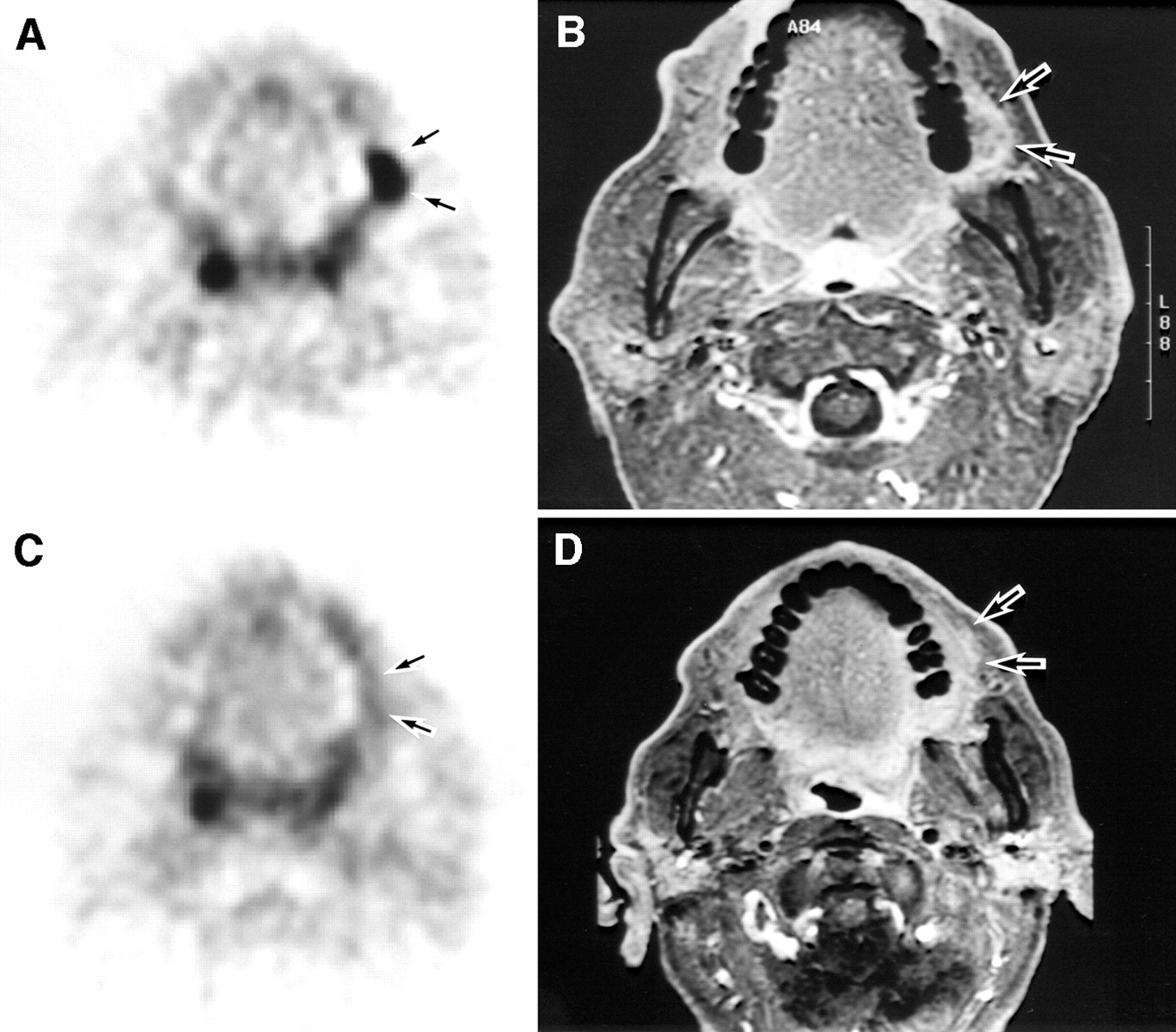
  • FIGURE 2.
    • Download figure
    • Open in new tab
    • Download powerpoint
    FIGURE 2.

    Pretreatment (A and B) and posttreatment (C and D) images of 60-y-old man with large squamous cell carcinoma (T4 N2a M0) on left mandibular gingiva. Pretreatment 18F-FDG PET image (A) shows focus of high 18F-FDG accumulation (SUV = 12.77 mg/mL) on left mandible (arrows), consistent with postcontrast CT (arrows, B) findings. After chemoradiotherapy, tumor disappeared on visual inspection with slight induration. (C) Posttreatment 18F-FDG PET image shows no abnormal 18F-FDG accumulation (arrows; SUV = 3.11 mg/mL), consistent with histologic finding (PCR). (D) CT image shows remarkable reduction in tumor size but does not exclude residual tumor because of contrast enhancement (arrows). According to 18F-FDG PET findings, patient successfully underwent functional neck dissection and marginal resection of mandible, requiring neither continuous resection of mandible nor reconstructive surgery.
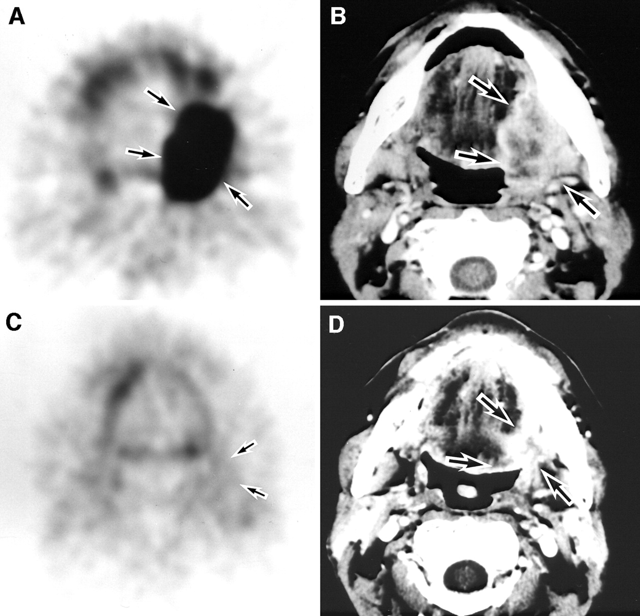
  • FIGURE 3.
    • Download figure
    • Open in new tab
    • Download powerpoint
    FIGURE 3.

    Pretreatment (A and B) and posttreatment (C and D) neck images of 78-y-old man with squamous cell carcinoma (T2 N0 M0) on floor of mouth. Pretreatment PET (A) and corresponding postcontrast CT (B) images demonstrate no metastasis to neck lymph nodes. (C) After chemoradiotherapy, neck lymph nodes were not palpable and posttreatment PET image shows abnormal 18F-FDG uptake in neck region (arrows, false-positive) probably due to inflammatory and reactive change. (D) Posttreatment CT image shows small lymph node (<1 cm; arrows, true-negative) in neck region, suggesting no metastasis. Patient was confirmed to have no metastasis in neck by clinical follow-up for >3 y.

Tables

  • Figures
    • View popup
    TABLE 1

    Patient Characteristics and Imaging Results Evaluated for Primary Tumors Before Treatment

    Patient no.Age (y)SexLocationTNMPre-SUV*Interpretation confidence rating
    67Ga scintigraphyCTMRIPET
    163FTongueT4 N2b M07.850444
    285FTongueT2 N0 M04.170004
    371MTongueT2 N0 M010.560004
    450MTongueT4 N1 M014.123444
    563MFloor of mouthT2 N0 M1†5.924444
    666MBuccal mucosaT3 N2b M05.073444
    770MMaxillary gingivaT2 N0 M07.960434
    873FMaxillary gingivarT1 N0 M05.291334
    971FTongueT2 N0 M04.070034
    1047MLower lipT2 N1 M011.220124
    1151MMandibleT4 N1 M07.283444
    1266MTongueT2 N0 M05.150123
    1348MMandibular gingivaT2 N1 M07.704224
    1474MFloor of mouthT3 N1 M014.544444
    1560MMandibular gingivaT4 N2a M012.773444
    1668MPalatal mucosaT1 N0 M09.760—34
    1763MBuccal mucosaT3 N0 M05.590034
    1851FTongueT2 N0 M08.540444
    1964MMandibular gingivaT4 N0 M08.403434
    2071MFloor of mouthT2 N0 M06.60—334
    2158MMandibular gingivaT4 N0 M011.34—444
    2256MFloor of mouthT4 N0 M026.100434
    2378MFloor of mouthT2 N0 M010.46—334
    Average score1.42.83.04.0
    • ↵* Pre-SUV = pretreatment standardized uptake value.

    • ↵† Lung metastasis.

    • Grading system: grade 0 = definitely no tumor; grade 1 = probably no tumor; grade 2 = equivocal; grade 3 = probable tumor; grade 4 = definite tumor.

    • View popup
    TABLE 2

    Sensitivity of Pretreatment Images for Primary Tumors

    Pretreatment images67Ga scintigraphyCTMRIPET
    True-positive (n)8151823
    False-negative (n)12750
    Sensitivity (%)406878100
     95% CI* (%)20–6445–8556–9282–100
    • ↵* 95% Confidence interval (15).

    • View popup
    TABLE 3

    Histologic Findings and Imaging Results Evaluated for Primary Tumors and Lymph Nodes After Treatment

    Patient no.Histologic evaluation for primary tumors after chemoradiotherapyPost-SUV*Interpretation confidence rating for images after chemoradiotherapy
    CT (primary)MRI (primary)PET (primary)CT (LN)MRI (LN)PET (LN)
    1RD4.413—32—2
    2PCR4.84——1——2
    3PCR3.16101223
    4RD4.39233120
    5PCR4.90433323
    6PCR4.25141234
    7PCR2.76320110
    8PCR4.77333211
    9PCR1.81220000
    10PCR1.12010210
    11RD5.02433320
    12PCR2.87——1——1
    13PCR3.52230222
    14PCR2.80331222
    15PCR3.11332332
    16PCR3.80000210
    17PCR2.80130210
    18RD8.32334333
    19PCR3.53431013
    20PCR1.61111100
    21PCR2.76430311
    22PCR4.61230220
    23PCR1.56010213
    • ↵* Post-SUV = posttreatment SUV.

    • LN = lymph nodes; RD = residual disease.

    • Grading system: grade 0 = definitely no tumor; grade 1 = probably no tumor; grade 2 = equivocal; grade 3 = probable tumor; grade 4 = definite tumor.

    • View popup
    TABLE 4

    Sensitivity and Specificity of Posttreatment Images for Primary Tumors and Lymph Nodes

    ParameterCT (primary)MRI (primary)PET (primary)CT (LN)MRI (LN)PET (LN)
    True-positive (n)334
    False-negative (n)100
    True-negative (n)10717161717
    False-positive (n)7102536
    Sensitivity (%)75100100
     95% CI* (%)22–9931–10040–100
    Specificity (%)594189768574
     95% CI* (%)33–8119–6765–9852–9161–9651–89
    • ↵* 95% Confidence interval (15).

    • LN = lymph nodes.

PreviousNext
Back to top

In this issue

Journal of Nuclear Medicine
Vol. 44, Issue 2
February 1, 2003
  • Table of Contents
  • Index by author
Print
Download PDF
Article Alerts
Sign In to Email Alerts with your Email Address
Email Article

Thank you for your interest in spreading the word on Journal of Nuclear Medicine.

NOTE: We only request your email address so that the person you are recommending the page to knows that you wanted them to see it, and that it is not junk mail. We do not capture any email address.

Enter multiple addresses on separate lines or separate them with commas.
Prospective Comparison of 18F-FDG PET with Conventional Imaging Modalities (MRI, CT, and 67Ga Scintigraphy) in Assessment of Combined Intraarterial Chemotherapy and Radiotherapy for Head and Neck Carcinoma
(Your Name) has sent you a message from Journal of Nuclear Medicine
(Your Name) thought you would like to see the Journal of Nuclear Medicine web site.
Citation Tools
Prospective Comparison of 18F-FDG PET with Conventional Imaging Modalities (MRI, CT, and 67Ga Scintigraphy) in Assessment of Combined Intraarterial Chemotherapy and Radiotherapy for Head and Neck Carcinoma
Yoshimasa Kitagawa, Sadahiko Nishizawa, Kazuo Sano, Toshiyuki Ogasawara, Mikiko Nakamura, Norihiro Sadato, Masanori Yoshida, Yoshiharu Yonekura
Journal of Nuclear Medicine Feb 2003, 44 (2) 198-206;

Citation Manager Formats

  • BibTeX
  • Bookends
  • EasyBib
  • EndNote (tagged)
  • EndNote 8 (xml)
  • Medlars
  • Mendeley
  • Papers
  • RefWorks Tagged
  • Ref Manager
  • RIS
  • Zotero
Share
Prospective Comparison of 18F-FDG PET with Conventional Imaging Modalities (MRI, CT, and 67Ga Scintigraphy) in Assessment of Combined Intraarterial Chemotherapy and Radiotherapy for Head and Neck Carcinoma
Yoshimasa Kitagawa, Sadahiko Nishizawa, Kazuo Sano, Toshiyuki Ogasawara, Mikiko Nakamura, Norihiro Sadato, Masanori Yoshida, Yoshiharu Yonekura
Journal of Nuclear Medicine Feb 2003, 44 (2) 198-206;
Twitter logo Facebook logo LinkedIn logo Mendeley logo
  • Tweet Widget
  • Facebook Like
  • Google Plus One
Bookmark this article

Jump to section

  • Article
    • Abstract
    • MATERIALS AND METHODS
    • RESULTS
    • DISCUSSION
    • CONCLUSION
    • Acknowledgments
    • Footnotes
    • REFERENCES
  • Figures & Data
  • Info & Metrics
  • PDF

Related Articles

  • No related articles found.
  • PubMed
  • Google Scholar

Cited By...

  • Computed Tomography of Lymph Node Metastasis Before and After Radiation Therapy: Correlations With Residual Tumour
  • The Value of PET Compared to MRI in Malignant Head and Neck Tumors
  • Added Value of Baseline 18F-FDG Uptake in Serial 18F-FDG PET for Evaluation of Response of Solid Extracerebral Tumors to Systemic Cytotoxic Neoadjuvant Treatment: A Meta-Analysis
  • Fluorodeoxyglucose-Positron-Emission Tomography Imaging of Head and Neck Squamous Cell Cancer
  • 18F-FDG PET as a Routine Posttreatment Surveillance Tool in Oral and Oropharyngeal Squamous Cell Carcinoma: A ProspectiveStudy
  • Does 18F-FDG PET/CT Improve the Detection of Posttreatment Recurrence of Head and Neck Squamous Cell Carcinoma in Patients Negative for Disease on Clinical Follow-up?
  • False-positive positron emission tomography appearance with 18F-fluorodeoxyglucose after definitive radiotherapy for cancer of the mobile tongue
  • Clinical Role of 18F-FDG PET/CT in the Management of Squamous Cell Carcinoma of the Head and Neck and Thyroid Carcinoma
  • Google Scholar

More in this TOC Section

  • Cardiac Presynaptic Sympathetic Nervous Function Evaluated by Cardiac PET in Patients with Chronotropic Incompetence Without Heart Failure
  • Validation and Evaluation of a Vendor-Provided Head Motion Correction Algorithm on the uMI Panorama PET/CT System
  • Left Ventricular Strain from Myocardial Perfusion PET Imaging: Method Development and Comparison to 2-Dimensional Echocardiography
Show more Clinical Investigations

Similar Articles

SNMMI

© 2025 SNMMI

Powered by HighWire