


Article Figures & Data
Figures
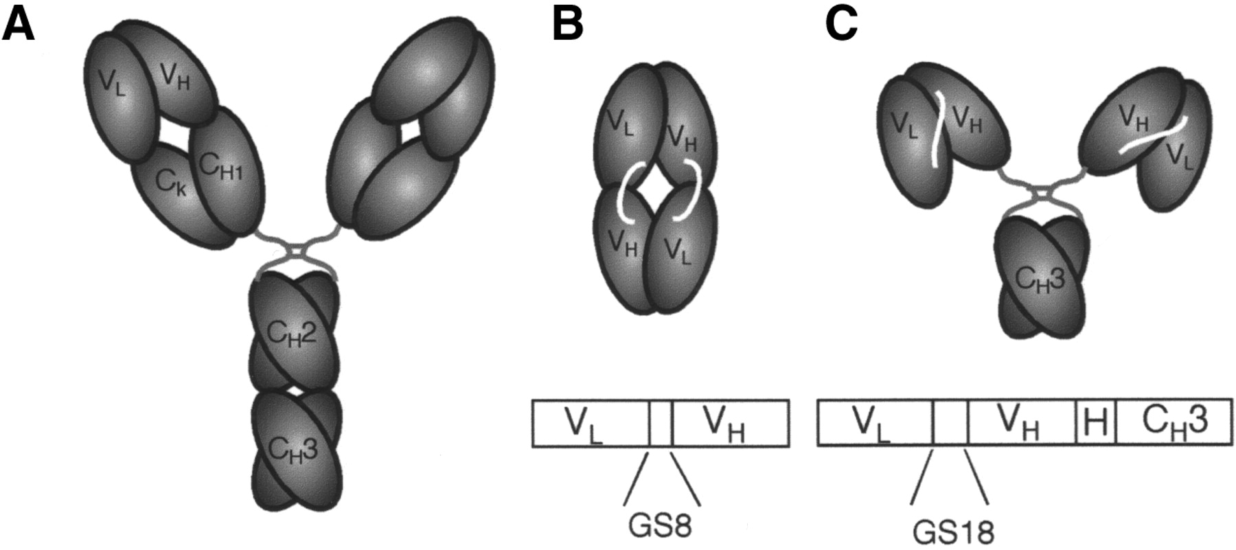
- FIGURE 1.
Schematic drawing shows domain structure of parental intact T84.66 antibody (A), T84.66 diabody (B), and T84.66 minibody (C). Anti-CEA minibody (80 kDa) and diabody (55 kDa) were derived from T84.66, a high-affinity and highly specific antibody that recognizes an epitope on A3 domain of CEA. Minibody (C) has glycine-serine-rich 18-amino-acid linker (GS18) between variable light (VL) and variable heavy chains (VH) and human IgG1 hinge (H) joining them to CH3 domain, also derived from human IgG1. Gene encoding diabody (B) encodes glycine-serine-rich 8-amino-acid linker (GS8) between VL and VH regions. Amino acid sequence is GSTSGGGSGGGSGGGGSS for GS18 and GGGSGGGG for GS8.
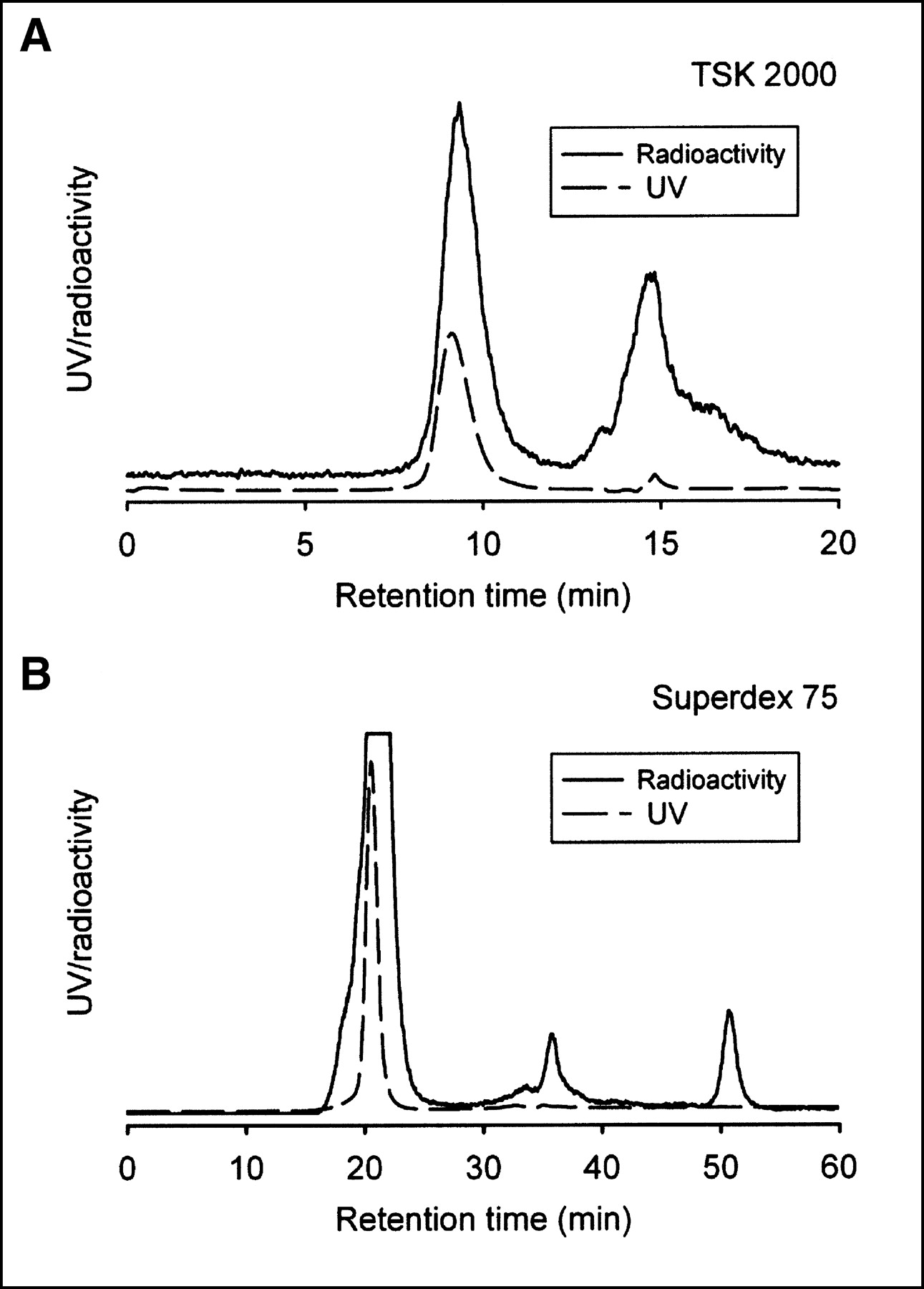
- FIGURE 2.
Size-exclusion HPLC analysis of 124I-radiolabeled anti-CEA minibody (A) and diabody (B). 124I was conjugated to T84.66 minibody and T84.66 diabody as described in Materials and Methods. Radiolabeling efficiency was determined by integrating areas on HPLC trace and determining radioactivity associated with 80-kDa protein peak for minibody or 55-kDa peak for diabody as percentage of total radioactivity eluted. Labeling efficiencies were 33% (not shown) or 46% for minibody (A) and 88% for diabody (B). Peak fractions (based on protein absorbance) were pooled for animal studies. Smaller peaks represent unincorporated label and low-molecular-weight components.
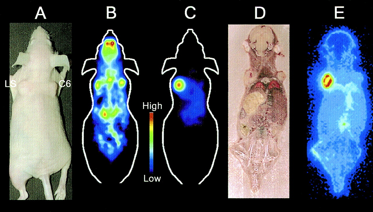
- FIGURE 3.
(A) Subcutaneous LS174T (left shoulder) and C6 glioma (right shoulder) xenografts were established in nude mouse. (B) PET imaging with 18F-FDG was done 2 d before injection of 124I (10/64 planes shown). (C) Mouse was injected with 3.1 MBq (85 μCi) 124I-minibody and imaged at 18 h by PET (10/64 planes shown). Both these images (B and C) are on same color scale. (D) After 18-h scan, mouse was euthanized and frozen, and whole-body coronal sections were cut in cryostat and processed for DWBA. (E) DWBA confirms specific localization of 124I minibody to CEA-positive tumor and low levels of background activity.
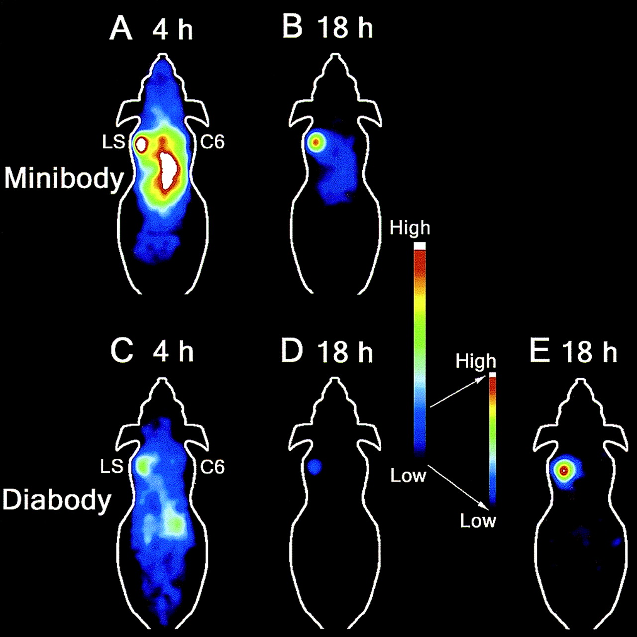
- FIGURE 4.
Comparison of 124I minibody and 124I diabody by serial PET imaging at 4 and 18 h. Mice bearing LS174T (LS) and C6 rat glioma (C6) xenografts were injected via tail vein with 1.9–3.1 MBq (65–85 μCi) 124I minibody (A and B) or diabody (C–E) and imaged at 4 and 18 h. At 18 h, background activity in central region was minimal in 124I diabody (D and E)–injected animal, resulting in high contrast, which can be seen in these maximum a posteriori reconstructed images (10/64 planes shown). A–D are on common scale. D was rescaled to E to illustrate excellent contrast achieved with 124I diabody at 18 h.


Tables
- TABLE 1
Ratios Derived from PET Data Comparing Target-to-Background Levels of 124I Activity
Ratio Minibody (n = 8) Diabody (n = 4) 4 h 18 h 4 h 18 h LS174T to C6 3.46 ± 0.72 10.85 ± 2.03* 3.99 ± 0.98 —† LS174T to background 0.78 ± 0.10 3.48 ± 0.48* 1.83 ± 0.40 6.11 ± 1.49‡ LS174T to soft tissue 3.05 ± 0.72 11.03 ± 2.35* 3.95 ± 1.27 10.93 ± 2.21‡ - TABLE 2
%ID/g Values and Target-to-Background (LS174T/Organ) Ratios Derived from Ex Vivo Counting of Weighed Tissues in γ-Counter After 18-Hour Uptake Time
Tissue %ID/g Ratio Minibody (n = 5) Diabody (n = 4) Minibody (n = 5) Diabody (n = 4) LS174T 20.55 ± 2.74 4.53 ± 0.50* C6 1.87 ± 0.38† 0.23 ± 0.03*‡ 26.19 ± 11.16 21.31 ± 4.47 Heart 1.78 ± 0.21† 0.07 ± 0.01*‡ 18.93 ± 6.10 61.57 ± 7.84* Liver 1.18 ± 0.18† 0.15 ± 0.01*‡ 31.05 ± 9.91 31.16 ± 3.46 Kidney 1.51 ± 0.22† 0.37 ± 0.05*‡ 25.73 ± 9.87 12.66 ± 2.17
In this issue


Jump to section
Related Articles
Cited By...
- Preclinical Evaluation of an Engineered Single-Chain Fragment Variable-Fragment Crystallizable Targeting Human CD44
- In Vivo Imaging of the Programmed Death Ligand 1 by 18F PET
- Anti-MET ImmunoPET for Non-Small Cell Lung Cancer Using Novel Fully Human Antibody Fragments
- Development of 124I Immuno-PET Targeting Tumor Vascular TEM1/Endosialin
- A Novel Engineered Anti-CD20 Tracer Enables Early Time PET Imaging in a Humanized Transgenic Mouse Model of B-cell Non-Hodgkins Lymphoma
- Advances in Immuno-Positron Emission Tomography: Antibodies for Molecular Imaging in Oncology
- Anti-carcinoembryonic Antigen Single-chain Variable Fragment Antibody Variants Bind Mouse and Human Neonatal Fc Receptor with Different Affinities That Reveal Distinct Cross-species Differences in Serum Half-life
- Evaluation of the Anti-HER2 C6.5 Diabody as a PET Radiotracer to Monitor HER2 status and Predict Response to Trastuzumab Treatment
- Cytotoxic Enhancement of a Bispecific Diabody by Format Conversion to Tandem Single-chain Variable Fragment (taFv): THE CASE OF THE hEx3 DIABODY
- Identification of Internalizing Human Single-Chain Antibodies Targeting Brain Tumor Sphere Cells
- Monodispersed DOTA-PEG-Conjugated Anti-TAG-72 Diabody Has Low Kidney Uptake and High Tumor-to-Blood Ratios Resulting in Improved 64Cu PET
- Recombinant Anti-CD20 Antibody Fragments for Small-Animal PET Imaging of B-Cell Lymphomas
- PET Imaging of Prostate Cancer Xenografts with a Highly Specific Antibody against the Prostate-Specific Membrane Antigen
- Antibodies and Antimatter: The Resurgence of Immuno-PET
- Humanized Radioiodinated Minibody For Imaging of Prostate Stem Cell Antigen-Expressing Tumors
- Comprehensive Analysis of the Factors Contributing to the Stability and Solubility of Autonomous Human VH Domains
- Immuno-PET: A Navigator in Monoclonal Antibody Development and Applications
- Imaging of Weak-Source Distributions in LSO-Based Small-Animal PET Scanners
- Bispecific Antibody Pretargeting of Radionuclides for Immuno-Single-Photon Emission Computed Tomography and Immuno-Positron Emission Tomography Molecular Imaging: An Update
- PET Imaging of Colorectal Cancer in Xenograft-Bearing Mice by Use of an 18F-Labeled T84.66 Anti-Carcinoembryonic Antigen Diabody
- Radioiodinated versus Radiometal-Labeled Anti-Carcinoembryonic Antigen Single-Chain Fv-Fc Antibody Fragments: Optimal Pharmacokinetics for Therapy
- Radioimaging of Light Chain Amyloid with a Fibril-Reactive Monoclonal Antibody
- Bispecific Antibody Pretargeting PET (ImmunoPET) with an 124I-Labeled Hapten-Peptide
- The Progress and Promise of Molecular Imaging Probes in Oncologic Drug Development
- Optimizing Radiolabeled Engineered Anti-p185HER2 Antibody Fragments for In vivo Imaging
- Radiolabeled Small-Molecule Ligands for Prostate-Specific Membrane Antigen: In vivo Imaging in Experimental Models of Prostate Cancer
- Quantitative Immuno-Positron Emission Tomography Imaging of HER2-Positive Tumor Xenografts with an Iodine-124 Labeled Anti-HER2 Diabody
- Tailoring the Pharmacokinetics and Positron Emission Tomography Imaging Properties of Anti-Carcinoembryonic Antigen Single-Chain Fv-Fc Antibody Fragments
- The Promise of Immuno-PET in Radioimmunotherapy
- Tumor imaging by means of proteolytic activation of cell-penetrating peptides
- Genetically Engineered Antibody Fragments and PET Imaging: A New Era of Radioimmunodiagnosis