


Article Figures & Data
Figures
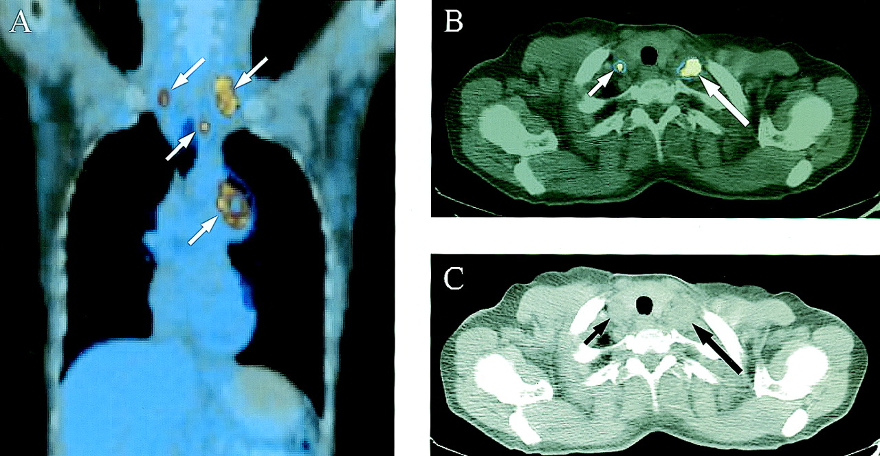
- FIGURE 1.
Coronal (A) and transaxial (B) PET/CT scans of chest and thoracic aperture in 47-y-old female patient show left central LD SCLC with ipsilateral paratracheal and bilateral supraclavicular lymph node metastases (white arrows). (C) Transaxial CT scan at level of thorax aperture shows pathologically enlarged ipsilateral (large black arrow) and normal-sized contralateral (small black arrow) supraclavicular lymph nodes.
- FIGURE 2.
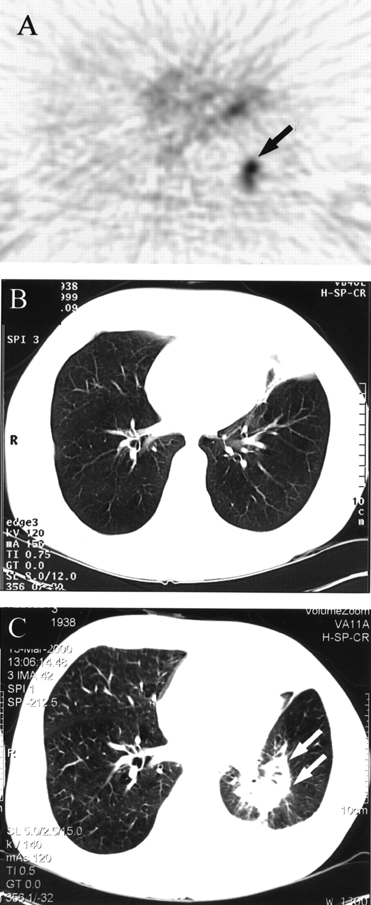
Transaxial thoracic 18F-FDG PET scan (A) in 64-y-old male patient shows pathologic 18F-FDG uptake at lower lobe of left lung consistent with residual disease after 5 chemotherapy cycles (black arrow) with no corresponding CT scan abnormality (B). Follow-up CT scan at 7 mo (C) shows local recurrence at lower lobe of left lung (white arrows).
- FIGURE 3.
Transaxial thoracic slices obtained by 18F-FDG PET (A), CT (B), and PET/CT (C) image fusion in 59-y-old female patient show residual disease after induction chemotherapy (arrows).


Tables
- TABLE 1
Performance of 18F-FDG PET vs. Conventional Imaging Modalities Among Initial Staging Patient Group (n = 24)
Patient Stage Pathology CI PET Incongruence PET impact on management 1 LD SCLC* ED LD False-positive contralateral lung metastasis (CT) Curative surgical resection 2–3 LD SCLC LD LD Contralateral supraclavicular lymph node metastasis (PET) Radiotherapy field extension 4 LD SCLC† LD LD Contralateral cervical lymph node metastasis (PET) Radiotherapy field extension 5 LD SCLC LD LD Contralateral mediastinal lymph node metastasis (PET) Radiotherapy volume change 6 LD SCLC* LD LD Additional ipsilateral lung metastasis (PET) Radiotherapy volume change 7–14 LD SCLC LD LD — — 15 LD SCLC† LD LD — — 16–18 ED SCLC LD ED Occult distant metastases (PET) Palliative chemotherapy 19 ED SCLC ED ED Additional visceral metastases (PET) — 20 ED SCLC ED ED False-positive adrenal metastases (CT) — 21–22 ED SCLC ED ED False-negative brain metastases (PET) — 23–24 ED SCLC ED ED — — - TABLE 2
Performance of 18F-FDG PET vs. Conventional Imaging Modalities Among Restaging Patient Group (n = 20)
Patient State Pathology CI PET Incongruence PET impact on management 1–2 CR SCLC RD CR False-positive mediastinal lymph nodes (CT) Chemotherapy cessation 3–5 CR SCLC CR CR — — 6 RD SCLC RD CR False-negative subcarinal lymph node (PET) — 7 RD SCLC CR RD Active parenchymal lung lesion (PET) Chemotherapy reinstitution 8 RD SCLC RD RD False-positive sternal metastasis (CT, BS) — 9 RD SCLC RD RD True-positive soft-tissue metastases (PET) — 10–17 RD SCLC RD RD — — 18–20 PD SCLC PD PD — — CI = conventional imaging modalities; RD = residual disease; CR = complete response; BS = bone scan; PD = progressive disease.
In this issue


Jump to section
Related Articles
Cited By...
- Small Cell Lung Cancer
- Modern Staging of Small Cell Lung Cancer
- Small Cell Lung Cancer
- Guidelines on the radical management of patients with lung cancer
- NCCN Task Force: Clinical Utility of PET in a Variety of Tumor Types
- Evaluation of Dual-Time-Point 18F-FDG PET for Staging in Patients with Lung Cancer
- Partnerships in Oncology and Radiology: The Role of Radiology in the Detection, Staging, and Follow-up of Lung Cancer
- PET Evaluation of Lung Cancer
- Significance of Incidental 18F-FDG Accumulations in the Gastrointestinal Tract in PET/CT: Correlation with Endoscopic and Histopathologic Results
- Positron Emission Tomography in Limited-Stage Small-Cell Lung Cancer: A Prospective Study
- Why Most PET of Lung and Head-and-Neck Cancer Will Be PET/CT