


Article Figures & Data
Figures
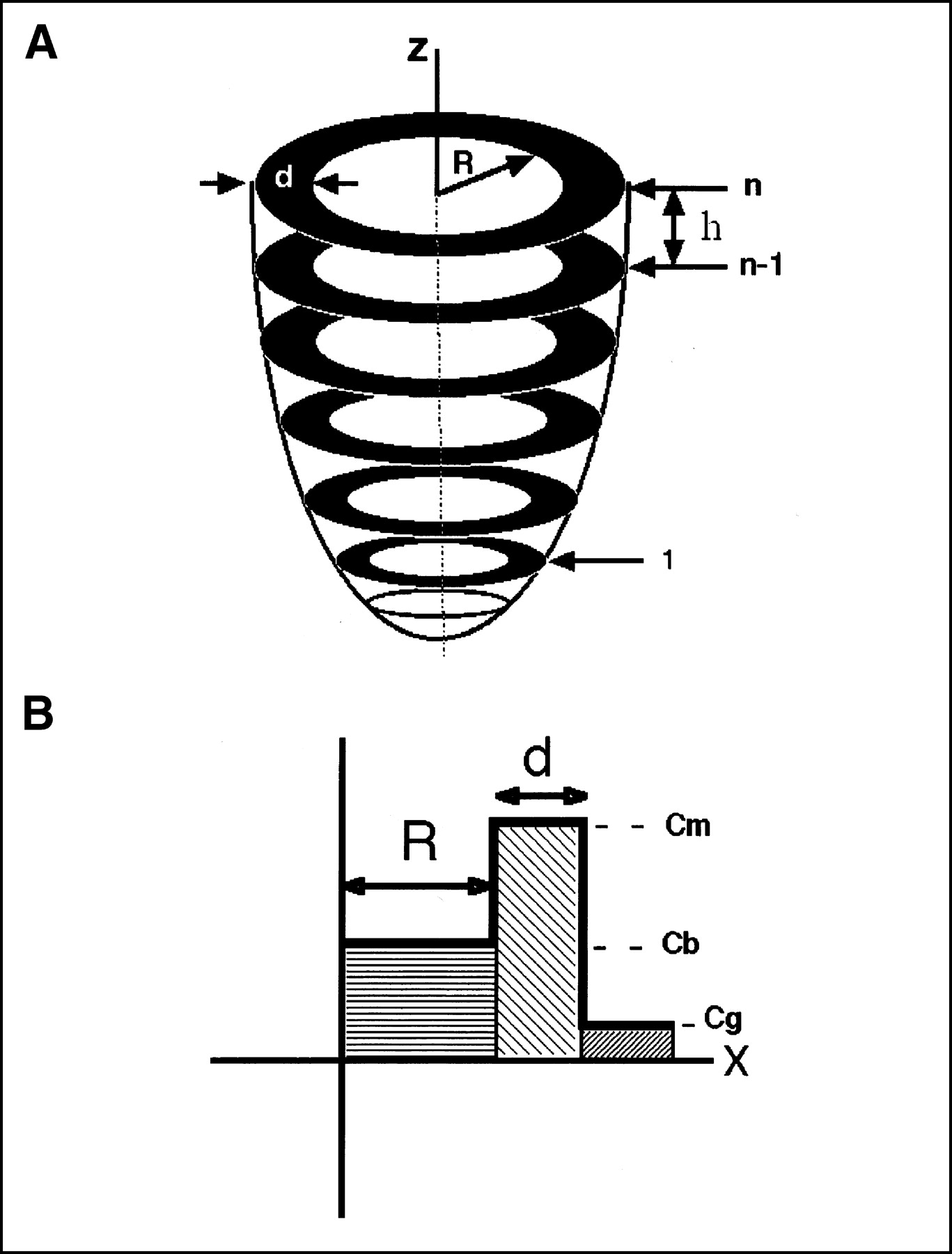
- FIGURE 1.
(A) Three-dimensional model of left ventricle as used for calculation of volume and mass. (B) One-dimensional model of radial tracer profile originating from center of left ventricle and extending through myocardium into background. Cb = blood-pool activity; Cg = background activity; Cm = myocardial activity; d = wall thickness; h = slice thickness; n = slice number; R = left ventricular radius; z = left ventricular long axis.
- FIGURE 2.
Example of model-based parameter estimation of cardiac geometry. Short-axis images obtained at basal (left), midventricular (middle), and apical (right) level by MRI (first and fourth rows) and PET (second and fifth rows) for diastolic (top 3 rows) and systolic (bottom 3 rows) time points. Third and sixth rows depict short-axis views with overlay of estimated endocardial and epicardial contours.
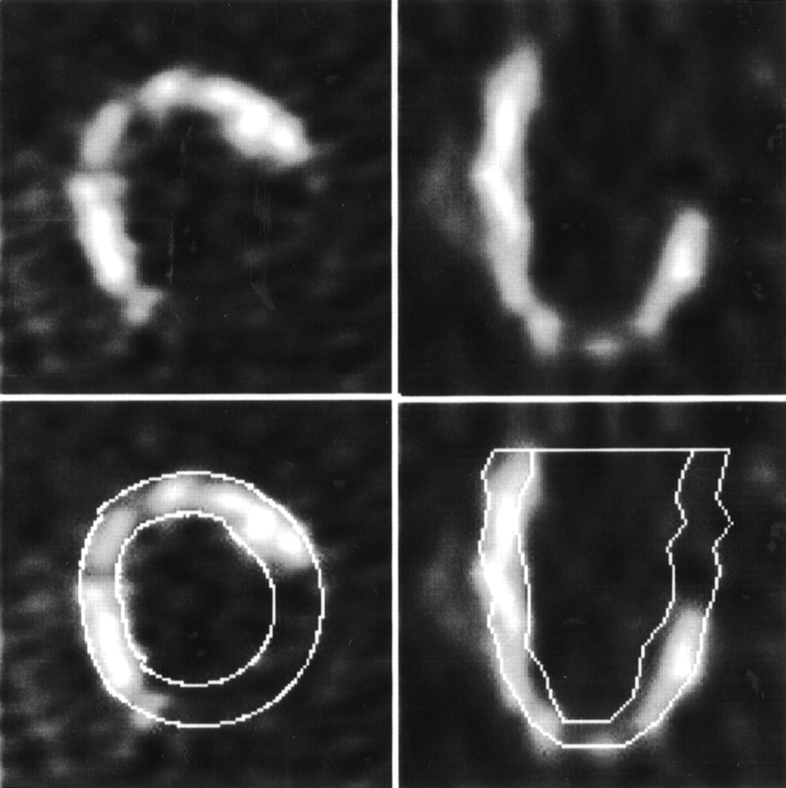
- FIGURE 3.
Example of model-based parameter estimation in patient with large defect in tracer uptake due to myocardial infarction: short-axis (left) and horizontal long-axis (right) images with estimated endocardial and epicardial contours (bottom row).
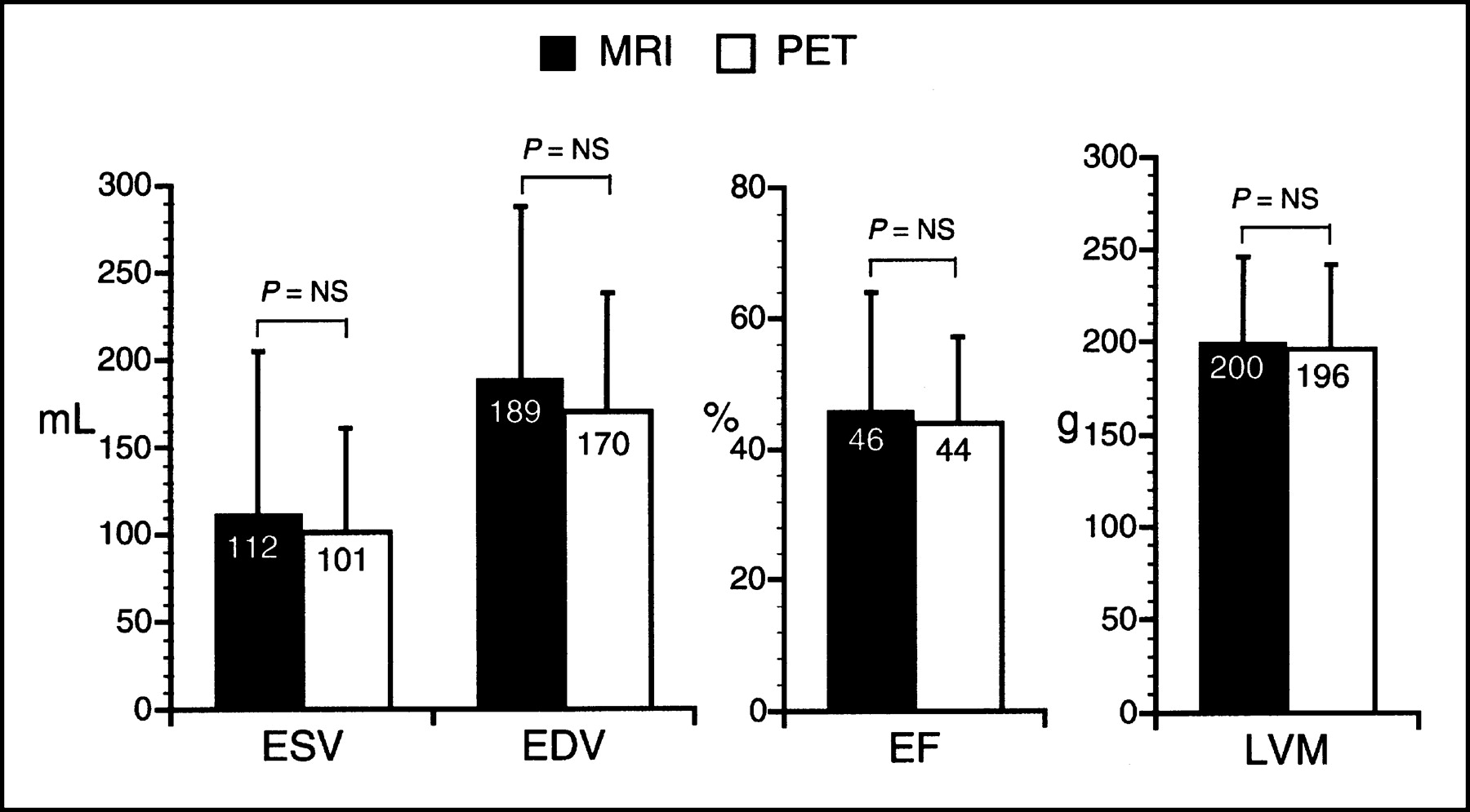
- FIGURE 4.
End-systolic (ESV) and end-diastolic (EDV) volumes of left ventricle, ejection fraction (EF), and left ventricular mass (LVM) as obtained from MRI and PET (P = not statistically significant [NS], paired t test).
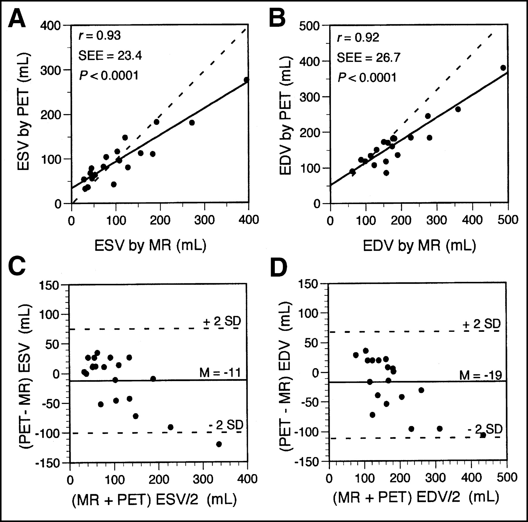
- FIGURE 5.
Regression analysis and Bland-Altman plots of end-systolic (ESV) (A and C) and end-diastolic (EDV) (B and D) volumes measured by PET and MRI. Dashed lines in A and B are lines of identity (PET value = MRI value). Mean difference (M) with 95% confidence limits (± 2 SDs) in C and D are presented by solid and dashed lines, respectively.
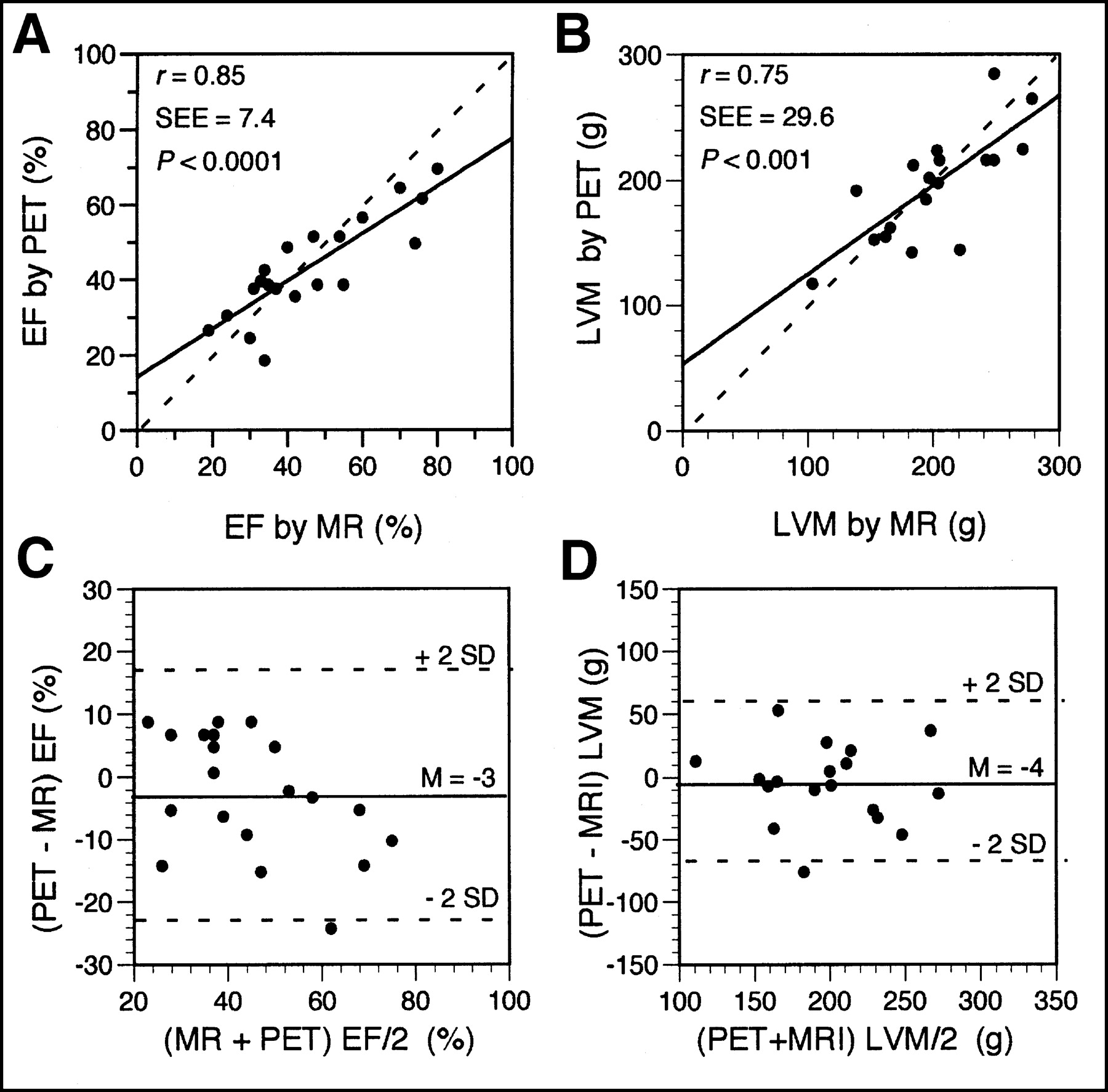
- FIGURE 6.
Regression analysis and Bland-Altman plots of ejection fraction (EF) (A and C) and left ventricular mass (LVM) (B and D) measured by PET and MRI. Dashed lines in A and B are lines of identity (PET value = MRI value). Mean difference (M) with 95% confidence limits (± 2 SDs) in C and D are presented by solid and dashed lines, respectively.


