
Main menu

  • Home
  • Content
    • Current
    • Ahead of print
    • Past Issues
    • JNM Supplement
    • SNMMI Annual Meeting Abstracts
    • Continuing Education
    • JNM Podcasts
  • Subscriptions
    • Subscribers
    • Institutional and Non-member
    • Rates
    • Journal Claims
    • Corporate & Special Sales
  • Authors
    • Submit to JNM
    • Information for Authors
    • Assignment of Copyright
    • AQARA requirements
  • Info
    • Reviewers
    • Permissions
    • Advertisers
  • About
    • About Us
    • Editorial Board
    • Contact Information
  • More
    • Alerts
    • Feedback
    • Help
    • SNMMI Journals
  • SNMMI
    • JNM
    • JNMT
    • SNMMI Journals
    • SNMMI

User menu

  • Subscribe
  • My alerts
  • Log in
  • My Cart

Search

  • Advanced search
Journal of Nuclear Medicine
  • SNMMI
    • JNM
    • JNMT
    • SNMMI Journals
    • SNMMI
  • Subscribe
  • My alerts
  • Log in
  • My Cart
Journal of Nuclear Medicine

Advanced Search

  • Home
  • Content
    • Current
    • Ahead of print
    • Past Issues
    • JNM Supplement
    • SNMMI Annual Meeting Abstracts
    • Continuing Education
    • JNM Podcasts
  • Subscriptions
    • Subscribers
    • Institutional and Non-member
    • Rates
    • Journal Claims
    • Corporate & Special Sales
  • Authors
    • Submit to JNM
    • Information for Authors
    • Assignment of Copyright
    • AQARA requirements
  • Info
    • Reviewers
    • Permissions
    • Advertisers
  • About
    • About Us
    • Editorial Board
    • Contact Information
  • More
    • Alerts
    • Feedback
    • Help
    • SNMMI Journals
  • View or Listen to JNM Podcast
  • Visit JNM on Facebook
  • Join JNM on LinkedIn
  • Follow JNM on Twitter
  • Subscribe to our RSS feeds
OtherClinical Investigations

Human-Observer Receiver-Operating-Characteristic Evaluation of Attenuation, Scatter, and Resolution Compensation Strategies for 99mTc Myocardial Perfusion Imaging

Manoj V. Narayanan, Michael A. King, P. Hendrik Pretorius, Seth T. Dahlberg, Frederick Spencer, Ellen Simon, Eric Ewald, Edward Healy, Kirk MacNaught and Jeffrey A. Leppo
Journal of Nuclear Medicine November 2003, 44 (11) 1725-1734;
Manoj V. Narayanan
  • Find this author on Google Scholar
  • Find this author on PubMed
  • Search for this author on this site
Michael A. King
  • Find this author on Google Scholar
  • Find this author on PubMed
  • Search for this author on this site
P. Hendrik Pretorius
  • Find this author on Google Scholar
  • Find this author on PubMed
  • Search for this author on this site
Seth T. Dahlberg
  • Find this author on Google Scholar
  • Find this author on PubMed
  • Search for this author on this site
Frederick Spencer
  • Find this author on Google Scholar
  • Find this author on PubMed
  • Search for this author on this site
Ellen Simon
  • Find this author on Google Scholar
  • Find this author on PubMed
  • Search for this author on this site
Eric Ewald
  • Find this author on Google Scholar
  • Find this author on PubMed
  • Search for this author on this site
Edward Healy
  • Find this author on Google Scholar
  • Find this author on PubMed
  • Search for this author on this site
Kirk MacNaught
  • Find this author on Google Scholar
  • Find this author on PubMed
  • Search for this author on this site
Jeffrey A. Leppo
  • Find this author on Google Scholar
  • Find this author on PubMed
  • Search for this author on this site
  • Article
  • Figures & Data
  • Info & Metrics
  • PDF
Loading

Article Figures & Data

Figures

  • Tables
  • FIGURE 1.
    • Download figure
    • Open in new tab
    • Download powerpoint
    FIGURE 1.

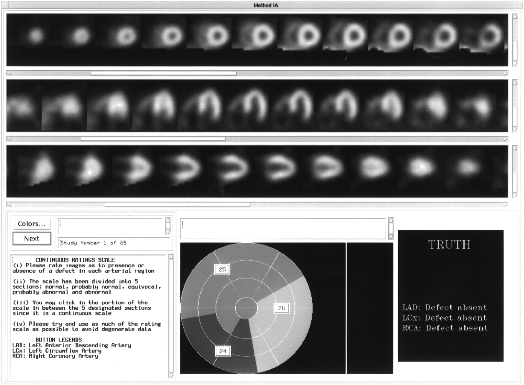
    GUI used in ROC studies. Top 3 panels display SA, HLA, and VLA slices, from top to bottom, for interpretation by observer. Three pull-down buttons shown on polar map marking 3 arterial territories (middle panel at bottom) allowed user to assign score to each vascular territory (LAD, LCx, and RCA). During training, feedback regarding presence or absence of defect would appear in right panel at bottom of interface after observer completed assigning scores as described in text. Instructions for observer were given in left panel at bottom.

  • FIGURE 2.
    • Download figure
    • Open in new tab
    • Download powerpoint
    FIGURE 2.

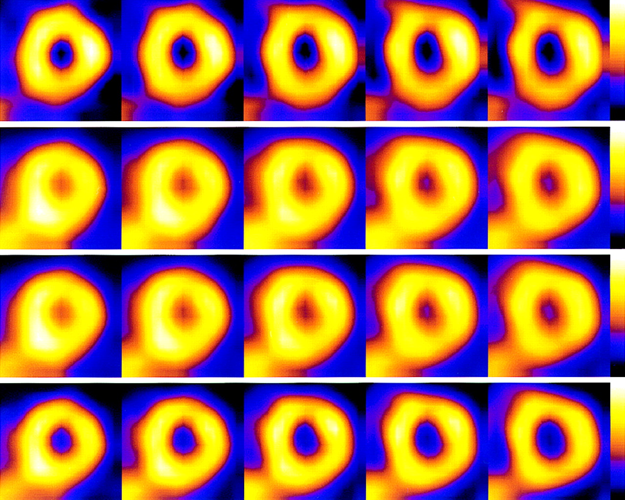
    Comparison of SA slices of FBP (first row), OSEM with AC (second row), OSEM with AC + SC (third row), and OSEM with AC + SC + RC (fourth row) reconstructions for patient with low likelihood of CAD. Note that inferior wall cooling artifact associated with subdiaphragmatic attenuation and presence of extracardiac activity, although present in FBP images, is absent in corrected images. Enhanced levels of extracardiac activity were seen in OSEM images with AC alone, in contrast to OSEM images with both AC + SC and AC + SC + RC.

  • FIGURE 3.
    • Download figure
    • Open in new tab
    • Download powerpoint
    FIGURE 3.

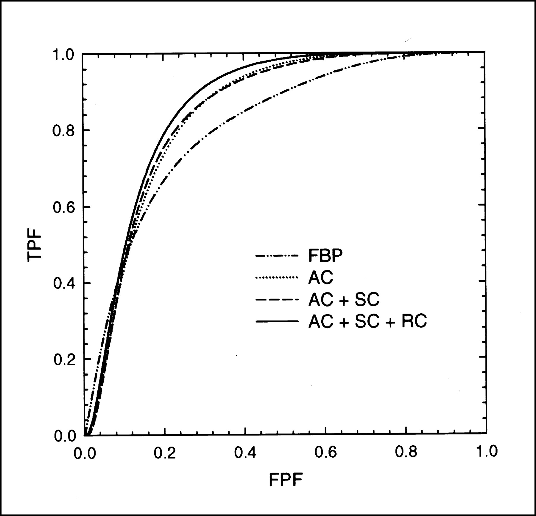
    Comparison of ROC curves for overall detection of CAD (averaged over 7 observers) for FBP, OSEM with AC, OSEM with AC + SC, and OSEM with AC + SC + RC. TPF = true-positive fraction; FPF = false-positive fraction.

  • FIGURE 4.
    • Download figure
    • Open in new tab
    • Download powerpoint
    FIGURE 4.

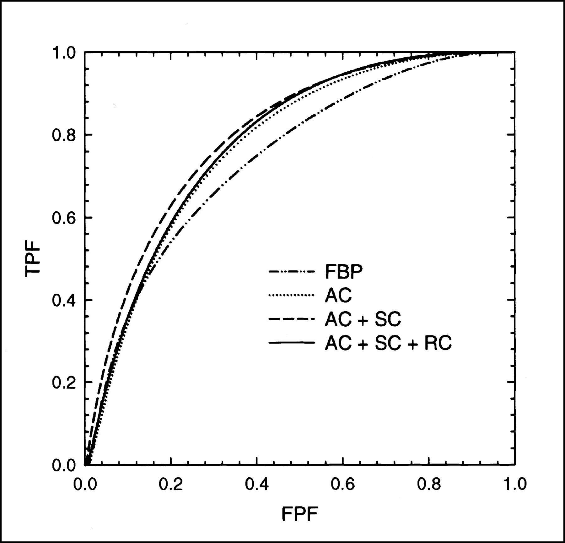
    Average ROC curves for LAD localization for FBP, OSEM with AC, OSEM with AC + SC, and OSEM with AC + SC + RC. TPF = true-positive fraction; FPF = false-positive fraction.

  • FIGURE 5.
    • Download figure
    • Open in new tab
    • Download powerpoint
    FIGURE 5.

    Average ROC curves for LCx localization for FBP, OSEM with AC, OSEM with AC + SC, and OSEM with AC + SC + RC. TPF = true-positive fraction; FPF = false-positive fraction.

  • FIGURE 6.
    • Download figure
    • Open in new tab
    • Download powerpoint
    FIGURE 6.

    Average ROC curves for RCA localization for FBP, OSEM with AC, OSEM with AC + SC, and OSEM with AC + SC + RC. TPF = true-positive fraction; FPF = false-positive fraction.

Tables

  • Figures
    • View popup
    TABLE 1

    Aggregate Az Values for Detecting CAD and Arterial Defects and Significance of Differences Between Reconstruction Methods

    Disease or defect locationMean ± SD Az (P value*) for:
    FBPACAC + SCAC + SC + RC
    CAD0.808 ± 0.0230.845 ± 0.010 (0.011)0.868 ± 0.009 (<0.001)0.894 ± 0.014 (<0.001†)
    LAD0.746 ± 0.0260.784 ± 0.0240.781 ± 0.0250.803 ± 0.024 (0.007)
    LCx0.809 ± 0.0130.837 ± 0.0240.845 ± 0.0220.863 ± 0.021 (0.036)
    RCA0.744 ± 0.0320.775 ± 0.0230.801 ± 0.019 (0.022)0.785 ± 0.014
    • ↵* P values are for comparisons between indicated method and FBP, unless indicated otherwise. Only significant and nonredundant P values are shown.

    • ↵† P value is for comparisons between AC + SC + RC and FBP, and between AC + SC + RC and AC.

    • View popup
    TABLE 2

    Aggregate Sensitivity for Detecting CAD and Arterial Defects at 80% Specificity and Significance of Differences Between Reconstruction Methods

    Disease or defect locationMean ± SD % sensitivity (P value*) for:
    FBPACAC + SCAC + SC + RC
    CAD65.9 ± 4.974.8 ± 2.6 (0.034)77.0 ± 2.1 (0.007)82.2 ± 2.7 (<0.001)
    LAD57.1 ± 4.560.1 ± 5.661.9 ± 4.864.9 ± 4.8 (0.025)
    LCx64.6 ± 3.070.3 ± 5.769.5 ± 5.775.5 ± 4.8
    RCA56.4 ± 6.157.4 ± 5.364.3 ± 3.858.3 ± 3.1
    • ↵* P values are for comparisons between indicated method and FBP. Only significant and nonredundant P values are shown. No significant differences were noted for LCx and RCA territories.

    • View popup
    TABLE 3

    Aggregate Specificity for Detecting CAD and Arterial Defects at 80% Sensitivity and Significance of Differences Between Reconstruction Methods

    Disease or defect locationMean ± SD % specificity (P value*) for:
    FBPACAC + SCAC + SC + RC
    CAD66.7 ± 4.573.1 ± 2.3 (0.016)78.0 ± 1.6 (<0.001)82.6 ± 3.0 (0.001†)
    LAD52.5 ± 4.863.0 ± 4.0 (0.019)61.2 ± 5.166.5 ± 4.9 (0.0017)
    LCx67.2 ± 2.276.1 ± 4.276.9 ± 3.579.2 ± 3.2 (0.0164)
    RCA53.4 ± 6.162.1 ± 3.8 (0.024)65.5 ± 3.6 (0.002)63.6 ± 2.4 (0.007)
    • ↵* P values are for comparisons between indicated method and FBP, unless indicated otherwise. Only significant and nonredundant P values are shown.

    • ↵† P value is for comparisons between AC + SC + RC and FBP, and between AC + SC + RC and AC.

    • View popup
    TABLE 4

    Comparison of Average Az Values Obtained by Attending Physicians (AP) and Cardiology Fellows (CF)

    Disease or defect locationObserversMean ± SD Az for:
    FBPACAC + SCAC + SC + RC
    CADAP0.819 ± 0.0100.852 ± 0.0050.870 ± 0.0050.892 ± 0.011
    CF0.800 ± 0.0150.840 ± 0.0100.867 ± 0.0120.896 ± 0.006
    LADAP0.729 ± 0.0230.799 ± 0.0190.775 ± 0.0090.799 ± 0.007
    CF0.759 ± 0.0040.772 ± 0.0120.786 ± 0.0130.807 ± 0.019
    LCxAP0.829 ± 0.0160.866 ± 0.0160.849 ± 0.0100.840 ± 0.023
    CF0.793 ± 0.0150.815 ± 0.0120.842 ± 0.0090.880 ± 0.012
    RCAAP0.759 ± 0.0040.769 ± 0.0260.817 ± 0.0100.790 ± 0.007
    CF0.733 ± 0.0170.779 ± 0.0220.790 ± 0.0180.781 ± 0.007
PreviousNext
Back to top

In this issue

Journal of Nuclear Medicine
Vol. 44, Issue 11
November 1, 2003
  • Table of Contents
  • Index by author
Print
Download PDF
Article Alerts
Sign In to Email Alerts with your Email Address
Email Article

Thank you for your interest in spreading the word on Journal of Nuclear Medicine.

NOTE: We only request your email address so that the person you are recommending the page to knows that you wanted them to see it, and that it is not junk mail. We do not capture any email address.

Enter multiple addresses on separate lines or separate them with commas.
Human-Observer Receiver-Operating-Characteristic Evaluation of Attenuation, Scatter, and Resolution Compensation Strategies for 99mTc Myocardial Perfusion Imaging
(Your Name) has sent you a message from Journal of Nuclear Medicine
(Your Name) thought you would like to see the Journal of Nuclear Medicine web site.
Citation Tools
Human-Observer Receiver-Operating-Characteristic Evaluation of Attenuation, Scatter, and Resolution Compensation Strategies for 99mTc Myocardial Perfusion Imaging
Manoj V. Narayanan, Michael A. King, P. Hendrik Pretorius, Seth T. Dahlberg, Frederick Spencer, Ellen Simon, Eric Ewald, Edward Healy, Kirk MacNaught, Jeffrey A. Leppo
Journal of Nuclear Medicine Nov 2003, 44 (11) 1725-1734;

Citation Manager Formats

  • BibTeX
  • Bookends
  • EasyBib
  • EndNote (tagged)
  • EndNote 8 (xml)
  • Medlars
  • Mendeley
  • Papers
  • RefWorks Tagged
  • Ref Manager
  • RIS
  • Zotero
Share
Human-Observer Receiver-Operating-Characteristic Evaluation of Attenuation, Scatter, and Resolution Compensation Strategies for 99mTc Myocardial Perfusion Imaging
Manoj V. Narayanan, Michael A. King, P. Hendrik Pretorius, Seth T. Dahlberg, Frederick Spencer, Ellen Simon, Eric Ewald, Edward Healy, Kirk MacNaught, Jeffrey A. Leppo
Journal of Nuclear Medicine Nov 2003, 44 (11) 1725-1734;
Twitter logo Facebook logo LinkedIn logo Mendeley logo
  • Tweet Widget
  • Facebook Like
  • Google Plus One
Bookmark this article

Jump to section

  • Article
    • Abstract
    • MATERIALS AND METHODS
    • RESULTS
    • DISCUSSION
    • CONCLUSION
    • Acknowledgments
    • Footnotes
    • REFERENCES
  • Figures & Data
  • Info & Metrics
  • PDF

Related Articles

  • No related articles found.
  • PubMed
  • Google Scholar

Cited By...

  • Three-Dimensional Ordered-Subset Expectation Maximization Iterative Protocol for Evaluation of Left Ventricular Volumes and Function by Quantitative Gated SPECT: A Dynamic Phantom Study
  • A Postprocessing Method for Compensation of Scatter and Collimator Blurring in SPECT: A Proof-of-Concept Study
  • Half-Time SPECT Myocardial Perfusion Imaging with Attenuation Correction
  • Recent Advances in SPECT Imaging
  • Evaluation of 3D Monte Carlo-Based Scatter Correction for 201Tl Cardiac Perfusion SPECT
  • Evaluation of 3D Monte Carlo-Based Scatter Correction for 99mTc Cardiac Perfusion SPECT
  • The year in cardiac imaging
  • Google Scholar

More in this TOC Section

  • Feasibility of Ultra-Low-Activity 18F-FDG PET/CT Imaging Using a Long–Axial-Field-of-View PET/CT System
  • Cardiac Presynaptic Sympathetic Nervous Function Evaluated by Cardiac PET in Patients with Chronotropic Incompetence Without Heart Failure
  • Validation and Evaluation of a Vendor-Provided Head Motion Correction Algorithm on the uMI Panorama PET/CT System
Show more Clinical Investigations

Similar Articles

SNMMI

© 2025 SNMMI

Powered by HighWire