


Article Figures & Data
Figures
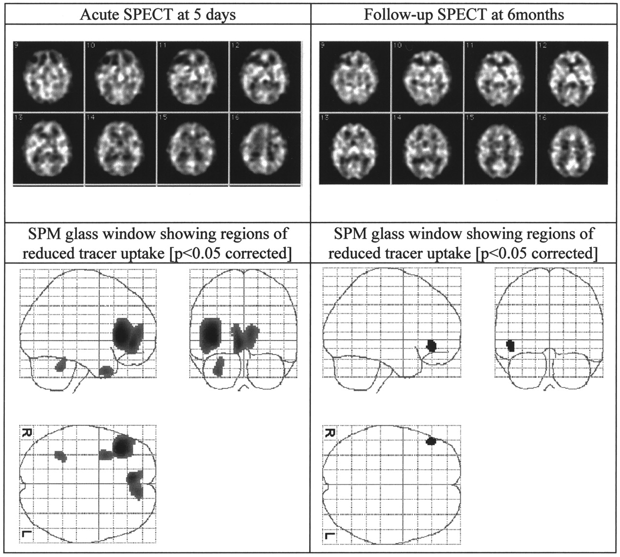
- FIGURE 1.
SPECT (midsections) and SPECT SPM images of 33-y-old man after sports injury. He had GCS rating of 15 on admission. Patient was from focal injury group.
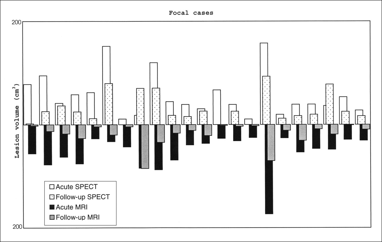
- FIGURE 2.
Total lesion volume (cm3) detected for each of 22 patients with focal injuries.
- FIGURE 3.
Total lesion volume (cm3) detected for each of 22 patients with diffuse injuries.
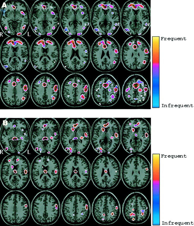
- FIGURE 4.
Frequency distribution of blood flow abnormalities in focal patients (n = 22). (A) Acute. (B) Follow-up. Yellow areas signify more frequent blood flow abnormalities and blue areas signify less frequent abnormalities.
- FIGURE 5.
Frequency distribution of blood flow abnormalities in diffuse patients (n = 22). (A) Acute. (B) Follow-up. Yellow areas signify more frequent blood flow abnormalities and blue areas signify less frequent abnormalities.


Tables
- TABLE 1
Mean Lesion Volumes in Patients with Focal, Diffuse, and All Types of Injury at Acute and Follow-Up Stages and Percentage of Total Lesion Changes from Acute to Follow-Up
Type of injury SPECT MRI Mean lesion volume (cm3) Follow-up/acute (%) Mean lesion volume (cm3) Follow-up/acute (%) Acute Follow-up Acute Follow-up Focal (n = 22) 56.31 30.39 54 53.93 18.82 35 Diffuse (n = 22) 12.61 7.59 60 5.68 0.76 13 All (n = 61) 31.41 18.37 58 28.34 9.78 35 Focal acute Focal follow-up L and R medial frontal gyrus L and R medial frontal gyrus L and R middle frontal gyrus L and R middle frontal gyrus L and R inferior frontal gyrus L and R superior frontal gyrus L and R superior frontal gyrus R inferior frontal gyrus L and R frontal precentral gyrus R frontal precentral gyrus L and R anterior cingulate L and R anterior cingulate L and R cingulate gyrus L and R cingulate gyrus R superior temporal gyrus L and R superior temporal gyrus R middle temporal gyrus R middle temporal gyrus L parietal angular gyrus L parietal supramarginal gyrus L inferior parietal lobule L inferior parietal lobule R parietal postcentral gyrus L and R corpus callosum R parietal lobe, precuneus L and R caudate R inferior occipital gyrus R middle occipital gyrus L caudate R corpus callosum L lentiform nucleus, putamen -
n = 22 patients.
-
Diffuse acute Diffuse follow-up L and R medial frontal gyrus R superior frontal gyrus L and R frontal lobe, precentral gyrus L frontal precentral gyrus L and R superior frontal gyrus L medial frontal gyrus L inferior frontal gyrus L and R anterior cingulate L and R anterior cingulate L superior temporal gyrus L and R cingulate gyrus L insula L inferior temporal gyrus L inferior parietal lobule L middle temporal gyrus L parietal postcentral gyrus L superior temporal gyrus R parietal lobe, precuneus L insula R inferior occipital gyrus L parietal angular gyrus L and R middle occipital gyrus L parietal postcentral gyrus R occipital lobe, cuneus R inferior parietal lobule R caudate L occipital lobe, cuneus L and R thalamus L and R middle occipital gyrus L and R brain stem, midbrain -
n = 22 patients.
-
In this issue


Jump to section
Related Articles
Cited By...
- Traumatic Brain Injury Imaging Research Roadmap
- Imaging Evidence and Recommendations for Traumatic Brain Injury: Advanced Neuro- and Neurovascular Imaging Techniques
- Cognitive Impairment in Mild Traumatic Brain Injury: A Longitudinal Diffusional Kurtosis and Perfusion Imaging Study
- Relationship between regional cerebral metabolism and consciousness disturbance in traumatic diffuse brain injury without large focal lesions: an FDG-PET study with statistical parametric mapping analysis
- Evaluation of Methods to Detect Interhemispheric Asymmetry on Cerebral Perfusion SPECT: Application to Epilepsy