
Main menu

  • Home
  • Content
    • Current
    • Ahead of print
    • Past Issues
    • JNM Supplement
    • SNMMI Annual Meeting Abstracts
    • Continuing Education
    • JNM Podcasts
  • Subscriptions
    • Subscribers
    • Institutional and Non-member
    • Rates
    • Journal Claims
    • Corporate & Special Sales
  • Authors
    • Submit to JNM
    • Information for Authors
    • Assignment of Copyright
    • AQARA requirements
  • Info
    • Reviewers
    • Permissions
    • Advertisers
  • About
    • About Us
    • Editorial Board
    • Contact Information
  • More
    • Alerts
    • Feedback
    • Help
    • SNMMI Journals
  • SNMMI
    • JNM
    • JNMT
    • SNMMI Journals
    • SNMMI

User menu

  • Subscribe
  • My alerts
  • Log in
  • My Cart

Search

  • Advanced search
Journal of Nuclear Medicine
  • SNMMI
    • JNM
    • JNMT
    • SNMMI Journals
    • SNMMI
  • Subscribe
  • My alerts
  • Log in
  • My Cart
Journal of Nuclear Medicine

Advanced Search

  • Home
  • Content
    • Current
    • Ahead of print
    • Past Issues
    • JNM Supplement
    • SNMMI Annual Meeting Abstracts
    • Continuing Education
    • JNM Podcasts
  • Subscriptions
    • Subscribers
    • Institutional and Non-member
    • Rates
    • Journal Claims
    • Corporate & Special Sales
  • Authors
    • Submit to JNM
    • Information for Authors
    • Assignment of Copyright
    • AQARA requirements
  • Info
    • Reviewers
    • Permissions
    • Advertisers
  • About
    • About Us
    • Editorial Board
    • Contact Information
  • More
    • Alerts
    • Feedback
    • Help
    • SNMMI Journals
  • View or Listen to JNM Podcast
  • Visit JNM on Facebook
  • Join JNM on LinkedIn
  • Follow JNM on Twitter
  • Subscribe to our RSS feeds
OtherClinical Investigations

Usefulness of Brain SPECT to Evaluate Brain Tolerance and Hemodynamic Changes During Temporary Balloon Occlusion Test and After Permanent Carotid Occlusion

Yoshifumi Sugawara, Takanori Kikuchi, Toshihiro Ueda, Mamoru Nishizaki, Shigeru Nakata, Teruhito Mochizuki and Junpei Ikezoe
Journal of Nuclear Medicine December 2002, 43 (12) 1616-1623;
Yoshifumi Sugawara
  • Find this author on Google Scholar
  • Find this author on PubMed
  • Search for this author on this site
Takanori Kikuchi
  • Find this author on Google Scholar
  • Find this author on PubMed
  • Search for this author on this site
Toshihiro Ueda
  • Find this author on Google Scholar
  • Find this author on PubMed
  • Search for this author on this site
Mamoru Nishizaki
  • Find this author on Google Scholar
  • Find this author on PubMed
  • Search for this author on this site
Shigeru Nakata
  • Find this author on Google Scholar
  • Find this author on PubMed
  • Search for this author on this site
Teruhito Mochizuki
  • Find this author on Google Scholar
  • Find this author on PubMed
  • Search for this author on this site
Junpei Ikezoe
  • Find this author on Google Scholar
  • Find this author on PubMed
  • Search for this author on this site
  • Article
  • Figures & Data
  • Info & Metrics
  • PDF
Loading

Article Figures & Data

Figures

  • Tables
  • FIGURE 1.
    • Download figure
    • Open in new tab
    • Download powerpoint
    FIGURE 1.

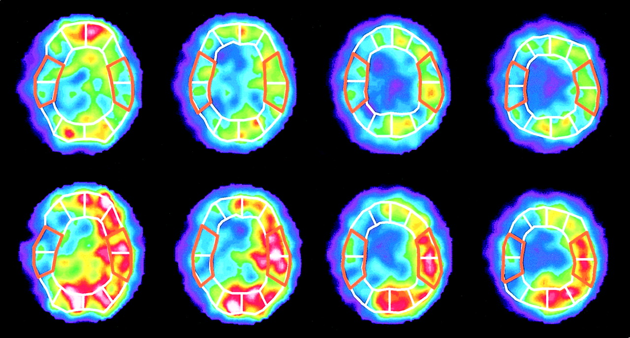
    133Xe CBF images at rest (top row) and after ACZ enhancement (bottom row) with placement of regions of interest. Mean CBF is calculated in MCA territories on 4 slices from basal ganglia through centrum semiovale. CPR is defined as percentage increase in CBF after ACZ enhancement, or CPR = [(CBF [ACZ] − CBF [rest])/CBF (rest)] × 100 (%).

  • FIGURE 2.
    • Download figure
    • Open in new tab
    • Download powerpoint
    FIGURE 2.

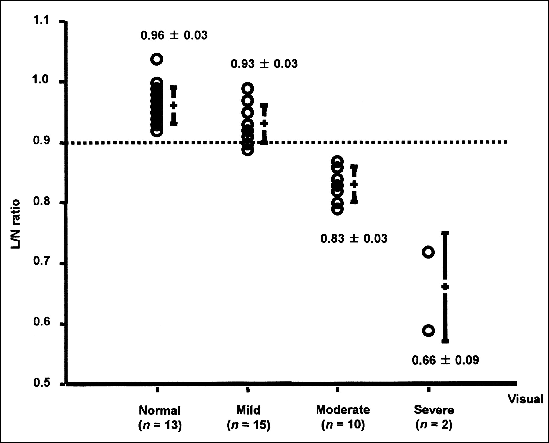
    Comparisons of visual analysis and L/N ratio. Visual interpretation agrees well with L/N ratio. All cases of moderate or severe hypoperfusion show L/N ratios less than 0.9.

  • FIGURE 3.
    • Download figure
    • Open in new tab
    • Download powerpoint
    FIGURE 3.

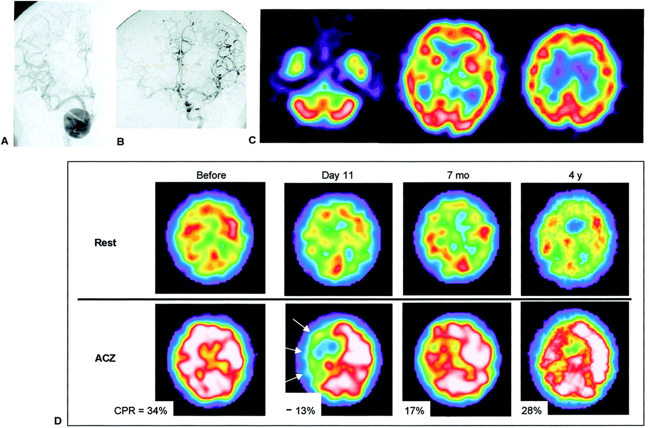
    A 63-y-old woman (patient 4) with giant aneurysm of right ICA. (A) Right ICA angiogram shows giant aneurysm in carotid siphon. (B) Left ICA angiogram during BOT of right ICA shows good collateral cross-flow. (C) 99mTc-ECD SPECT images demonstrate symmetric cerebral uptake (L/N of 0.96), and no neurologic symptoms were observed. This patient was considered to be tolerant of carotid occlusion. However, after proximal occlusion of right ICA, she showed transient left hemiparesis when her blood pressure dropped. Her hemiparesis disappeared immediately after injection of vasoconstrictor, and no infarction developed. (D) 133Xe-CBF SPECT revealed normal CBF at rest but decreased CPR in right cerebral hemisphere (mean CPR in right MCA territory was −13% on day 11) (arrows). On follow-up, impaired CPR gradually improved.

  • FIGURE 4.
    • Download figure
    • Open in new tab
    • Download powerpoint
    FIGURE 4.

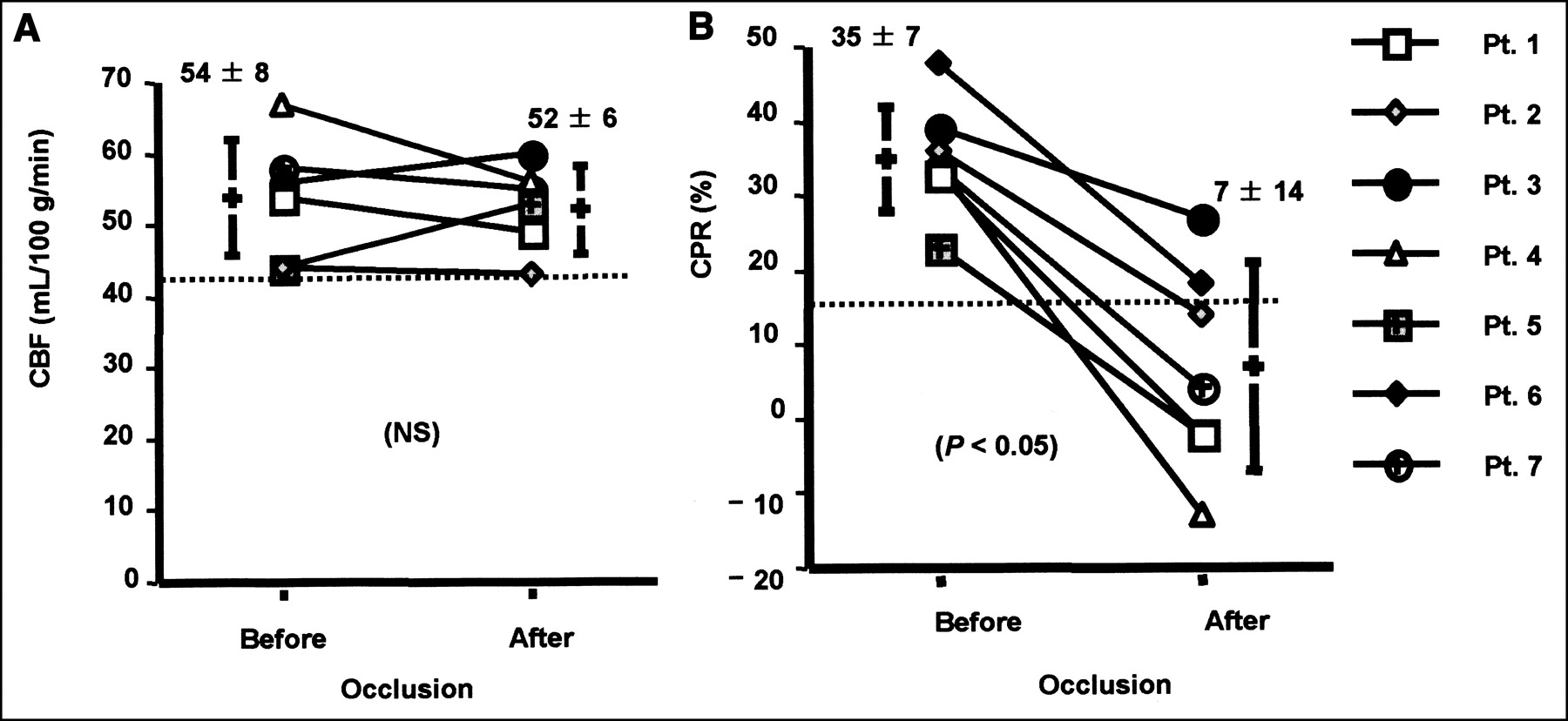
    Comparisons of CBF and CPR before and after permanent carotid occlusion. (A) CBF changed slightly (54 ± 8 mL/100 g/min vs. 52 ± 6 mL/100 g/min, not significant (NS)). (B) CPR significantly decreased in all patients after permanent carotid occlusion (35% ± 7% vs. 7% ± 14%, P < 0.05). In 5 of 7 patients, CPR decreased below normal range (CPR < 15%), and steal phenomenon was observed in 3 patients.
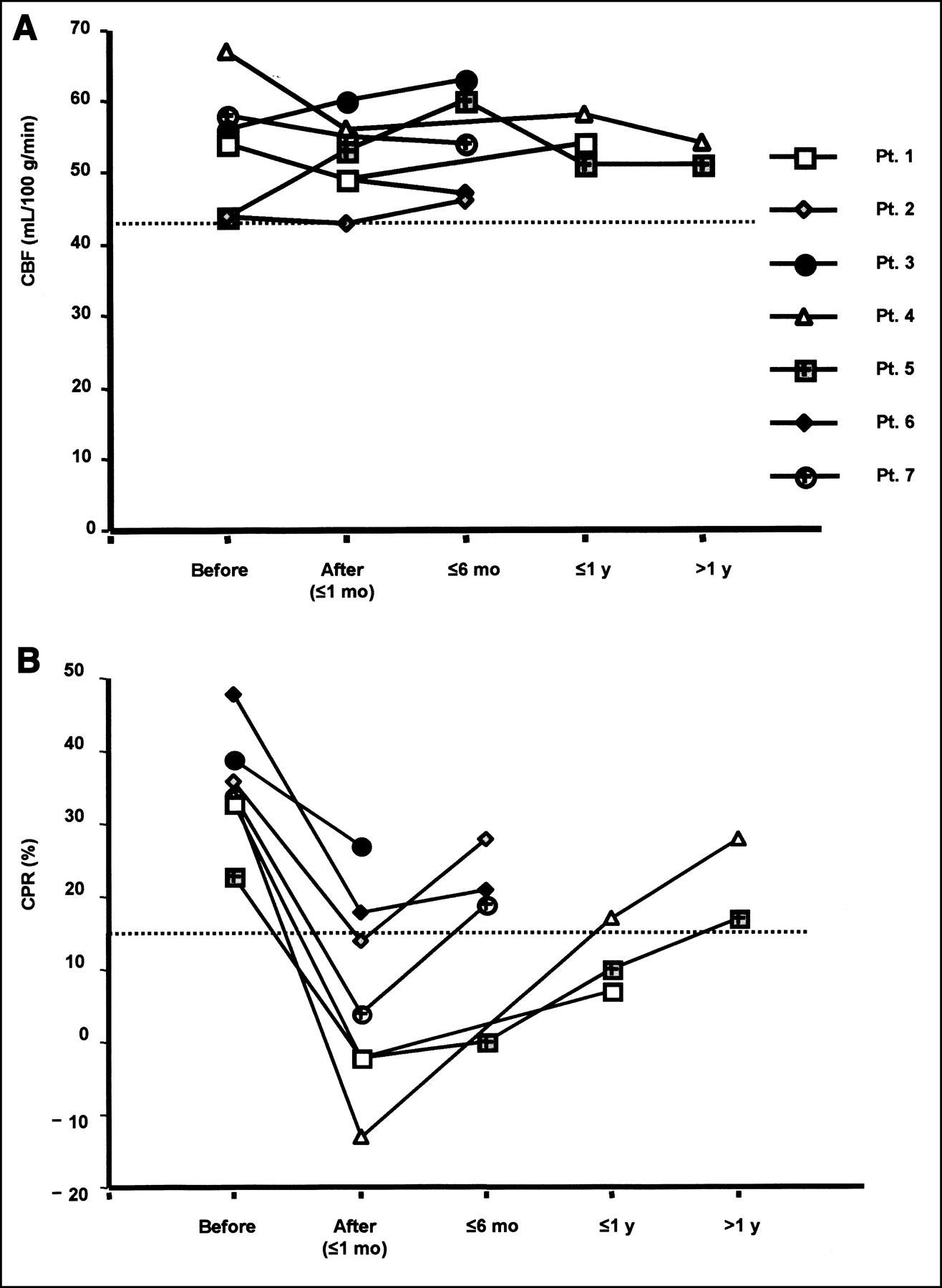
  • FIGURE 5.
    • Download figure
    • Open in new tab
    • Download powerpoint
    FIGURE 5.

    Changes in CBF and CPR on follow-up after permanent carotid occlusion. (A) CBF changed within normal range (CBF ≥ 43 mL/100 g/min). (B) In contrast, decreased CPR gradually improved after permanent carotid occlusion.

Tables

  • Figures
    • View popup
    TABLE 1

    Symptoms and SPECT Findings During Balloon Occlusion Test

    Symptoms (BOT)Hypoperfusion findings on 99mTc-ECD SPECTTotal
    NormalMildModerateSevere
    Absent11159136
    Present2*0114
    Total131510240
    • ↵* 99mTc-ECD was injected after balloon deflation.

    • View popup
    TABLE 2

    Patients with Permanent Carotid Occlusion

    Patient no.AgeSexDiseaseSite of occlusionTreatmentCBF (mL/ 100 g/min)CPR (%)Symptoms (BOT)Infarction (CT/MRI)
    BeforeAfterBeforeAfter
    164FAneurysmR ICALigation544933−2AbsentAbsent
    259FAneurysmR ICABalloon occlusion44433614AbsentNA
    349FAneurysmR ICABalloon occlusion56603927AbsentAbsent
    463FAneurysmR ICACoil embolization675634−13Present*Absent
    570FAneurysmR ICABalloon occlusion445323−2AbsentPresent†
    666MAneurysmR ICABalloon occlusion54494818AbsentAbsent
    736FAneurysmL ICATrapping5855344AbsentAbsent
    • ↵* Transient left hemiparesis during hypotension.

    • ↵† Small infarction probably due to thromboembolism.

    • NA = not available.

PreviousNext
Back to top

In this issue

Journal of Nuclear Medicine
Vol. 43, Issue 12
December 1, 2002
  • Table of Contents
  • Index by author
Print
Download PDF
Article Alerts
Sign In to Email Alerts with your Email Address
Email Article

Thank you for your interest in spreading the word on Journal of Nuclear Medicine.

NOTE: We only request your email address so that the person you are recommending the page to knows that you wanted them to see it, and that it is not junk mail. We do not capture any email address.

Enter multiple addresses on separate lines or separate them with commas.
Usefulness of Brain SPECT to Evaluate Brain Tolerance and Hemodynamic Changes During Temporary Balloon Occlusion Test and After Permanent Carotid Occlusion
(Your Name) has sent you a message from Journal of Nuclear Medicine
(Your Name) thought you would like to see the Journal of Nuclear Medicine web site.
Citation Tools
Usefulness of Brain SPECT to Evaluate Brain Tolerance and Hemodynamic Changes During Temporary Balloon Occlusion Test and After Permanent Carotid Occlusion
Yoshifumi Sugawara, Takanori Kikuchi, Toshihiro Ueda, Mamoru Nishizaki, Shigeru Nakata, Teruhito Mochizuki, Junpei Ikezoe
Journal of Nuclear Medicine Dec 2002, 43 (12) 1616-1623;

Citation Manager Formats

  • BibTeX
  • Bookends
  • EasyBib
  • EndNote (tagged)
  • EndNote 8 (xml)
  • Medlars
  • Mendeley
  • Papers
  • RefWorks Tagged
  • Ref Manager
  • RIS
  • Zotero
Share
Usefulness of Brain SPECT to Evaluate Brain Tolerance and Hemodynamic Changes During Temporary Balloon Occlusion Test and After Permanent Carotid Occlusion
Yoshifumi Sugawara, Takanori Kikuchi, Toshihiro Ueda, Mamoru Nishizaki, Shigeru Nakata, Teruhito Mochizuki, Junpei Ikezoe
Journal of Nuclear Medicine Dec 2002, 43 (12) 1616-1623;
Twitter logo Facebook logo LinkedIn logo Mendeley logo
  • Tweet Widget
  • Facebook Like
  • Google Plus One
Bookmark this article

Jump to section

  • Article
    • Abstract
    • MATERIALS AND METHODS
    • RESULTS
    • DISCUSSION
    • CONCLUSION
    • Acknowledgments
    • Footnotes
    • REFERENCES
  • Figures & Data
  • Info & Metrics
  • PDF

Related Articles

  • No related articles found.
  • PubMed
  • Google Scholar

Cited By...

  • The Acetazolamide Challenge: Techniques and Applications in the Evaluation of Chronic Cerebral Ischemia
  • Google Scholar

More in this TOC Section

  • Feasibility of Ultra-Low-Activity 18F-FDG PET/CT Imaging Using a Long–Axial-Field-of-View PET/CT System
  • Cardiac Presynaptic Sympathetic Nervous Function Evaluated by Cardiac PET in Patients with Chronotropic Incompetence Without Heart Failure
  • Validation and Evaluation of a Vendor-Provided Head Motion Correction Algorithm on the uMI Panorama PET/CT System
Show more Clinical Investigations

Similar Articles

SNMMI

© 2025 SNMMI

Powered by HighWire