


Article Figures & Data
Figures
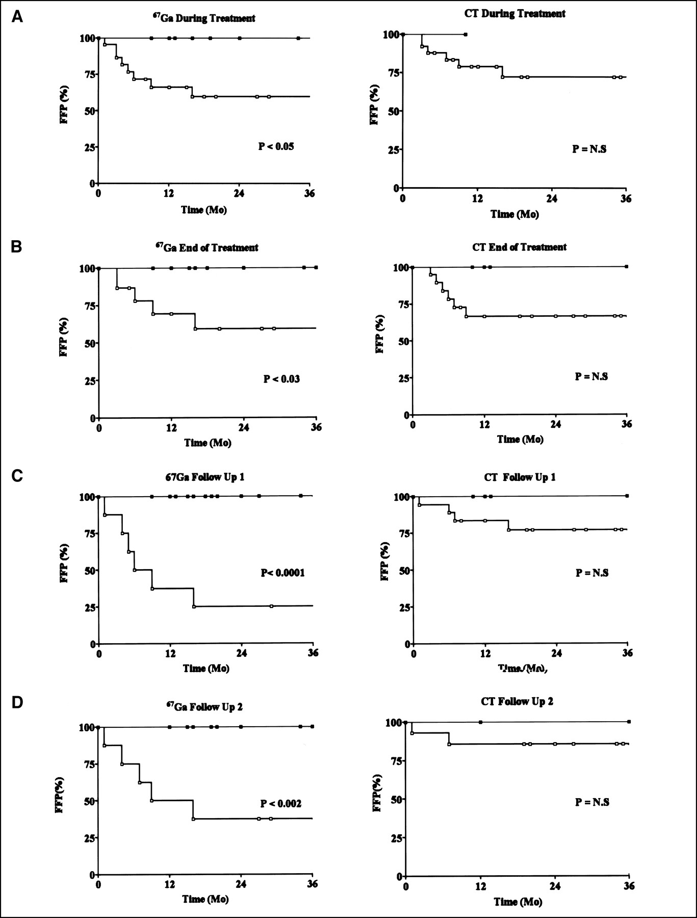
- FIGURE 1.
Three-year FFP in patients with positive (□) and negative (▪) 67Ga scintigraphy and CT findings. Graphs show findings during treatment (A), at end of treatment (B), at FU1 (C), and at FU2 (D).
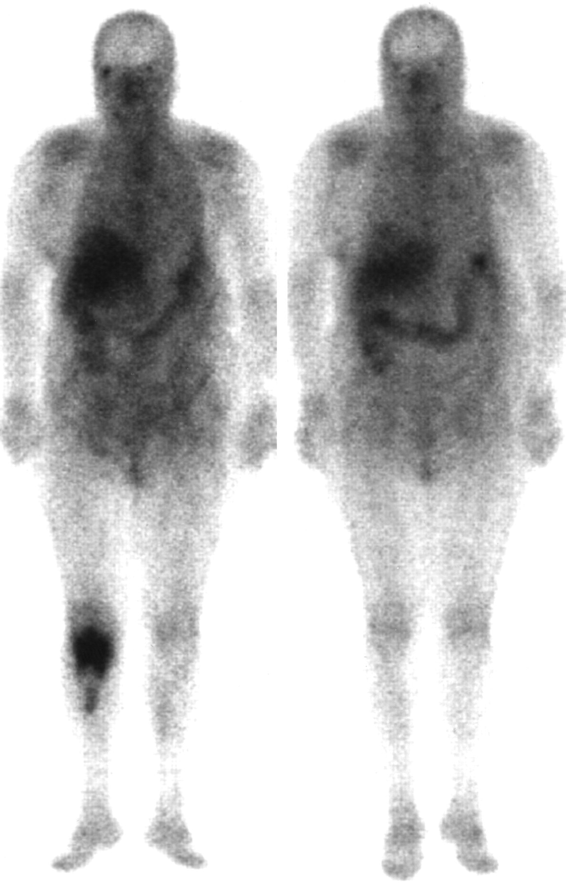
- FIGURE 2.
Negative 67Ga scintigraphy and abnormal CT findings at end of treatment in 37-y-old man with diffuse large cell B-type NHL, stage I EA, involving posterior arch of 9th and 10th right ribs and T9 and T10 vertebrae. (A) 67Ga scintigraphy at diagnosis (left) shows pathologic uptake at involved sites of disease. Marked improvement is seen at mid treatment (center), with residual abnormal 67Ga activity in lower thoracic spine. 67Ga scintigraphy findings at end of treatment (right) are negative. (B) CT at diagnosis shows infiltrative osteolytic lesion with cortical disruption of posterior arch of 9th right rib (arrows) and surrounding soft-tissue involvement. Moth-eaten pattern is seen in vertebral body of T9. (C) CT at end of treatment shows mixed sclerotic and lytic pattern in 9th rib and T9 vertebral body (arrows). Patient had no evidence of disease for 30 mo.
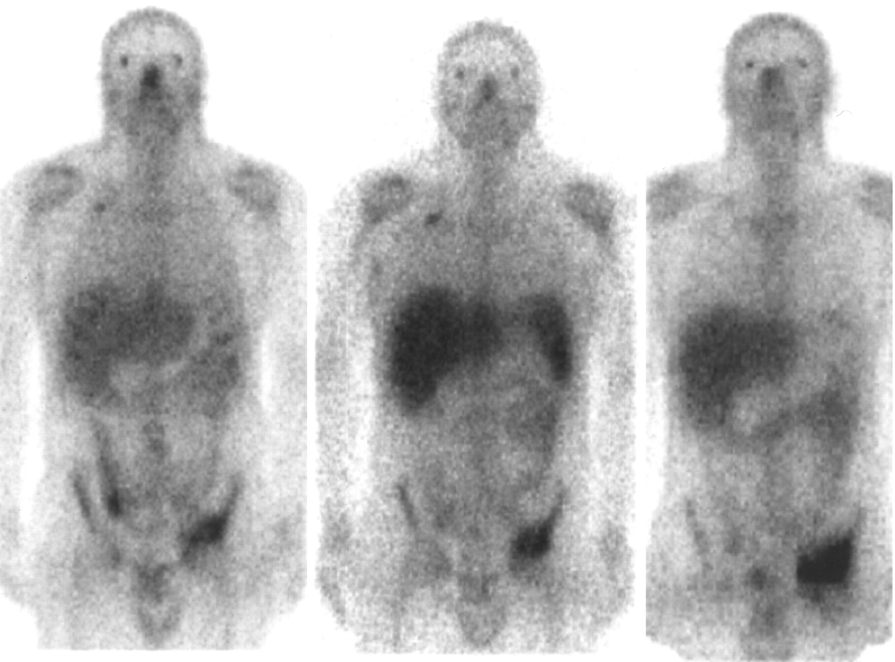
- FIGURE 3.
Negative 67Ga scintigraphy findings during chemotherapy in 68-y-old woman with follicular mixed low-grade lymphoma involving right inguinal lymph nodes and right tibia. 67Ga scintigraphy at baseline (left) shows abnormal uptake in proximal right tibia. Repeated 67Ga scintigraphy after 4 cycles of chemotherapy (right) shows negative findings, which remained unchanged at end of treatment and during follow-up. Disease has been in complete remission for 12 mo.
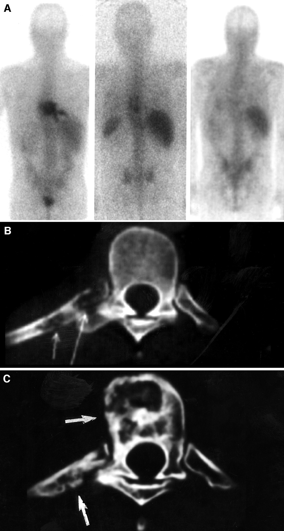
- FIGURE 4.
Abnormal 67Ga scintigraphy findings during and after chemotherapy in 33-y-old man with biopsy-proven recurrent HD involving left acetabulum. 67Ga scintigraphy before treatment (left) shows area of pathologic uptake in left acetabulum. Chemotherapy including dexamethasone, ifosfamide, cisplatin, and etoposide was initiated. 67Ga scintigraphy during treatment (center) and at completion of chemotherapy (right) shows increase in size and intensity of pathologic uptake. Repeated bone biopsy indicated presence of active HD. (Note also abnormal 67Ga uptake in involved right supraclavicular lymph node.) Tumor progression was diagnosed, and patient died 6 mo later.

Tables
Site n NHL HD Spine 32 25 7 Pelvis 18 15 3 Thoracic cage 11 9 2 Skull 8 8 — Long bones 22 21 1 Total 91 78 13 - TABLE 2
Number of Patients with Negative and Positive 67Ga Scintigraphy and CT Findings During, at End of, and After Treatment
Time of evaluation Test Total no. patients No. patients with negative test No. patients with positive test During treatment 67Ga 36 9 (25) 27 (75) CT 30 1 (3) 29 (97) End of treatment 67Ga 31 13 (42) 18 (58) CT 28 5 (18) 23 (82) FU1 67Ga 31 21 (68) 10 (32) CT 24 4 (17) 20 (83) FU2 67Ga 23 14 (61) 9 (39) CT 19 4 (21) 15 (79) Numbers in parentheses are percentages.
- TABLE 3
Three-Year FFP Rate in Lymphoma Patients with Negative and Positive 67Ga Scintigraphy and CT Findings
Time of test 67Ga CT Negative Positive P Negative Positive P During treatment 100 48 0.05 100 72 NS End of treatment 100 59 0.03 100 67 NS FU1 100 25 0.0001 100 77 NS FU2 100 38 0.002 100 86 NS NS = not statistically significant.
P is based on Kaplan-Meier curves and shows significance of difference in 3-y FFP rate between patients with negative and positive 67Ga scintigraphy and CT findings.
Data are percentages.
- TABLE 4
Treatment-Related Distribution of CT Patterns and Their Correlation with 67Ga Avidity
CT pattern Baseline During treatment End of treatment FU1 FU2 Osteolysis* CT sites 70 49 26 31 21 67Ga-positive lytic sites 68 32 63 30 — Osteosclerosis* CT sites 23 27 29 25 38 67Ga-positive sclerotic sites 83 50 67 13 11 Mixed† CT sites 7 21 27 24 20 67Ga-positive mixed-pattern sites 75 38 33 38 20 Normal CT sites — 3 18 20 21 67Ga-positive normal-CT sites — — — —


