

FIGURE 2.

FIGURE 2.
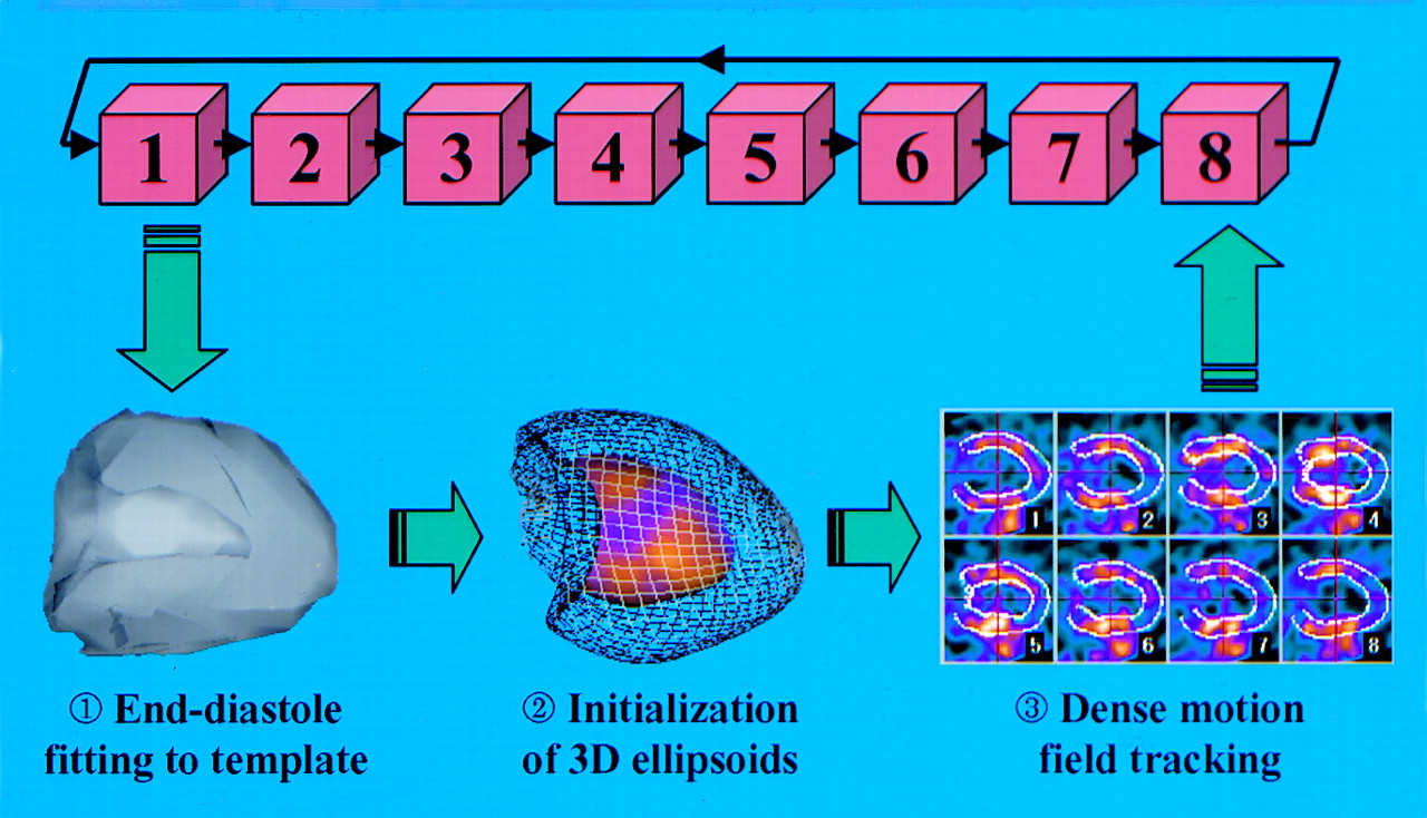
Description of MTK algorithm for calculation of ejection fraction. Unique myocardial segmentation is performed on first time-frame volume (1, end-diastole) with matching to reference template to obtain morphological constraints of perfusion distribution, particularly for determination of valve plane. This procedure generates two 3D elastic ellipsoids (2, endocardial surface is represented mapped with perfusion polar map and epicardial surface is represented as wire grid). Node positions of these ellipsoids are tracked throughout 8 frames of cardiac cycle (3).
In this issue


Related Articles
Cited By...
- No citing articles found.