
Main menu

  • Home
  • Content
    • Current
    • Ahead of print
    • Past Issues
    • JNM Supplement
    • SNMMI Annual Meeting Abstracts
    • Continuing Education
    • JNM Podcasts
  • Subscriptions
    • Subscribers
    • Institutional and Non-member
    • Rates
    • Journal Claims
    • Corporate & Special Sales
  • Authors
    • Submit to JNM
    • Information for Authors
    • Assignment of Copyright
    • AQARA requirements
  • Info
    • Reviewers
    • Permissions
    • Advertisers
  • About
    • About Us
    • Editorial Board
    • Contact Information
  • More
    • Alerts
    • Feedback
    • Help
    • SNMMI Journals
  • SNMMI
    • JNM
    • JNMT
    • SNMMI Journals
    • SNMMI

User menu

  • Subscribe
  • My alerts
  • Log in
  • Log out
  • My Cart

Search

  • Advanced search
Journal of Nuclear Medicine
  • SNMMI
    • JNM
    • JNMT
    • SNMMI Journals
    • SNMMI
  • Subscribe
  • My alerts
  • Log in
  • Log out
  • My Cart
Journal of Nuclear Medicine

Advanced Search

  • Home
  • Content
    • Current
    • Ahead of print
    • Past Issues
    • JNM Supplement
    • SNMMI Annual Meeting Abstracts
    • Continuing Education
    • JNM Podcasts
  • Subscriptions
    • Subscribers
    • Institutional and Non-member
    • Rates
    • Journal Claims
    • Corporate & Special Sales
  • Authors
    • Submit to JNM
    • Information for Authors
    • Assignment of Copyright
    • AQARA requirements
  • Info
    • Reviewers
    • Permissions
    • Advertisers
  • About
    • About Us
    • Editorial Board
    • Contact Information
  • More
    • Alerts
    • Feedback
    • Help
    • SNMMI Journals
  • View or Listen to JNM Podcast
  • Visit JNM on Facebook
  • Join JNM on LinkedIn
  • Follow JNM on Twitter
  • Subscribe to our RSS feeds
OtherBASIC SCIENCE INVESTIGATIONS

Establishment and Characterization of a Breast Cancer Cell Line Expressing Na+/I− Symporters for Radioiodide Concentrator Gene Therapy

Yuji Nakamoto, Tsuneo Saga, Takashi Misaki, Hisataka Kobayashi, Noriko Sato, Takayoshi Ishimori, Shinji Kosugi, Harumi Sakahara and Junji Konishi
Journal of Nuclear Medicine November 2000, 41 (11) 1898-1904;
Yuji Nakamoto
  • Find this author on Google Scholar
  • Find this author on PubMed
  • Search for this author on this site
Tsuneo Saga
  • Find this author on Google Scholar
  • Find this author on PubMed
  • Search for this author on this site
Takashi Misaki
  • Find this author on Google Scholar
  • Find this author on PubMed
  • Search for this author on this site
Hisataka Kobayashi
  • Find this author on Google Scholar
  • Find this author on PubMed
  • Search for this author on this site
Noriko Sato
  • Find this author on Google Scholar
  • Find this author on PubMed
  • Search for this author on this site
Takayoshi Ishimori
  • Find this author on Google Scholar
  • Find this author on PubMed
  • Search for this author on this site
Shinji Kosugi
  • Find this author on Google Scholar
  • Find this author on PubMed
  • Search for this author on this site
Harumi Sakahara
  • Find this author on Google Scholar
  • Find this author on PubMed
  • Search for this author on this site
Junji Konishi
  • Find this author on Google Scholar
  • Find this author on PubMed
  • Search for this author on this site
  • Article
  • Figures & Data
  • Info & Metrics
  • PDF
Loading

Article Figures & Data

Figures

  • Tables
  • FIGURE 1.
    • Download figure
    • Open in new tab
    • Download powerpoint
    FIGURE 1.

    Time course of iodide uptake by MCF3B without perchlorate, MCF3B with 1 mmol/L perchlorate, and MCF7 without perchlorate. All data are expressed as means (pmol/106 cells) of duplicate wells. Iodide uptake by MCF3B was rapid and became maximal within 30 min, whereas uptake by MCF3B with perchlorate and by MCF7 remained low.
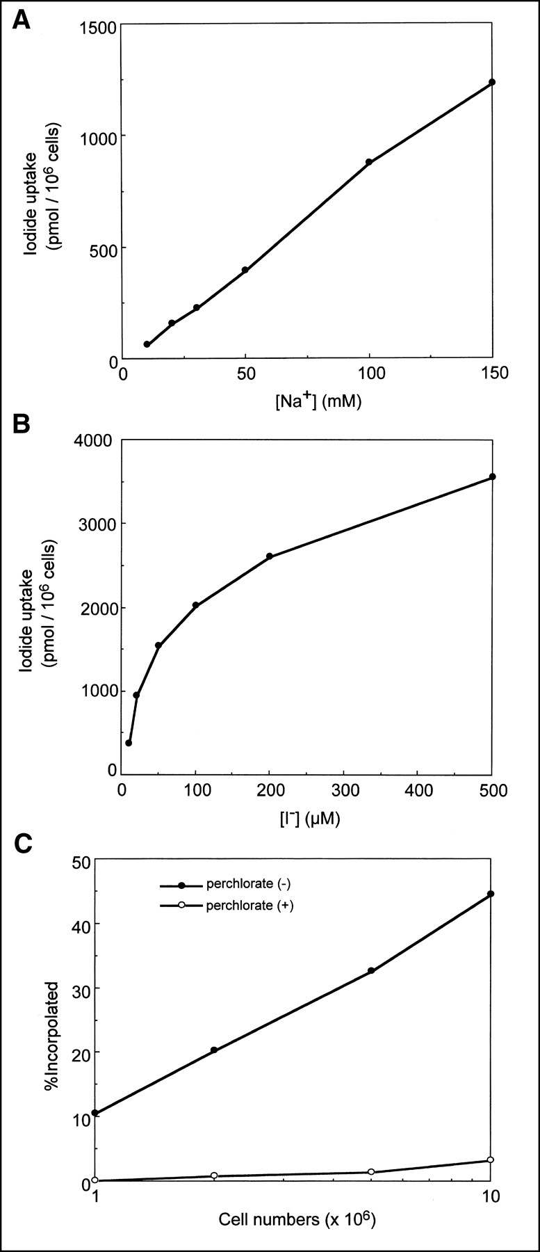
  • FIGURE 2.
    • Download figure
    • Open in new tab
    • Download powerpoint
    FIGURE 2.

    Iodide uptake plotted against Na+ concentration (A), I− concentration (B), and cell numbers (C).
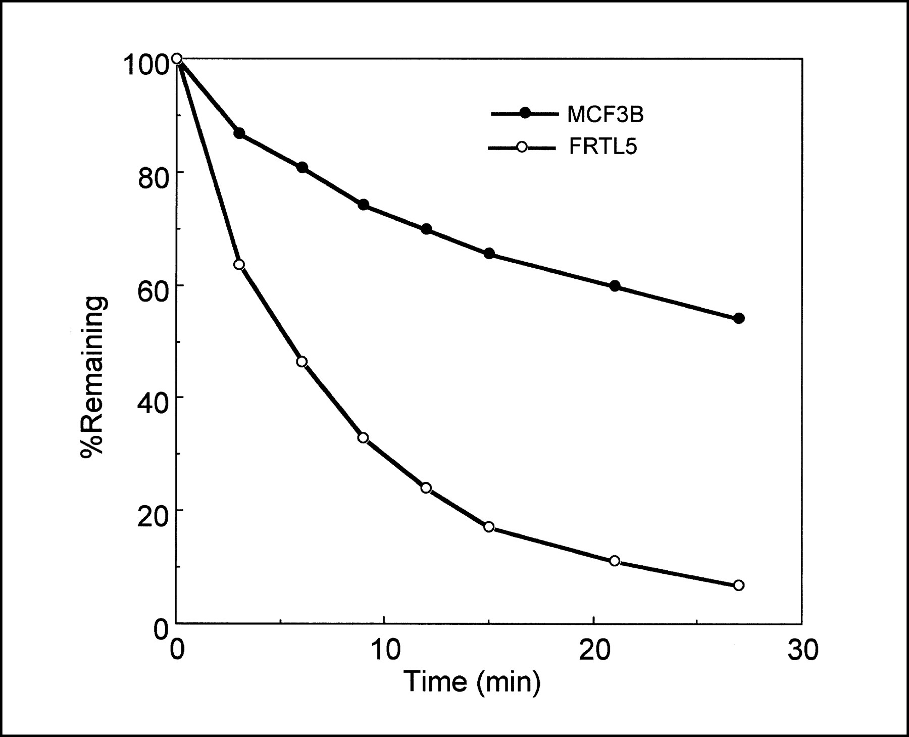
  • FIGURE 3.
    • Download figure
    • Open in new tab
    • Download powerpoint
    FIGURE 3.

    Iodide efflux from MCF3B and FRTL5 cells. All data are expressed as means (remaining percentage of 125I−) of duplicate wells. Although iodide that accumulated in FRTL5 cells was rapidly exchangeable, MCF3B cells released 125I− more slowly.
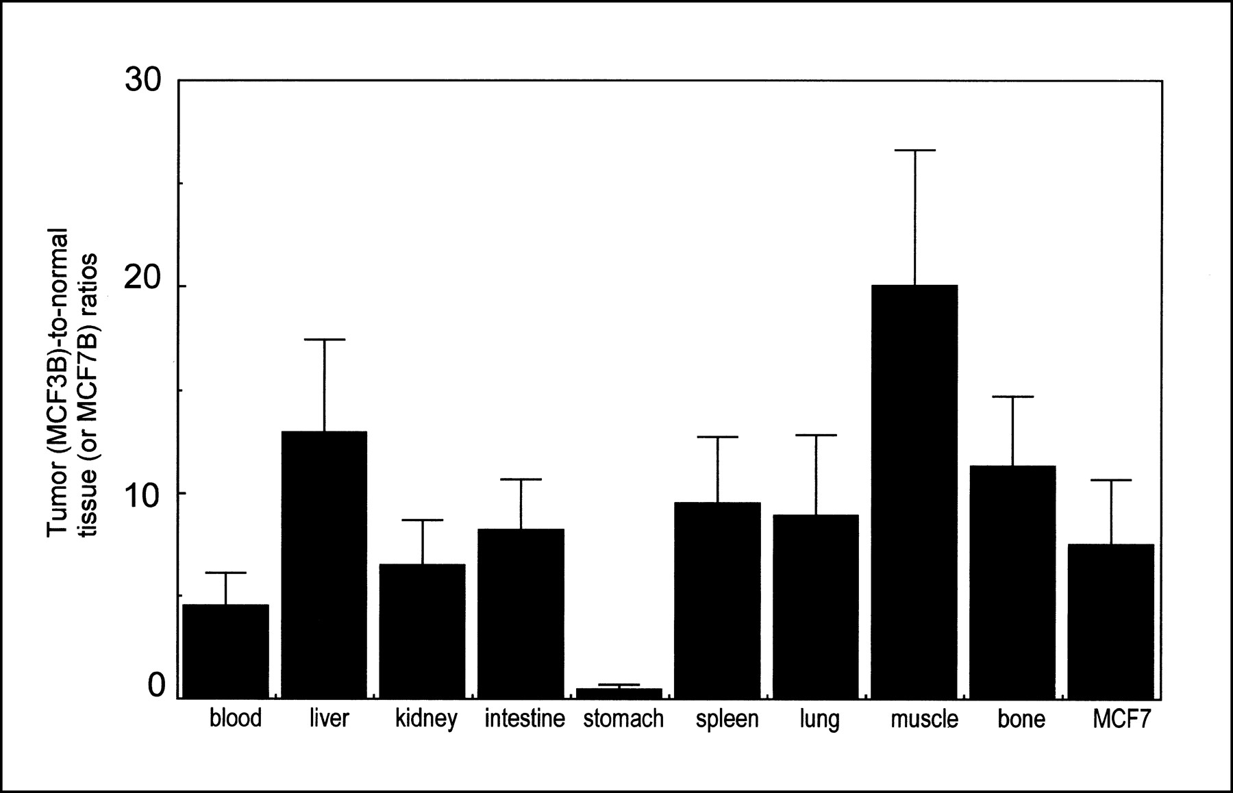
  • FIGURE 4.
    • Download figure
    • Open in new tab
    • Download powerpoint
    FIGURE 4.

    Tumor-to-normal tissue ratios. High ratios were obtained as early as 1 h after injection, except in stomach.
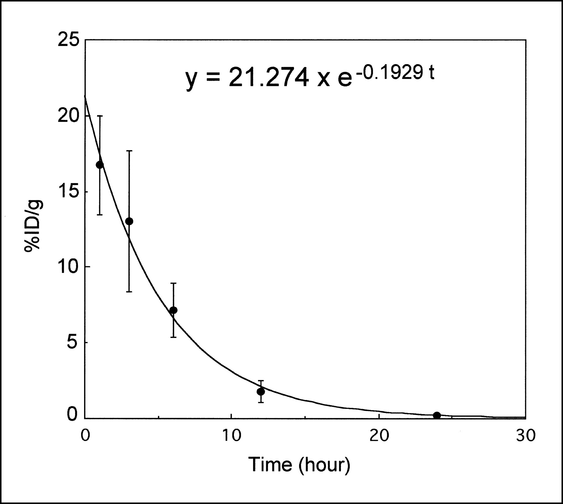
  • FIGURE 5.
    • Download figure
    • Open in new tab
    • Download powerpoint
    FIGURE 5.

    Time course of decay-corrected iodide accumulation in subcutaneous tumors. At 24 h after administration of 125I−, uptake in tumor was reduced to 0.2%. Monoexponential curve fitted for each point was also shown on identical graph. Calculated effective T1/2 was 3.59 h.

  • FIGURE 6.
    • Download figure
    • Open in new tab
    • Download powerpoint
    FIGURE 6.

    Imaging of MCF3B tumors by gamma camera. Two hours after injection of Na131I (11.1 MBq) intravenously, planar image (B) of mouse was obtained. Note visualized NIS-transduced MCF3B tumor (T). Thyroid, stomach, and bladder were also detected by physiologic uptake. Planar image is to scale with photograph (A).
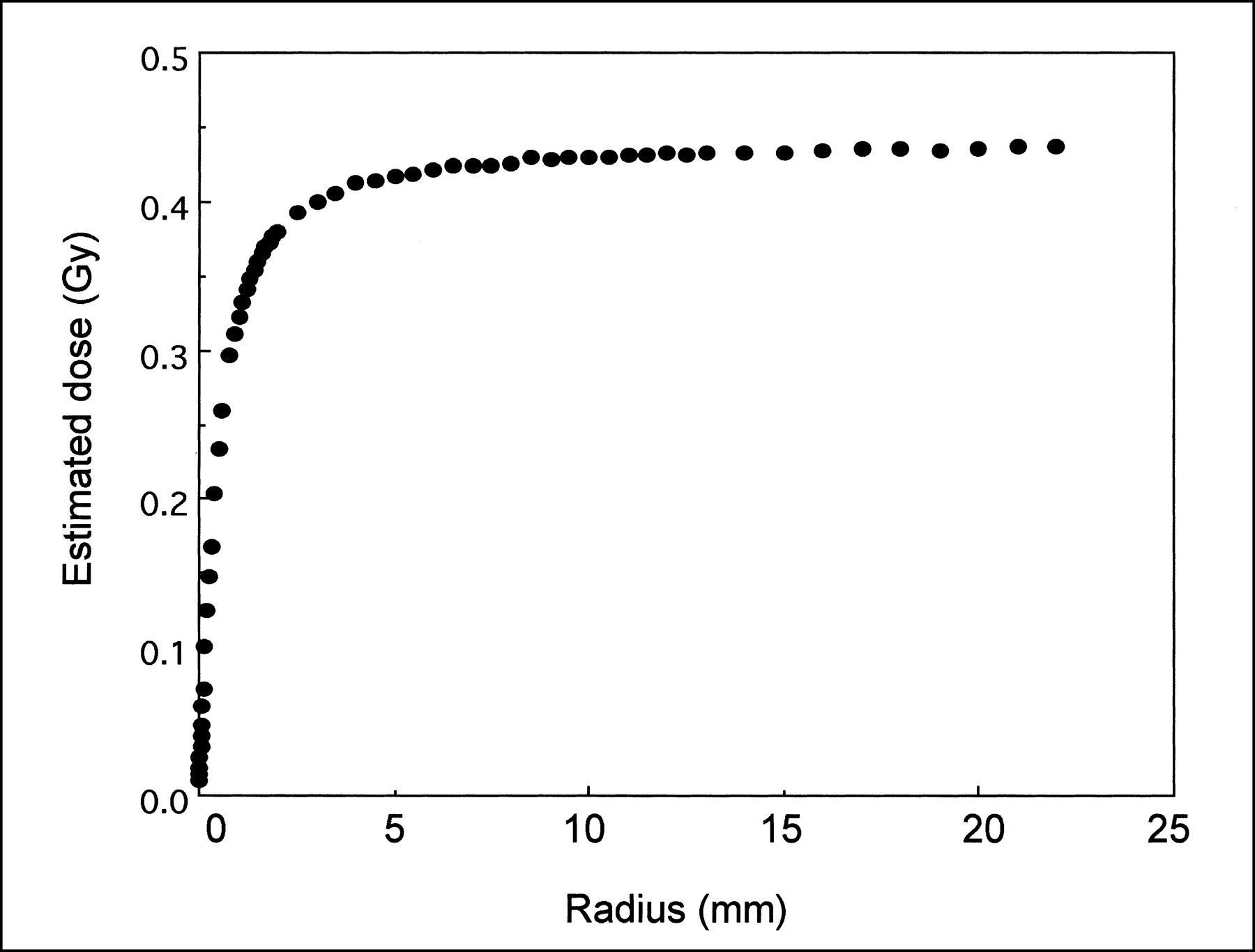
  • FIGURE 7.
    • Download figure
    • Open in new tab
    • Download powerpoint
    FIGURE 7.

    Relationship between radius of tumor and estimated dose for 3.7 MBq 131I administration. Dose increased with increase in radius of tumor but was considered to be therapeutically insufficient.

Tables

  • Figures
    • View popup
    TABLE 1.

    Biodistribution Data for Tumor-Bearing Nude Mice

    Site1 h3 h6 h12 h24 h
    Blood3.61 ± 0.701.40 ± 0.240.56 ± 0.090.14 ± 0.070.04 ± 0.01
    Liver1.25 ± 0.200.55 ± 0.080.27 ± 0.040.09 ± 0.030.05 ± 0.01
    Kidney2.52 ± 0.401.01 ± 0.110.56 ± 0.080.18 ± 0.210.04 ± 0.01
    Intestine1.90 ± 0.200.93 ± 0.120.71 ± 0.150.25 ± 0.090.10 ± 0.06
    Stomach39.25 ± 10.2720.91 ± 1.0020.06 ± 7.941.12 ± 0.530.39 ± 0.20
    Spleen1.79 ± 0.450.72 ± 0.100.32 ± 0.050.08 ± 0.030.04 ± 0.02
    Lung1.89 ± 0.470.79 ± 0.140.32 ± 0.080.09 ± 0.040.04 ± 0.01
    Muscle0.81 ± 0.110.70 ± 0.490.25 ± 0.100.08 ± 0.070.02 ± 0.01
    Bone1.49 ± 0.220.63 ± 0.120.38 ± 0.210.06 ± 0.030.03 ± 0.02
    Tumor (MCF3B)16.73 ± 3.2613.02 ± 4.657.17 ± 1.811.78 ± 0.710.22 ± 0.13
    Tumor (MCF7)2.45 ± 0.800.69 ± 0.170.37 ± 0.030.10 ± 0.080.02 ± 0.01
    T/B ratio4.84 ± 1.529.54 ± 4.1612.93 ± 2.5312.97 ± 1.094.75 ± 2.12
    T/K ratio6.90 ± 2.1712.87 ± 4.2213.06 ± 3.9915.29 ± 6.025.67 ± 2.15
    • Data are mean ± SD of %ID/g and its tumor (MCF3B)-to-blood (T/B) or tumor (MCF3B)-to-kidney (T/K) ratio in 4–5 mice.

PreviousNext
Back to top

In this issue

Journal of Nuclear Medicine
Vol. 41, Issue 11
November 1, 2000
  • Table of Contents
  • Table of Contents (PDF)
  • Index by author
Print
Download PDF
Article Alerts
Sign In to Email Alerts with your Email Address
Email Article

Thank you for your interest in spreading the word on Journal of Nuclear Medicine.

NOTE: We only request your email address so that the person you are recommending the page to knows that you wanted them to see it, and that it is not junk mail. We do not capture any email address.

Enter multiple addresses on separate lines or separate them with commas.
Establishment and Characterization of a Breast Cancer Cell Line Expressing Na+/I− Symporters for Radioiodide Concentrator Gene Therapy
(Your Name) has sent you a message from Journal of Nuclear Medicine
(Your Name) thought you would like to see the Journal of Nuclear Medicine web site.
Citation Tools
Establishment and Characterization of a Breast Cancer Cell Line Expressing Na+/I− Symporters for Radioiodide Concentrator Gene Therapy
Yuji Nakamoto, Tsuneo Saga, Takashi Misaki, Hisataka Kobayashi, Noriko Sato, Takayoshi Ishimori, Shinji Kosugi, Harumi Sakahara, Junji Konishi
Journal of Nuclear Medicine Nov 2000, 41 (11) 1898-1904;

Citation Manager Formats

  • BibTeX
  • Bookends
  • EasyBib
  • EndNote (tagged)
  • EndNote 8 (xml)
  • Medlars
  • Mendeley
  • Papers
  • RefWorks Tagged
  • Ref Manager
  • RIS
  • Zotero
Share
Establishment and Characterization of a Breast Cancer Cell Line Expressing Na+/I− Symporters for Radioiodide Concentrator Gene Therapy
Yuji Nakamoto, Tsuneo Saga, Takashi Misaki, Hisataka Kobayashi, Noriko Sato, Takayoshi Ishimori, Shinji Kosugi, Harumi Sakahara, Junji Konishi
Journal of Nuclear Medicine Nov 2000, 41 (11) 1898-1904;
Twitter logo Facebook logo LinkedIn logo Mendeley logo
  • Tweet Widget
  • Facebook Like
  • Google Plus One
Bookmark this article

Jump to section

  • Article
    • Abstract
    • MATERIALS AND METHODS
    • RESULTS
    • DISCUSSION
    • CONCLUSION
    • Footnotes
    • REFERENCES
  • Figures & Data
  • Info & Metrics
  • PDF

Related Articles

  • No related articles found.
  • PubMed
  • Google Scholar

Cited By...

  • Transfection of the Human Sodium/Iodide Symporter (NIS) Gene with Liposomes and the Expression of the NIS Protein in Human Lung A549 Cancer Cells
  • Radioiodide imaging and radiovirotherapy of multiple myeloma using VSV({Delta}51)-NIS, an attenuated vesicular stomatitis virus encoding the sodium iodide symporter gene
  • Enhanced Expression of Adenovirus-Mediated Sodium Iodide Symporter Gene in MCF-7 Breast Cancer Cells with Retinoic Acid Treatment
  • A perspective view of sodium iodide symporter research and its clinical implications.
  • Development of a Sodium/Iodide Symporter (NIS)-Transgenic Mouse for Imaging of Cardiomyocyte-Specific Reporter Gene Expression
  • In vivo Radioiodide Imaging and Treatment of Breast Cancer Xenografts after MUC1-Driven Expression of the Sodium Iodide Symporter
  • Long-Term Radioiodine Retention and Regression of Liver Cancer after Sodium Iodide Symporter Gene Transfer in Wistar Rats
  • Establishment of a Human Hepatocellular Carcinoma Cell Line Highly Expressing Sodium Iodide Symporter for Radionuclide Gene Therapy
  • Iodide Kinetics and Dosimetry In Vivo After Transfer of the Human Sodium Iodide Symporter Gene in Rat Thyroid Carcinoma Cells
  • Kinetics of Perrhenate Uptake and Comparative Biodistribution of Perrhenate, Pertechnetate, and Iodide by NaI Symporter-Expressing Tissues In Vivo
  • Systemic Retinoic Acid Treatment Induces Sodium/Iodide Symporter Expression and Radioiodide Uptake in Mouse Breast Cancer Models
  • In Vitro Cytotoxicity of 211At-Astatide and 131I-Iodide to Glioma Tumor Cells Expressing the Sodium/Iodide Symporter
  • Radioiodide Treatment after Sodium Iodide Symporter Gene Transfer Is a Highly Effective Therapy in Neuroendocrine Tumor Cells
  • Sodium Iodide Symporter: Its Role in Nuclear Medicine
  • The Endogenous Mammary Gland Na+/I- Symporter May Mediate Effective Radioiodide Therapy in Breast Cancer
  • Google Scholar

More in this TOC Section

  • How Sensitive Is the Upper Gastrointestinal Tract to 90Y Radioembolization? A Histologic and Dosimetric Analysis in a Porcine Model
  • 11C-Methionine PET of Myocardial Inflammation in a Rat Model of Experimental Autoimmune Myocarditis
  • Counting Rate Characteristics and Image Distortion in Preclinical PET Imaging During Radiopharmaceutical Therapy
Show more Basic Science Investigations

Similar Articles

SNMMI

© 2025 SNMMI

Powered by HighWire