


Abstract
The sodium/iodide symporter (NIS) has been identified as an attractive target for cancer therapy. The efficacy of 131I-iodide for NIS-expressing tumor therapy may be limited by a combination of poor cellular retention and unfavorable physical characteristics (long physical half-life and low linear-energy-transfer [LET] radiative emissions). On the other hand, 211At-astatide is also transported by NIS and offers several therapeutic advantages over 131I-iodide due to its physical characteristics (short half-life, high LET α-particle emissions). The objective of this study was to directly compare the radiotoxicity of both radionuclides using a NIS-transfected cultured cell model. Methods: Cytotoxicity was determined by colony-forming assays. Also, a first-order pharmacokinetic model was used to simulate the closed compartmental system between the medium and cells. Experimental data were then fitted to this model and used to estimate the transfer coefficients between medium and cells, kmc, and between cells and medium, kcm. Using the pharmacokinetic model, the cumulated activity concentrations in the medium and cells were calculated. Monte Carlo transport methods were then used to assess absorbed doses from 131I and 211At. Results: 211At-Astatide was significantly more cytotoxic than 131I-iodide in this closed compartmental system. For 211At-astatide, absorbed doses per unit administered activity were 54- to 65-fold higher than for 131I-iodide. Both NIS-expressing and control cells showed increased sensitivity to 211At over 131I, with significantly lower D0 (absorbed dose required to reduce the survival fraction to e−1) and SF2 (2-Gy survival fraction) values, highlighting the higher intrinsic cytotoxicity of α-particles. However, NIS-independent (nonspecific) binding of 211At-astatide was higher than that of 131I-iodide, therefore, yielding a lower absorbed dose ratio between NIS-transfected and -nontransfected cells. Conclusion: Treatment of NIS-expressing cells with 211At-astatide resulted in higher absorbed doses and increased cytotoxicity per unit administered activity than that observed with 131I-iodide. These results suggest that 211At-astatide may be a promising treatment strategy for the therapy of NIS-expressing tumors.
It is now well established that the accumulation of iodide by the thyroid gland is mediated by the sodium/iodide symporter (NIS), an integral membrane protein expressed on the basolateral surface of the thyrocyte. Iodide is transported across the cell membrane against a concentration gradient in a sodium-dependent manner, in a process electrochemically coupled to the energy-dependent Na+/K+-adenosinetriphosphatase (1). The cloning of the NIS gene has allowed investigations into the effect of plasmid- or viral-mediated NIS expression on the iodide-concentrating capacity in a variety of thyroid and nonthyroid cell types (2). These studies have indicated that NIS transgene expression, under the control of a variety of promoters, confers iodide uptake capacity in a wide range of cell lines, allowing the possibility of radioiodide therapy for many tumor types. However, several of these studies also report that the efflux of iodide from NIS-transfected cells, including anaplastic thyroid cells, is extremely rapid (3–6). This observation is of fundamental importance for any endoradiotherapeutic strategy, as radiation-absorbed dose (and thus treatment efficacy) is a function of the cellular residence time and physical half-life (t1/2) of the radionuclide (7), with optimal conditions reached when the radiologic t1/2 matches the biologic t1/2 (i.e., cellular retention). Dosimetric calculations and subsequent in vivo studies, using a murine xenograft model, have indicated that successful treatment of low-retention NIS-expressing tumor deposits with 131I-iodide (t1/2 = 8.04 d) may not possible without the administration of prohibitively high activities of radioiodide (8–10). In addition, the effectiveness of 131I in curing micrometastatic disease decreases with tumor size, due to the relatively long path length of the β-particle (11), resulting in an increased probability of disease recurrence.
Astatine (astatide), the heaviest of the halogens, exhibits a pronounced accumulation in the thyroid gland in halide form and in the stomach (12). We have previously demonstrated NIS-mediated accumulation of astatide, with uptake and efflux characteristics almost identical to those of iodide (4). Fortunately, 211At has a physical t1/2 of 7.214 h, which closely matches the biologic t1/2 reported for iodide in a variety of NIS-expressing xenograft models (9,13,14), unlike the physical t1/2 of 131I. In addition, the decay of 211At results in the emission of α-particles with a mean linear energy transfer (LET) of 97 keV/μm. This value, representing the ionizing energy deposited by the particulate emission per unit distance, is approximately 500 times that of the therapeutic β-emitting radionuclides 90Y and 131I. Upon interaction with DNA, this high density of ionization events result in a high incidence of generally irreparable double-strand DNA breaks, which accounts for the extreme radiotoxicity of this radionuclide (15). Also, the short range of 211At α-particles in tissue (maximum range, 70 μm) makes such emissions more effective for the sterilization of micrometastatic tumor clusters than β-particles. Another advantage of α-particle therapy is that cell survival is independent of dose rate, which is of particular significance in relation to the rapid uptake and efflux kinetics of radiohalides in NIS-expressing cells lacking an organification mechanism.
This article describes in vitro studies directed at evaluating the hypothesized cytotoxic advantages of 211At-astatide over 131I-iodide for the treatment of NIS-expressing cells lacking a halide organification mechanism. Experiments were performed using the human glioma cell line UVW, transfected with human NIS complementary DNA (cDNA) (called NIS6 hereafter), using the parental cell line (UVW) as a negative control. The sensitivity of each cell line to external beam irradiation was established by clonogenic assay, allowing comparison of intrinsic radiosensitivity in both cell lines and also of the cellular sensitivity to both low and high LET particle-emitting radionuclides. Sensitivity of both cell lines to varying activity concentrations of 131I-iodide and 211At-astatide was also established by colony-forming assay. A pharmacokinetic model was derived to describe the closed compartmental system between the medium and cells. Experimentally derived kinetic data were then fitted to this model and used to estimate transfer coefficients between medium and cells. Using the pharmacokinetic model, the cumulated activity concentrations in the medium and cells were calculated. Monte Carlo transport methods were then used to assess absorbed doses from 131I and 211At, which were correlated with the radionuclide sensitivity data, allowing the determination and comparison of radiobiologic parameters for both cell lines to each irradiation modality.
MATERIALS AND METHODS
Cell Culture
The human glioma cell line UVW (European Collection of Cell Cultures no. 86022703) was transfected with cDNA encoding the human NIS gene to create the NIS6 cell line with a method that has previously been described (4). Cells were maintained in Eagle’s minimal essential medium (Gibco), supplemented with 10% fetal bovine serum (Hyclone), penicillin/streptomycin (100 U/mL), and fungizone (2 mg/mL) (Gibco). Cells were cultured at 37°C in a 5% CO2 atmosphere. Transfected cells were maintained in geneticin G-418 sulfate (0.5 mg/mL) (Gibco) in addition to the conditions described above.
External Beam Irradiations
Cells were trypsinised, counted, and resuspended in sterile tubes containing 5 mL fresh medium to a final concentration of 5 × 10⁵ cells per milliliter. Irradiations (0–9 Gy) were performed using a 6-MV external photon beam accelerator at a dose rate of 0.5 Gy-min−1 with the appropriate bolus to ensure full buildup of the radiation beam. Cells were plated for colony-forming assay in 6-well plates (growth area, 9.6 cm2) and grown at 37°C in a 5% CO2 atmosphere for 7–10 d. Colonies were then stained and counted, with colonies comprising 50 or more cells scored as viable.
Radionuclides
The radionuclide 211At was produced at the Duke University Medical Center CS-30 cyclotron via the 209Bi(α, 2n)211At reaction using the MIT-1 internal target system as previously described (12). 211At was distilled from the molten bismuth target and trapped in a cooled condenser. It was then isolated from the condenser by washing with approximately 1 mL phosphate-buffered saline (PBS), pH 7.4. After isolation, Na2SO3 (Mallinckrodt) was added to a final concentration of 2 × 10⁻⁴ mol/L, to minimize the formation of higher oxidation state species. Sodium 131I-iodide (no carrier added) was purchased from Dupont–New England Nuclear.
Pharmacokinetics
Experiments were carried out to assess the uptake and efflux pharmacokinetics of radiohalides. Cells were plated in 24-well plates at approximately 5 × 10⁴ cells per well and allowed to attach overnight. For radionuclide uptake curves, cells were incubated for 1–30 min in wells with 0.5 mL PBS containing either 211At-astatide (100 kBq/mL) or 131I-iodide (50 kBq/mL). 211At-astatide uptake and 131I-iodide uptake were terminated by the removal of the radiohalide-containing incubation buffer, followed by 3 rapid washes in ice-cold PBS. Cells were then solubilized by the addition of 0.5 mL lysis buffer (0.1 mol/L NaOH, 1% sodium dodecyl sulfate, 2% Na2CO3) and assessed for radioactivity using an LKB 1282 γ-counter (LKB), in dual-channel mode. For radiohalide efflux studies, cells were plated as described above in PBS containing 211At-astatide (100 kBq/mL) and 131I-iodide (50 kBq/mL) for 30 min. Radioactive incubation buffer was then removed, and cells were incubated in PBS containing 1 × 10⁻³ mol/L thiocyanate ion (SCN−) to inhibit the reuptake of radiohalide for the various time intervals to ensure the same conditions used in the colony-forming assay. Cells were then solubilized in lysis buffer and assessed for radioactivity using a γ-counter.
To assess variation in total uptake as a function of cell density (number of cells per unit volume dc), cells were plated at various cell numbers between 1 × 10⁴ and 5 × 10⁵ per well and incubated as described above in 0.5 mL PBS containing either 211At-astatide (100 kBq/mL) or 131I-iodide (50 kBq/mL) for 30 min. Cells were then washed and counted for radioactivity with exact cell numbers determined with a hemocytometer. In this manner, we were able to estimate the uptake fraction as a function of cell density.
Cell Survival Fraction Assay
UVW and NIS6 cells were seeded in 24-well plates and allowed to attach for 24 h. Culture medium was then aspirated and replaced with fresh medium containing 131I-iodide (0–5 MBq/mL) or 211At-astatide (0–50 kBq/mL). For the cell survival fraction assay, cells were incubated for 30 min at 37°C in a 5% CO2 atmosphere. The total number of cells per well was determined by hemocytometer counting to assess the cell density dc. Cells were then given 3 rapid washes in PBS, trypsinized, and plated for colony-forming assay as described above. After 10–14 d, colonies were stained and counted, with colonies comprising 50 or more cells scored as viable. Absorbed dose calculations were then carried out to assess the survival fraction as a function of absorbed dose and estimate the radiotoxicity of 211At-astatide and 131I-iodide.
Pharmacokinetic Model of Halide Distribution
Figure 1 illustrates a closed system describing the distribution of halide anion between medium and cells, and the differential equations that describe this closed system, following Michaelis–Menten kinetics, are given by:
 Eq. 1 where nm is the average molar concentration of the nuclide in the medium; nc is the average molar concentration in a cell (mol/L); Vmax is the theoretic maximum transfer rate (mol/L s−1); and Km is the Michaelis–Menten constant for NIS in this system (mol/L); vc is the average volume of an adherent cell (cm3); dc = Nc/VT is the cell density (cell cm−3); and Nc is the total number of cells. We have previously estimated the Km for NIS in this system as 35 μmol/L with respect to iodide (4) but were unable to obtain a similar estimate using astatide due to the absence of a stable astatine isotope. However, we have previously demonstrated that astatide competes equally with iodide in this system (16) and, therefore, assume that the Km value for astatide is similar to that of iodide.
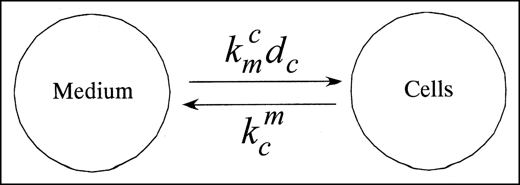
Compartmental model describing distribution of NIS substrate anions between medium and cell.
The maximum initial molar concentrations used in our clonogenic studies of either 211At-astatide or 131I-iodide in the medium (6.2 × 10⁻⁶ and 8.2 × 10⁻³ μmol/L, respectively) were several orders of magnitude smaller than the Km value estimated for iodide. Consequently, when nm ≪ Km, the above equations can be approximated as:
 Eq. 2 where kmc = Vmax/Km is the transfer constant from the medium to a cell (s−1) where the net transfer rate is given by Vmaxdcvc/Km, and kcm is the transfer constant from a cell to the medium (s−1); Nc is the total number of cells; and Vc = Ncvc is the volume of all cells (cm3); VT = Vm + Vc where VT is the total volume of the system (cm3); and Vm is the volume of the incubation medium (cm3). During uptake (u), the initial conditions are nm0 = cte, ncu = 0, and by conservation of mass Vmnm0 = Vmnmu + Ncvcncu, where dcu = Nc/VTu is the cell density and VTu is the volume of the system during the uptake period; thus, the solutions for nmu and ncu are:
 Eq. 3 where the decay-corrected fractional cell uptake F(dc) as a function of cell density dc for an incubation period tu is given by:
 Eq. 4 which tends to equilibrium conditions when t → ∞ thus, F(dc) → dcvckmc/(dcvckmc + kcm). This expression is used to estimate the values of kmc and kcm by means of nonlinear regression analysis from experimental observations. The solutions during efflux (e) for nme and nce after an incubation period tu and removal of the medium and dilution to a new cell density dce = Nc/VTe are:
 Eq. 5 where VTe is the total volume of system during efflux; nc0 = ncu(tu) is the initial molar concentration of cells; and by conservation of mass Ncvcnc0 = Vmnme + Ncvcnce any time afterwards. The activity concentration in the medium am and in cells ac are then given as:
 Eq. 6 where NA is Avogadro’s number; ρm and ρc are the densities of the medium and cells, respectively; and λ is the physical decay constant for the radionuclide in consideration. The cumulated activity concentration in the medium ãm and in the cells ãc are then given as:
 Eq. 7 where ãmu and ãcu are the cumulated activity concentration in the medium and a cell during uptake, respectively; and ãme and ãce are the corresponding cumulated activity concentrations for the medium and cells during efflux, respectively. The expressions for ãmu, ãme, ãcu, and ãce are given in Appendix A. The total cumulated activity per cell is then given as:
 Eq. 8 where vc is the average volume of a cell. Because this compartmental model is a closed system, the residence time of the radionuclide in the medium and cells during uptake is given by τu = τmu + τcu = λ−1(1 − e−λtu), and during efflux as τe = τme + τce = λ−1dcvc(1 − dcvc)−1ac0/am0, where ac0 is the cell activity concentration after an incubation period tu. A glossary of the terminology used is given in Appendix B.
Small-Scale Dosimetry of 131I-Iodide
Dosimetry of 131I-iodide was carried out using the EGS4 Monte Carlo transport code, which was estimated using the geometry utilized in the in vitro assays (17–19). The cell nucleus was considered to be the target in these calculations. UVW and NIS6 cells were stained using Cyto16 (Molecular Probes), which binds to nucleic acids. Phase-contrast and fluorescent images of cells were obtained and superimposed and used as a model for these calculations (Fig. 2). In these dosimetric calculations, 2 sources were identified: the medium where the activity was diluted, and the attached cells themselves, which take up the activity from the medium. The images provided the basic cell morphology that was used in these calculations. The nucleus of adherent cells had a semiellipsoid geometry. However, for simplification purposes, we used a spheric model of the cell and nucleus, where the volume of the semiellipsoid nucleus was that of the sphere nucleus. Thus, the estimated average cell and nuclear diameter used in these studies were 16 and 10 μm, respectively.

Superimposition of phase-contrast and fluorescent images of adherent cells labeled with Cyto16 nuclear stain to show nuclear and cell morphology of UVW and NIS6 cell lines. Colony-forming assays were performed under similar conditions. Images were used to derive cellular and nuclear geometry, which subsequently was used in calculations to determine cumulated absorbed dose. Strongly green regions identify cell nuclei.
The estimated dose conversion factor for 131I-iodide when the source was the medium and the target was the cells, S(m → c), was 1.48 × 10⁻¹¹ Gy g Bq−1 s−1, and the dose conversion factor when both the source and the target were the cells, S(c → c), was 5.5 × 10⁻⁴ Gy Bq−1 s−1. These dose conversion factors were used to assess the total absorbed dose to UVW and NIS6 cells from 131I-iodide. The total average absorbed dose to a cell is then given as:
 Eq. 9 where ãm is the cumulated activity concentration in the medium and q̃c is the average cumulated activity in a cell (Bq s).
Microdosimetry of 211At-Astatide
Microdosimetry of 211At-astatide was carried out using an α-particle Monte Carlo transport code as described elsewhere (20). The cell nucleus was considered to be the target in these calculations. The total absorbed dose is then given as:
 Eq. 10 where D is average absorbed dose (Gy); ãm is the total cumulated activity in the medium (Bq s g−1); hmc is the average number of hits from medium to cells (hits g Bq−1 s−1); z̄1mc is the average specific energy per event from medium to cells (Gy hit−1); q̃c is the cumulated activity per cell (Bq s cell−1); hcc is the average number of hits from cells to cells (hits Bq−1 s−1); and z̄1cc is the average specific energy per event from cells to cells (Gy hit−1). The values for hmc, hcc, z̄1mc, and z̄1cc were estimated as a function of cells per unit area to account for cross fire among cells (data not shown). As an example, a cylindric well (diameter, 16.5 mm) containing 1.4 × 10⁵ adherent cells resulted in a surface cell density of 655 cells mm−2, and the corresponding conversion factors for z̄1mc, z̄1cc, hmc, and hcc were 0.224 Gy hit−1, 0.114 Gy hit−1, 4.456 × 10⁻⁹ hit cm3 Bq−1 s−1, and 0.407 hit Bq−1 s−1, respectively.
RESULTS
External Beam Radiation
Data obtained from the external beam radiation dose/survival assay were fitted using the linear quadratic model of cell survival SF = exp(−αD − βD2), where SF is the surviving fraction and D is the absorbed dose (Fig. 3). Using the statistical analysis software SAS, we carried out the model fitting to assess the parameters α and β. The estimated 2-Gy survival fraction, SF2, for UVW and NIS6 cell lines was 0.71 (0.62–0.83, 95% confidence interval [CI]) and 0.61 (0.53–0.70, 95% CI), respectively. There was no statistically significant difference for the SF2 between the 2 cell lines based on a t test with equal variances (P = 0.3) (SAS).
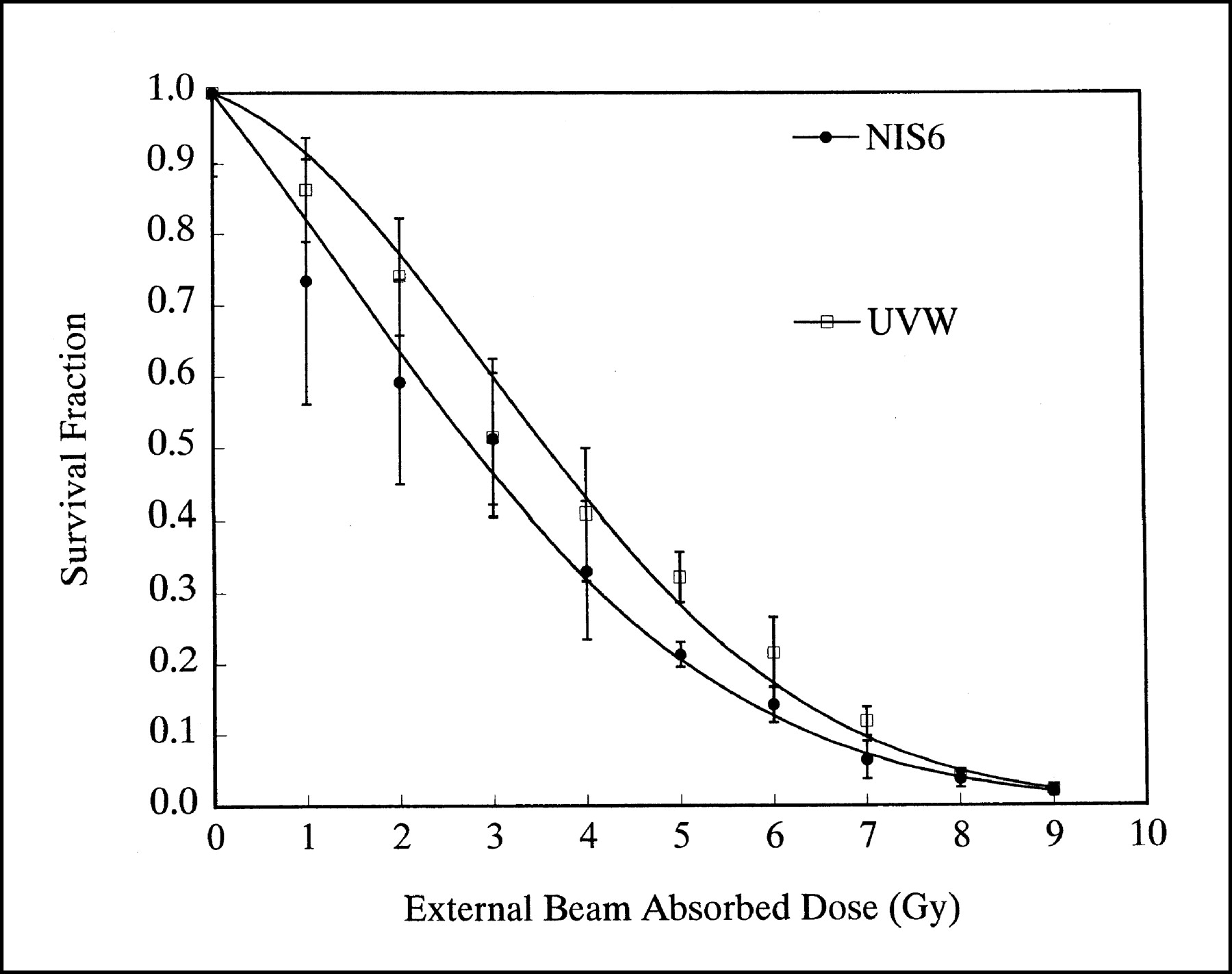
Survival fraction of NIS6 and UVW as function of external beam radiation-absorbed dose. Points represent average of triplicate determinations and corresponding SD. External beam dose rate was 0.5 Gy min−1. The estimated 2-Gy SF (95% confidence interval [CI]) was 0.71 (0.62–0.83, 95% CI) and 0.61 (0.53–0.70, 95% CI) for UVW and NIS6, respectively. There was no statistically significant difference between NIS6 and UVW cells lines (P = 0.296).
Radionuclide Uptake and Efflux Pharmacokinetics
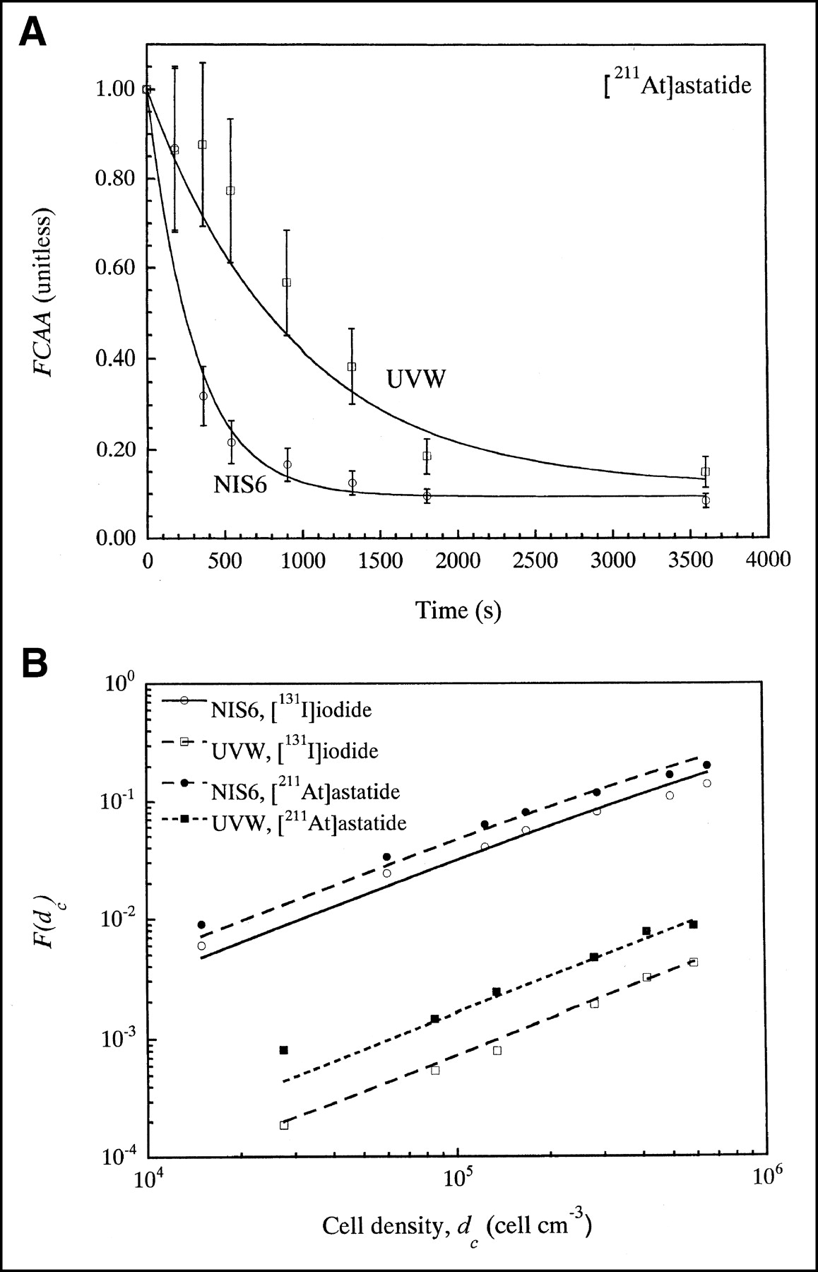
The decay-corrected fraction of administered activity taken up by the cells after a 30-min incubation period F(dc) was estimated as a function of cell density dc for 211At-astatide and 131I-iodide for UVW and NIS6 cell lines. Figure 4 shows the experimental data for NIS6 and UVW cell lines illustrating both specific and nonspecific cellular uptake. The uptake of 131I-iodide and 211At-astatide by NIS6 cells was approximately 60- and 27-fold higher than that of the nontransfected UVW (control), respectively. Also, the nonspecific (NIS independent) association of 211At-astatide was about 4-fold higher that that of 131I-iodide, possibly due to the presence of a higher oxidation state of astatine in the aqueous solution (16). Using nonlinear regression analysis, the transfer coefficients kmc and kcm were derived based on the observed efflux kinetics (Fig. 4A) and the expression for F(dc) (Fig. 4B). Table 1 shows the estimated values for kmc and kcm for UVW and NIS6 cell lines. In each instance, kmc was higher than kcm, indicating that, in the presence of an extracellular halide ion concentration, there is a net association of ion with the cell. The net rate of accumulation of astatide and iodide by NIS6 cells was 32.5- and 27.2-fold higher than that observed for UVW cells, respectively. We observed that astatide had a higher kmc and lower kcm value than iodide with respect to UVW cells, reflecting the higher nonspecific association of astatide previously reported (16).

(A) Fractional cell-associated activity (FCAA) of 211At-astatide for UVW and NIS6 cell lines during efflux as function of time. Similar curves were generated using 131I-iodide (data not shown). Data represent means ± SD from at least 2 triplicate determinations. (B) Fractional cell uptake F(dc) as function of cell density for NIS6 and UVW cell lines for incubation period of 30 min for 131I-iodide and 211At-astatide. Data represent means of 3 determinations. Experimental data were fitted to theoretic expression for F(dc) to estimate transfer constants kmc and kcm shown in Table 1. Uptake of 131I-iodide by NIS6 cells was approximately 60-fold higher than that of UVW (nonspecific binding). Total uptake of 211At-astatide to NIS6 cells was approximately 27-fold higher than that of UVW. Nonspecific binding of 211At-astatide was approximately 4-fold higher than that of 131I-iodide.
Estimated Pharmacokinetic Parameters for 131I-Iodide and 211At-Astatide for UVW and NIS6 Cell Lines
Pharmacokinetic Model of Halide Distribution
This model describes the distribution of 131I-iodide and 211At-astatide between the incubation medium and cells over time in the presence (uptake) and absence (efflux) of halide anions. For the purposes of these studies, we modeled a closed system to reproduce the incubation conditions used in the colony-forming assays. The uptake and efflux rate constants of the system (kmc and kcm, respectively) were determined from the experimental data as described above. Figure 5 is a representative example of the pharmacokinetics of 211At-astatide in NIS6 and UVW cells during uptake and efflux phases. This system is sensitive to cell density in the assay system (dc), as highlighted in Fig. 5B. During the efflux phase, the system reequilibrates as 211At-astatide moves from cells to medium and is then subject to reuptake (upper curve). Reduction of the cell density during the efflux phase, as occurs during plating for colony-forming assay, results in equilibration at a much lower cellular concentration (lower curve). This effect is more pronounced in NIS6 cells (Fig. 5B) than in UVW cells (Fig. 5A), as the total nonspecific cell-associated activity is many fold lower than NIS-mediated uptake.
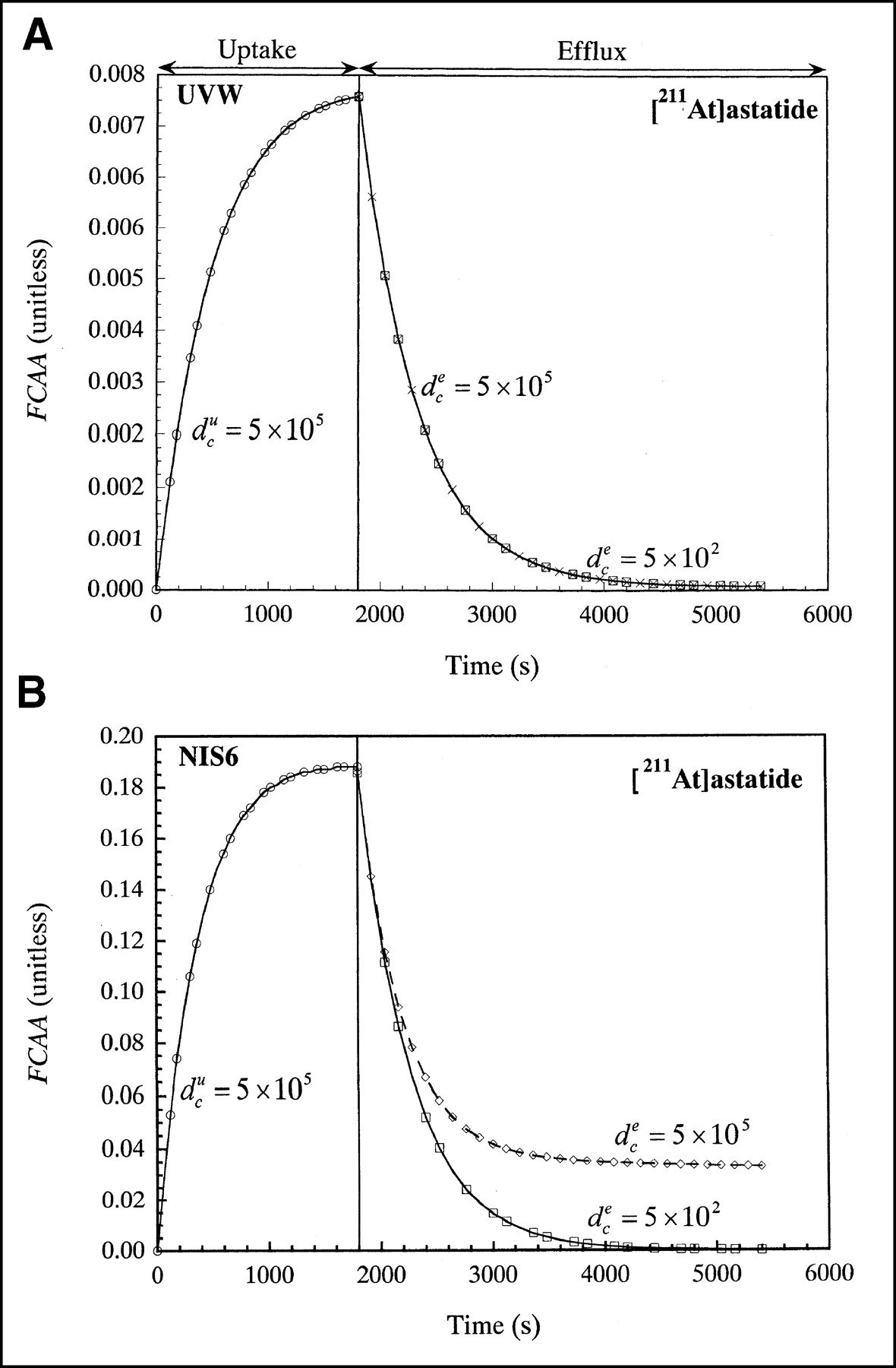
Calculated fractional cell-associated activity for UVW and NIS6 cell lines for 211At. Cell density during uptake was 5 × 10⁵. Cell density during efflux was reduced by factor of 1 × 10³ cells cm−3, reflecting conditions used during colony-forming assay. However, when cell density was left constant, a different efflux profile is observed as indicated in Equation 5. Uptake period was 30 min. Calculations were based upon estimated kmc and kcm coefficients that were derived from our experimental studies (Table 1). Area under the curve was calculated to assess cumulated activity concentration in cells and corresponding absorbed dose.
Administered Activity Concentration Versus Survival Fraction
The effect of increasing radionuclide concentration on clonogenic survival was assessed in UVW and NIS6 cells using both 131I-iodide and 211At-astatide. An activity concentration-dependent decrease in surviving clonogens was observed after treatment of NIS6 cells with both radionuclides (Fig. 6). For both radionuclides examined, the control parental UVW cell line was less sensitive than the NIS-transfected cell line, indicating the preferential therapeutic efficacy in NIS-expressing cells over control cells.

Survival fraction as function of activity concentration in medium for incubation period of 30 min. Survival fraction of NIS6 and UVW for 131I (A) and for 211At (B), respectively. Data represent means ± SD of at least 3 triplicate determinations. Therapeutic efficiency and toxicity ratios are given in Table 2.
The survival data were fitted using a single exponential and, for the purpose of comparison, we used this model to calculate A0, which indicates the initial activity concentrations of each radionuclide required to reduce the clonogenic capacity of UVW and NIS6 cells by 1 natural logarithm (e−1). These values are shown in Table 2. The A0 ratio between UVW and NIS6 cells was 13.2-fold for 131I-iodide and 11.5-fold for 211At-astatide. The similarity of these values is consistent with the similar pharmacokinetics for both radionuclides in this system. Comparison of the relative toxicity of each radionuclide (A0 for 131I divided by A0 for 211At) indicates that, in terms of radioactive concentration, 211At-astatide is between 600 and 700 times more radiotoxic that 131I-iodide in this model (Table 2). The higher nonspecific association of 211At-astatide in this system may account for the variation observed in both of these ratios. The cumulated activity concentrations in the medium and cells were estimated by integrating the uptake-efflux kinetics over time as a function of cell density dc and then were used to calculate the absorbed doses for UVW and NIS6 per unit administered activity (Table 3).
Effect of 131I-Iodide and 211At-Astatide Administration on Clonogenic Survival of UVW and NIS6 Cell Lines
Average Absorbed Dose per Unit Administered Activity In Vitro of 131I-Iodide and 211At-Astatide in UVW and NIS6 Cells Lines
Correlation of Survival Fraction and Absorbed Dose
Radionuclide sensitivity data from these experiments were correlated with estimates of absorbed dose. The cumulated activity concentrations were calculated by determining the area under the curve (AUC) derived from the pharmacokinetic model. Absorbed doses were estimated by calculating the cumulated activity concentration in the medium and cells and using the corresponding conversion factors as described in Equations 9 and 10 for 131I-iodide and 211At-astatide, respectively. These data, shown in Figure 7, demonstrate that both UVW and NIS6 cells have a greater sensitivity to an equivalent absorbed dose of high LET α-particles (Fig. 7A) than to low LET β-particles (Fig. 7B).
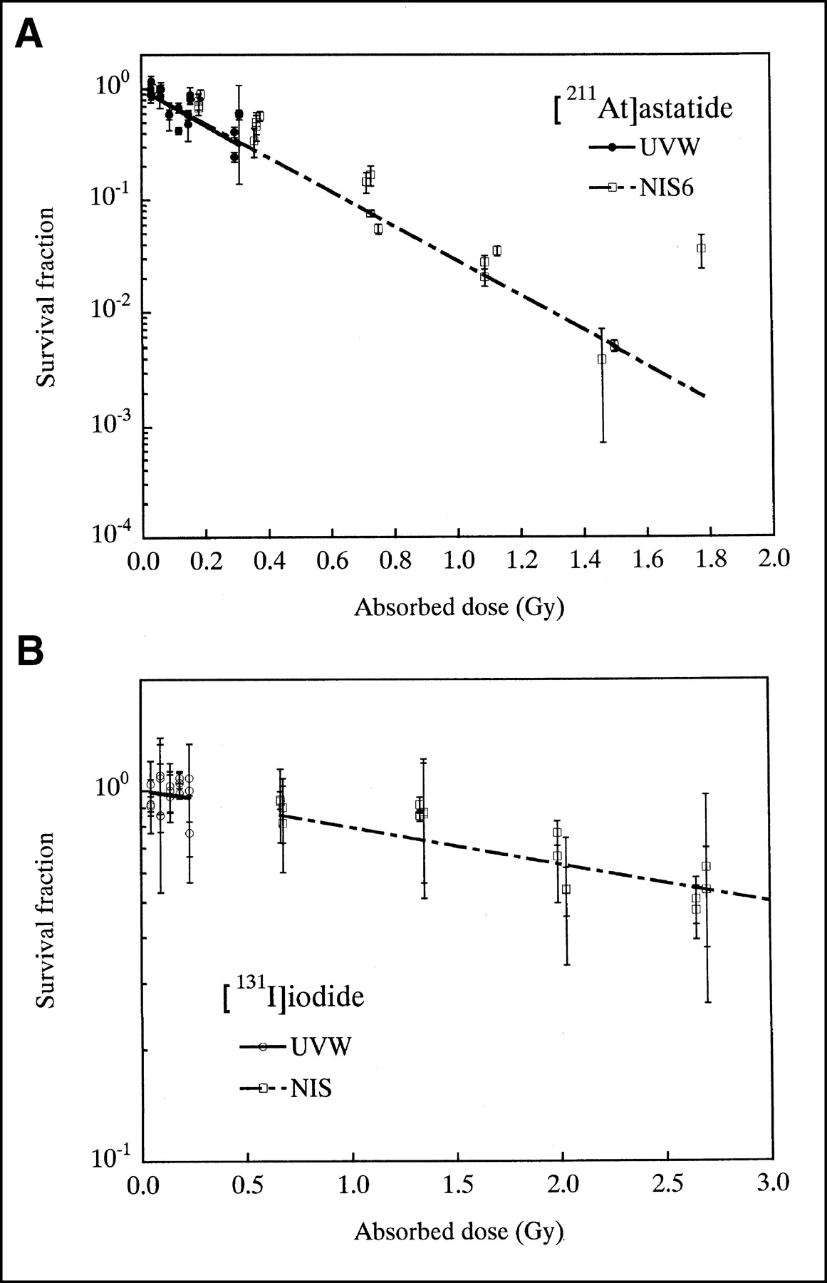
Survival fraction of UVW and NIS6 cell lines as function of absorbed dose from 211At-astatide (A) and 131I-iodide (B). Estimated D0 of UVW and NIS6 for 211At-astatide was 0.27 (0.19–0.46, 95% CI) Gy and 0.28 (0.26–0.30, 95% CI) Gy, respectively. 2-Gy SF was 6.3 × 10⁻⁴ (3.1 × 10⁻⁵ − 1.3 × 10⁻², 95% CI) and 7.4 × 10⁻⁴ (4.2 × 10⁻⁴ − 1.3 × 10⁻³, 95% CI), respectively. D0 of UVW and NIS6 for 131I-iodide was 5.27 (2.5–6.4, 95% CI) Gy and 4.31 (3.0–7.6, 95% CI) Gy, respectively, and 2-Gy SF for UVW and NIS6 was 0.68 (0.02–20.4, 95% CI) and 0.66 (0.35–1.22, 95% CI), respectively.
Single-exponential equations were fitted to these data and fitted coefficients were used to derive D0 and 2-Gy surviving fractions (SF2) for each cell line and radionuclide. These data are shown in Table 4. This similarity of D0 and SF2 values between NIS6 and UVW using each treatment modality cells confirms that plasmid-mediated NIS expression does not alter the intrinsic cellular radiosensitivity in this model system. The SF2 values obtained from external beam radiotherapy (Fig. 3) are similar to those derived from 131I-iodide treatment, which is in concordance with many previous observations. However, the SF2 values derived from 211At-astatide treatment were 900- to 1,100-fold higher (Table 4) than those from both external beam radiotherapy and 131I in both cell lines, reflecting the cytotoxicity of 211At-astatide observed in this system (Table 2). Comparison of D0 values for 211At-astatide and 131I-iodide demonstrates a 15.3- and 19.5-fold differential for UVW and NIS6, respectively, highlighting the therapeutic advantage, in terms of radiation quality, of high LET 211At α-particles over low LET 131I β-particles.
Dose Factors and 2-Gy Survival Fraction Associated with 131I-Iodide, 211At-Astatide, and External Beam Radiation in UVW and NIS6 Cells Lines
DISCUSSION
The transgene-mediated expression of the NIS has been demonstrated to confer iodide uptake capacity to a wide range of cell types that would not normally concentrate iodide (2–4). NIS gene transfer, followed by radioiodide administration, allows the possibility of treatment of tumors at many sites in a manner analogous to the highly efficacious treatment of differentiated thyroid carcinoma with radioiodide. However, the short retention time of iodide observed in nonthyroid xenografts, combined with the relatively long physical t1/2 of 131I, results in an insufficient absorbed dose for successful tumor eradication. The use of radionuclides with similar chemical properties and more favorable physical properties, such as a higher LET and shorter physical t1/2, should result in an increased radiation dose and thus enhanced efficacy.
We have previously reported that plasmid-mediated cellular expression of the NIS confers the ability to accumulate 211At-astatide in addition to iodide (16). Here we describe clonogenic survival experiments designed to directly compare the radiotoxicity of 131I-iodide and 211At-astatide on a cultured cell model and a pharmacokinetic model, which was used to estimate absorbed dose for each radionuclide in UVW and NIS6 cell lines. To determine whether expression of the NIS protein, or selection of NIS-positive clones, resulted in an alteration of cellular radiosensitivity, a comparison of the radiation sensitivity of UVW and NIS6 cell lines was performed by examining the clonogenic survival of both cell lines in response to external photon beam irradiation. The SF2 values obtained for both cell lines were not significantly different, 0.71 (0.62–0.83, 95% CI) and 0.61 (0.53–0.70, 95% CI) for UVW and NIS6, respectively (Fig. 3), indicating that expression of NIS has no effect on intrinsic radiosensitivity. The effect of activity concentration of both 131I-iodide and 211At-astatide on UVW and NIS6 cells was determined by colony-forming assay. An activity concentration-dependent decrease in clonogenic survival was observed in both cell lines with both radionuclides (Fig. 6), with NIS6 cells having increased sensitivity to each radionuclide when compared with control UVW cells. Using these data, values were calculated to indicate the activity concentration required to cause a reduction in survival by 1 natural logarithm (A0), using the incubation conditions described above. The ratio of A0 values between NIS-negative (UVW) and NIS-expressing (NIS6) cells (ratio 1, Table 2) was 13.2 for 131I-iodide and 11.5 for 211At-astatide (Table 2). The similarity of these values reflects the similar pharmacokinetics of cellular iodide and astatide association in this model system. The smaller therapeutic differential calculated for 211At may reflect the higher degree of nonspecific binding observed with aqueous astatide solutions (16,21). For this study, we define the terms “specific binding” as NIS-mediated, perchlorate-sensitive cellular uptake and “nonspecific binding” as non-NIS-mediated, perchlorate-insensitive cellular uptake of radiohalide. Detailed examination of the properties of specific and nonspecific uptake of both 131I-iodide and 211At-astatide in this model system have previously been reported (16). Comparison of A0 values for each radionuclide indicated an enhanced sensitivity to 211At of 686-fold in UVW cells and 598-fold in NIS6 cells compared with 131I. Similar enhancements in sensitivity (up to 1,400-fold) were observed previously when comparing the cytotoxicity of 211At- and 131I-labeled benzylguanidine compounds in neuroblastoma cells (22). These data, and our own observations, are in accordance with calculations that predict a 1,200-fold increase in cytotoxicity for α-particles on a single cell basis (23). It should be noted that A0 values, expressed in units of activity per unit volume (Bq mL−1) only indicate the relative toxicity of each radionuclide under the experimental conditions used for these studies. Calculation of cumulated absorbed dose from each radionuclide also requires that radiologic t1/2 and system geometry be taken into account. Absorbed dose calculations indicate that administration of 211At-astatide results in an increase (53.9- to 64.9-fold) over 131I-iodide in absorbed dose per administered activity concentration (Table 3).
The rapid efflux of iodide from NIS-expressing cells lacking an organification mechanism has been observed in several cell lines and rodent xenograft models (4,9,24). The biologic t1/2 of iodide in xenograft tumors is generally longer than that observed in 2-dimensional cultured cell models, presumably due to the continual reuptake of iodide by the tumor. Although this has allowed effective demonstrations of the potential of NIS expression for tumor imaging using NIS substrate molecules such as 123I−, 124I−, and 99mTcO4− (25–27), the successful sterilization of such tumors with 131I is likely to require the administration of prohibitively large quantities of radioactivity (9). However, studies performed using an in vivo prostate model have demonstrated, given sufficiently high administered radioactivity, that nonthyroid tumor xenografts with transgene-mediated NIS expression can be successfully treated with 131I (8). This demonstrates the principle that NIS-expressing tumor deposits can be successfully targeted and treated with an ionic radiohalide in vivo and that the therapeutic effect is dependent on delivering a sufficient radiation dose to tumor.
α-Particle-emitting radionuclides have considerable potential as cytotoxic agents in the treatment of malignant disease. The short range and high LET α-particle makes their use particularly suited to the treatment of micrometastatic and minimal residual disease, where the radiation dose to tumor is normally limited by the dose delivered to surrounding normal tissues. Tumor-specific targeting of α-particles can result in highly focal irradiation, high tumor absorbed doses, and minimization of surrounding normal tissue toxicity. The first clinical trial with an 211At-labeled therapeutic antibody (antitenascin monoclonal antibody 81C6) is currently underway, with encouraging initial observations of high radiation doses to the target area and low doses to surrounding normal tissue (28).
The data presented in this article indicate that the administration of 211At-astatide results in a significant increase (55- to 65.6-fold) over 131I-iodide in absorbed dose per administered activity concentration (Table 3). However, the nonspecific association of 211At-astatide with cells lacking NIS expression also contributes to the dose to these cells, resulting in a lower therapeutic differential than that observed with 131I-iodide at the same administered activity concentration (12.0 and 14.5, respectively) (Table 3).
A previous study evaluating the uptake of 211At-astatide by NIS transgene-expressing thyroid tumor cells (21) presented several observations similar to our current and previously published data (16). Uptake and efflux kinetics of both 211At-astatide and 131I-iodide are similar in each cultured cell model system, and uptake of both radiohalides was sensitive to excess perchlorate (ClO4−) and iodide (I−) concentration, demonstrating that 211At-astatide is transported by NIS with an efficiency approaching that of iodide. Using a murine xenograft model, the intraperitoneal administration of 211At-astatide resulted in a 14.4-fold increase in tumor absorbed dose over that predicted for 131I-iodide, with similar biologic half-lives observed for both anions (21). Although the difference in pharmacokinetics between in vitro and in vivo models precludes a direct comparison of absorbed dose, the conclusions of Petrich et al. (21), in accordance with this study, indicate that the administration of 211At-astatide results in a significantly higher absorbed dose to NIS-expressing cells than 131I-iodide per unit administered activity.
The pharmacokinetic model presented in this study describes the phenomenologic distribution of halide anions in a closed system, which accurately describes the conditions used in the cultured cell clonogenic assays described above. It is also possible to use this model to analyze the pharmacokinetics of other NIS substrate anions, such as 188ReO4− or 99mTcO4−, under similar experimental conditions. In addition, it can also be adapted, given appropriate experimental observations, to predict the effect of alteration of many system parameters (e.g., level of NIS expression, heterogeneous cell populations, pharmacologic inhibitors of halide efflux) on both pharmacokinetic distribution and cumulated absorbed dose. We intend to further develop this model to examine the pharmacokinetics of NIS-mediated anion transport in a 3-dimensional cultured cell system and, in combination with biodistribution data, to develop a model describing the pharmacokinetics in a dynamic extracellular system similar to that encountered in vivo, which will aid in the future design of NIS-based therapeutic strategies.
Given the similar pharmacokinetics of 131I-iodide and 211At-astatide in our model system, our observation of a higher absorbed dose using 211At-astatide can be explained by a combination of the higher energy α-particle (5.87 MeV) versus 131I β-particle emission (0.6 MeV) as well as the shorter physical t1/2 of 211At (7.2 h vs. 192 h for 131I). Similarly, a study into the potential of 188Re (in the form 188ReO4−) to sterilize NIS-expressing cells calculated a 4.5-fold higher absorbed dose, when compared with 131I (14). This enhancement is likely due to a combination of factors: 188Re emits β-particles with a higher energy than 131I (2.12 vs. 0.61 MeV), and ReO4− had higher tumor uptake than I− in a NIS-expressing xenograft model (14). However, much of the increase in absorbed dose results from the comparatively short physical t1/2 (16.7 h) of 188Re (14). These findings are of particular significance in relation to cell types lacking a peroxidase-based organification mechanism, as these cells generally exhibit a very short cellular retention of iodide (3,4), and matching of both the physical and biologic half-lives of the radioisotope is desirable for maximal radiobiologic effect.
In vivo, tumor cells may show a heterogeneous expression of NIS that can be predicted to result in a heterogeneous cumulated activity distribution with the tumor mass. However, depending on the range of the radiations emitted by the radionuclide used for therapy, there is a potential for cross fire among tumors cells. Although the range of α-particles is significantly shorter than that of β-particles, a degree of cross fire can be predicted. The proportion of cells receiving a highly toxic absorbed dose from 211At-astatide will therefore depend on a combination of the overall transfection efficiency, the spatial distribution of transgene expression and radionuclide within the tumor, and the degree of cross fire. The use of cultured cell models, such as the one described here, affords the opportunity for detailed analysis of these issues and may provide the basis for a clinical implementation of transgene-mediated radionuclide therapy in the future. It is also important to note that there are some tumor types, such as thyroid and invasive breast carcinoma, that may express sufficient NIS to facilitate 211At-astatide therapy without the need for transgene-mediated expression.
The use of ionic 211At-astatide for the treatment of thyroid carcinoma has previously been investigated in murine xenograft models, with high tumor uptakes reported for more differentiated thyroid tumor cells, which generally retain a higher level of NIS expression than anaplastic thyroid tumors (29–31). A study comparing the uptake of 125I-iodide and 211At-astatide in nude mice bearing human fetal thyroid and human malignant thyroid xenografts observed that the human tissue accumulated both halides with similar kinetics, suggesting that 211At-astatide may be an alternative radionuclide to 131I-iodide for the treatment of thyroid carcinoma (29). Another study examining the comparative transport of 125I-iodide and 211At-astatide using normal porcine thyrocytes in a polarized system also observed similarities in the basal membrane (i.e., NIS mediated) transport of both radiohalide anions (32). Interestingly, this study also reported the perchlorate- and ouabain-insensitive basal membrane transport of astatide, by normal thyrocytes, suggesting the possibility of another, NIS-independent astatide uptake mechanism in these cells. Further elucidation of this transport mechanism may provide some explanation as to the differences in biodistribution of astatide and iodide, in addition to providing another means of targeting astatine to tumor cells.
The biodistribution of systemically administered 211At-astatide, though similar to that of iodide, has several clinically relevant differences. In particular, a significantly higher uptake than iodide in both lung and spleen tissues is observed (12,33). At present, clinical trials of 211At-astatide for the treatment of thyroid carcinoma have not been initiated, possibly because of concerns over radiotoxicity in these tissues or due to the limited availability of the radionuclide. Previous investigations have revealed that pharmacologic intervention may modify the level of astatide uptake in normal tissues (12), and further studies into the possible mechanisms involved in the lung and spleen retention of astatide are currently underway. Although lung and spleen uptake currently precludes the systemic administration of 211At-astatide, combined gene transfer/radionuclide treatment of compartmentalized tumors such as those of brain, bladder, and ovary may be possible with minimal involvement of the systemic circulation. Further advances in the understanding of the mechanisms of astatine uptake and retention in lung and spleen will aid greatly in the clinical implementation of systemic 211At-astatide therapy for tumors with both endogenous and gene transfer–mediated NIS expression.
CONCLUSION
The data presented in this article demonstrate that 211At-astatide has potential as an alternative radionuclide to 131I-iodide for the treatment of NIS-expressing tumor cells. The direct comparison of 211At and 131I in this closed system highlights the enhancements in absorbed dose and radiotoxicity of high LET α-particles over low LET β-particle irradiation. 211At-Astatide administration results in increased cytotoxicity (686- to 598-fold) and higher absorbed doses (55- to 65.5-fold) per unit administered activity when compared with 131I-iodide. In addition, the physical t1/2 of 211At is closely matched to the radionuclide residence time observed in nonthyroid tumor types, providing a further therapeutic advantage over 131I. These observations imply that some of the limitations of 131I administration for the therapy of endogenous or transgene-mediated NIS-expressing tumors can be overcome. The pharmacokinetic model presented here is a valuable tool in predicting the influence on absorbed dose resulting from modulation of many experimental system parameters. Further development of this model to describe a 3-dimensional tumor within a dynamic environment will aid in the elucidation of the relationship between administered radioactivity and resulting absorbed doses in NIS-expressing tumors with different retention characteristics.
APPENDIX A
Pharmacokinetics
System description:
 The uptake solutions given conservation of mass Vmnm0 = Vmnm + Ncvcnc are:


 The efflux solutions given conservation of mass Ncvcnc0 = Vmnm + Ncvcnc are:


 Residence times:


APPENDIX B
Glossary
nm Average halide concentration in medium (mol/L)
nmu Average halide concentration in medium during uptake (mol/L)
nme Average halide concentration in medium during efflux (mol/L)
nc Average halide concentration in cells (mol/L)
ncu Average halide concentration in cells during uptake (mol/L)
nce Average halide concentration in cells during efflux (mol/L)
Vmax Theoretical maximum halide transfer rate (mol/L s−1)
Km Michaelis–Menten constant for NIS (mol/L)
vc Average volume of a cell (cm−3)
dc Average cell density (cell cm−3)
dcu Average cell density during uptake (cell cm−3)
dce Average cell density during efflux (cell cm−3)
Nc Total number of cells (cell)
kcm Transfer rate between a cell to medium (s−1)
kmc Transfer rate between medium to a cell (s−1)
Vm Volume of medium (cm3)
Vc Volume of cells (cm3)
VT Total volume of system (cm3)
VTu Total volume of system during uptake period (cm3)
VTe Total volume of system during efflux period (cm3)
F(dc) Fractional cell uptake (unitless)
FCAA Fractional cell-associated activity (unitless)
am Average activity concentration in medium (Bq cm−3)
ãmu Average cumulated activity concentration in medium during uptake (Bq s cm−3)
ãme Average cumulated activity concentration in medium during efflux (Bq s cm−3)
ac Average activity concentration in the cell (Bq cm−3)
ãcu Average cumulated activity concentration in a cell during uptake (Bq s cm−3)
ãce Average cumulated activity concentration in a cell during efflux (Bq s cm−3)
q̃c Average cumulated activity in a cell (Bq s)
τu Residence time of system during uptake (s)
τe Residence time of system during efflux (s)
τmu Residence time of halide in medium during uptake (s)
τcu Residence time of halide in cells during uptake (s)
τme Residence time of halide in medium during efflux (s)
τce Residence time of halide in cells during efflux (s)
D Average absorbed dose (Gy)
S(m → c) Dose conversion factor from medium (source) to cells (target) (Gy g Bq−1 s−1)
S(c → c) Dose conversion factor from cells (source) to cells (target) (Gy g Bq−1 s−1)
hmc Average number of hits from medium (source) to a cell (target) (hit)
hcc Average number of hits from cells (source) to a cell (target) (hit)
z̄1mc Specific energy per event from medium (source) to a cell (target) (Gy hit−1)
z̄1cc Specific energy per event from cells (source) to a cell (target) (Gy hit−1)
SF Survival fraction (unitless)
SF2 2-Gy survival fraction (unitless)
λ Physical decay constant of radionuclide (s−1)
D0 Absorbed dose required to reduce survival fraction to e−1 (Gy)
A0 Activity concentration required to reduce survival fraction to e−1 (MBq cm−3)
Acknowledgments
NIS cDNA was kindly provided by Dr. Sissy M. Jhiang (Ohio State University, Columbus, OH). This work was supported by grants CA42324, CA70164, and CA91927 from the National Institutes of Health and by an AstraZeneca Union Internationale Contre le Cancer Translational Cancer Research Fellowship (American Cancer Society Beginning Investigator 11/2001).
Footnotes
Received Apr. 11, 2003; revision accepted Jul. 22, 2003.
For correspondence or reprints contact: Michael R. Zalutsky, PhD, Department of Radiology, Box 3808, Duke University Medical Center, Durham, NC 27710.
E-mail: zalut001{at}mc.duke.edu
REFERENCES
In this issue


Jump to section
Related Articles
Cited By...
- PARP-1-Targeted Auger Emitters Display High-LET Cytotoxic Properties In Vitro but Show Limited Therapeutic Utility in Solid Tumor Models of Human Neuroblastoma
- A perspective view of sodium iodide symporter research and its clinical implications.
- Radioiodine Therapy of Hepatoma Using Targeted Transfer of the Human Sodium/Iodide Symporter Gene
- Effective Cancer Therapy with the {alpha}-Particle Emitter [211At]Astatine in a Mouse Model of Genetically Modified Sodium/Iodide Symporter-Expressing Tumors
- Establishment of a Human Hepatocellular Carcinoma Cell Line Highly Expressing Sodium Iodide Symporter for Radionuclide Gene Therapy