
Main menu

  • Home
  • Content
    • Current
    • Ahead of print
    • Past Issues
    • JNM Supplement
    • SNMMI Annual Meeting Abstracts
    • Continuing Education
    • JNM Podcasts
  • Subscriptions
    • Subscribers
    • Institutional and Non-member
    • Rates
    • Journal Claims
    • Corporate & Special Sales
  • Authors
    • Submit to JNM
    • Information for Authors
    • Assignment of Copyright
    • AQARA requirements
  • Info
    • Reviewers
    • Permissions
    • Advertisers
  • About
    • About Us
    • Editorial Board
    • Contact Information
  • More
    • Alerts
    • Feedback
    • Help
    • SNMMI Journals
  • SNMMI
    • JNM
    • JNMT
    • SNMMI Journals
    • SNMMI

User menu

  • Subscribe
  • My alerts
  • Log in
  • Log out
  • My Cart

Search

  • Advanced search
Journal of Nuclear Medicine
  • SNMMI
    • JNM
    • JNMT
    • SNMMI Journals
    • SNMMI
  • Subscribe
  • My alerts
  • Log in
  • Log out
  • My Cart
Journal of Nuclear Medicine

Advanced Search

  • Home
  • Content
    • Current
    • Ahead of print
    • Past Issues
    • JNM Supplement
    • SNMMI Annual Meeting Abstracts
    • Continuing Education
    • JNM Podcasts
  • Subscriptions
    • Subscribers
    • Institutional and Non-member
    • Rates
    • Journal Claims
    • Corporate & Special Sales
  • Authors
    • Submit to JNM
    • Information for Authors
    • Assignment of Copyright
    • AQARA requirements
  • Info
    • Reviewers
    • Permissions
    • Advertisers
  • About
    • About Us
    • Editorial Board
    • Contact Information
  • More
    • Alerts
    • Feedback
    • Help
    • SNMMI Journals
  • View or Listen to JNM Podcast
  • Visit JNM on Facebook
  • Join JNM on LinkedIn
  • Follow JNM on Twitter
  • Subscribe to our RSS feeds
Research ArticleCLINICAL INVESTIGATIONS

The Diagnostic Value of 123I-IMP SPECT in Non-Hodgkin's Lymphoma of the Central Nervous System

Yasuhiko Akiyama, Kouzo Moritake, Toshiki Yamasaki, Yoriyoshi Kimura, Akira Kaneko, Yoshiaki Yamamoto, Takeshi Miyazaki and Mitsuhiro Daisu
Journal of Nuclear Medicine November 2000, 41 (11) 1777-1783;
Yasuhiko Akiyama
  • Find this author on Google Scholar
  • Find this author on PubMed
  • Search for this author on this site
Kouzo Moritake
  • Find this author on Google Scholar
  • Find this author on PubMed
  • Search for this author on this site
Toshiki Yamasaki
  • Find this author on Google Scholar
  • Find this author on PubMed
  • Search for this author on this site
Yoriyoshi Kimura
  • Find this author on Google Scholar
  • Find this author on PubMed
  • Search for this author on this site
Akira Kaneko
  • Find this author on Google Scholar
  • Find this author on PubMed
  • Search for this author on this site
Yoshiaki Yamamoto
  • Find this author on Google Scholar
  • Find this author on PubMed
  • Search for this author on this site
Takeshi Miyazaki
  • Find this author on Google Scholar
  • Find this author on PubMed
  • Search for this author on this site
Mitsuhiro Daisu
  • Find this author on Google Scholar
  • Find this author on PubMed
  • Search for this author on this site
  • Article
  • Figures & Data
  • Info & Metrics
  • PDF
Loading

Article Figures & Data

Figures

  • Tables
  • FIGURE 1.
    • Download figure
    • Open in new tab
    • Download powerpoint
    FIGURE 1.

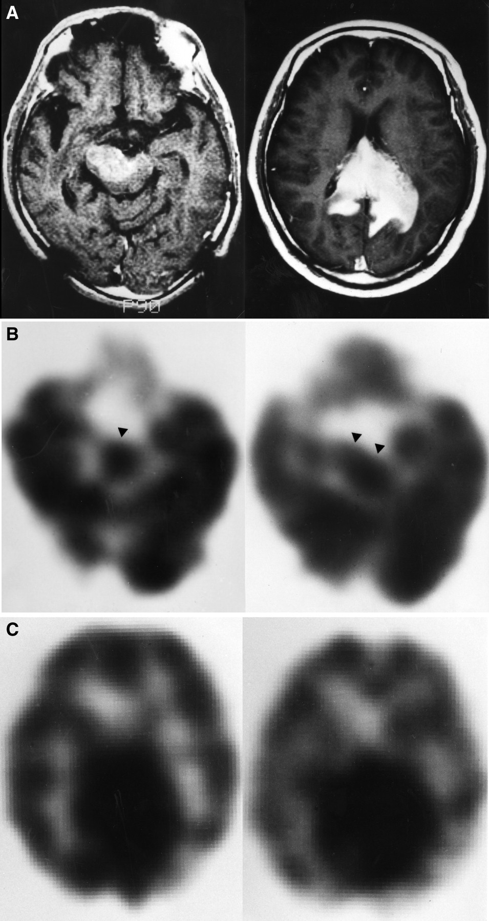
    A 69-y-old man with CNS lymphoma (patient 2). (A) T1-weighted MR image with gadolinium-diethylenetriaminepentaacetic acid shows homogenous enhancing lesions in midbrain and corpus callosum. (B) Midbrain tumor shows normal and high accumulation (arrowheads) on early (left) and delayed (right) SPECT images, respectively. (C) Early (left) and delayed (right) 123I-IMP SPECT images reveal increased uptake at corpus callosum.

  • FIGURE 2.
    • Download figure
    • Open in new tab
    • Download powerpoint
    FIGURE 2.

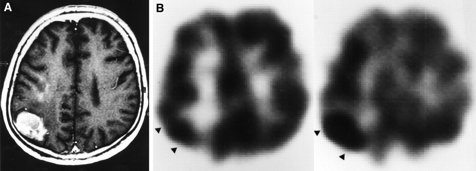
    A 52-y-old man with CNS lymphoma (patient 11). (A) T1-weighted MR image with gadolinium-diethylenetriaminepentaacetic acid shows heterogeneously enhancing mass in right parieto-occipital area. Mass is difficult to distinguish from high-grade glioma on MR image only. (B) 123I-IMP SPECT images show normal and increased accumulation (arrowheads) corresponding to tumor on early (left) and delayed (right) images, respectively.

  • FIGURE 3.
    • Download figure
    • Open in new tab
    • Download powerpoint
    FIGURE 3.

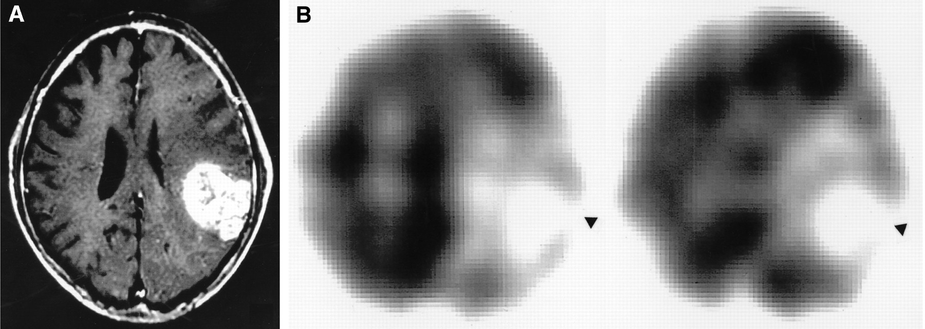
    A 72-y-old man with glioblastoma. (A) T1-weighted MR image with gadolinium-diethylenetriaminepentaacetic acid shows heterogeneously enhancing mass in left parietal region. (B) 123I-IMP SPECT images show defect (arrowheads) corresponding to tumor on both early (left) and delayed (right) images. T/Ns on early and delayed images are 0.23 and 0.21, respectively. T/Cs on early and delayed images are 0.18 and 0.20, respectively.

  • FIGURE 4.
    • Download figure
    • Open in new tab
    • Download powerpoint
    FIGURE 4.

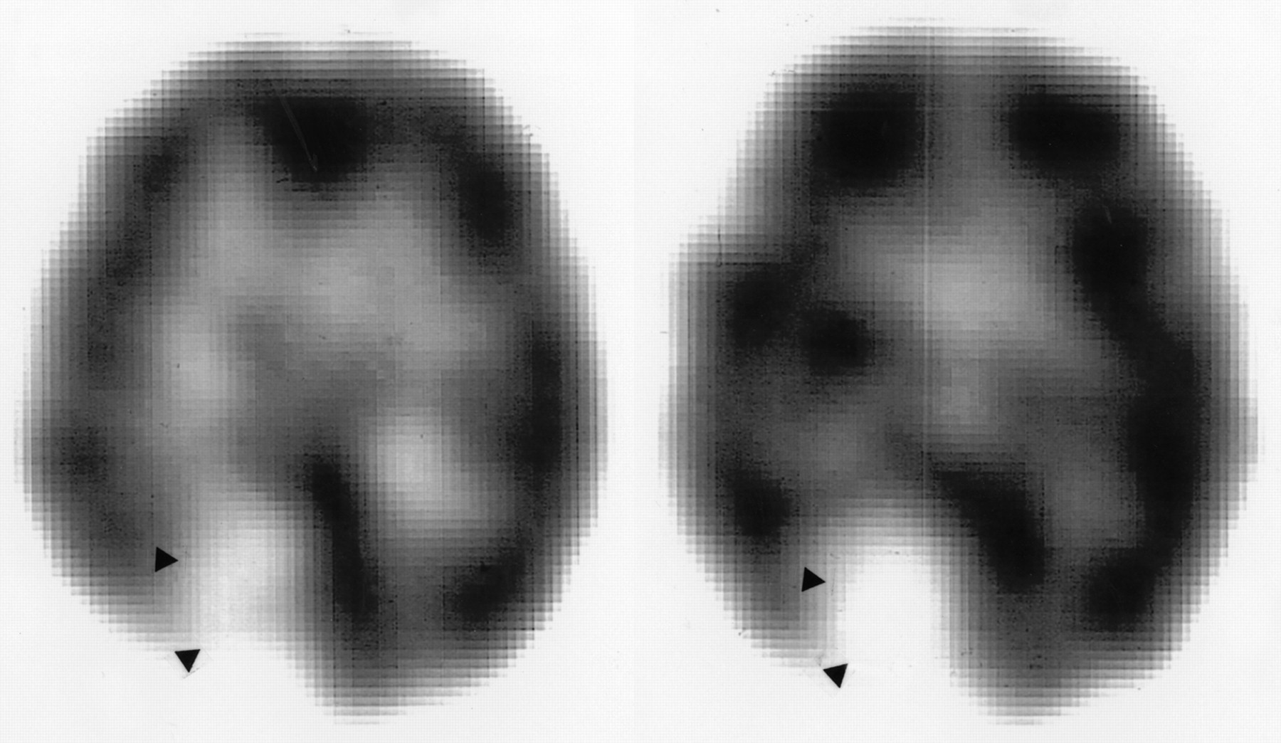
    A 71-y-old woman with right parasagittal meningioma. 123I-IMP SPECT images show low accumulation and defect (arrowheads) corresponding to tumor on early (left) and delayed (right) images, respectively. T/Ns on early and delayed images are 0.53 and 0.48, respectively. T/Cs on early and delayed images are 0.18 and 0.14, respectively.

Tables

  • Figures
    • View popup
    TABLE 1.

    Clinical Features and 123I-IMP SPECT Results for Patients with Non-Hodgkin's Lymphoma of Central Nervous System

    Patient no.Age (y)SexTumor typeTumor locationTumor volume (cm3)Visual imageT/NT/C
    EarlyDelayedEarlyDelayedEarlyDelayed
    177FMetastasisL thalamus to temporoparietal region6.2LowHigh0.771.300.511.36
    269MPrimaryMidbrain5.4NormalHigh1.161.250.800.92
    Splenium of corpus callosum12.0HighHigh1.92*2.25*1.061.23
    362MPrimaryR paraventricle4.0LowHigh0.62*1.31*0.530.95
    L paraventricle3.6LowHigh0.60*1.42*0.501.00
    472FPrimaryIntraventricular region7.4HighHigh2.012.720.750.95
    581FPrimaryL parietal region2.5LowNormal0.411.000.411.05
    673MPrimaryL temporal region3.2NormalHigh1.141.530.951.11
    774MPrimaryCorona radiata to centrum semiovale2.5NormalNormal1.001.140.700.84
    848MPrimaryR basal ganglia6.0LowHigh0.501.420.450.96
    Midbrain3.0NormalHigh0.851.200.741.04
    958FPrimaryR parietal region4.5NormalHighNDNDNDND
    L basal ganglia2.4NormalNormalNDNDNDND
    1043MPrimaryR parietal region5.0NormalHigh1.07*1.41*0.681.20
    L parietal region4.0NormalHigh1.03*1.35*0.671.10
    Pons3.4HighHigh1.761.541.351.05
    1152MPrimaryR parietooccipital region4.6NormalHigh1.001.400.961.00
    1279FPrimaryL frontal region9.4LowHigh0.591.520.431.42
    • ↵* Mirror image ROI using horizontal symmetry axis was used.

    • Metastasis = metastatic intracranial lymphoma from extracranial origin; primary = primary intracranial lymphoma; ND = no data.

    • Patient 1 had metastatic lymphoma from breast origin. Patients 2, 3, 8, 9, and 10 had 2 separated lesions. Patient 10 had recurrent lesion in pons.

    • View popup
    TABLE 2.

    Results of Semiquantitative Analysis of CNS Lymphoma, Gliomas, and Meningioma

    DiagnosisT/NT/C
    EarlyDelayedEarlyDelayed
    CNS lymphoma1.03 ± 0.471.48 ± 0.420.67 ± 0.211.08 ± 0.16
    Glioma*0.30 ± 0.10†0.30 ± 0.05†0.31 ± 0.09†0.31 ± 0.07†
    Meningioma‡0.56 ± 0.46§0.34 ± 0.10†0.59 ± 0.49‖0.41 ± 0.17†
    • ↵* Data were obtained from 4 glioblastomas, 4 anaplastic astrocytomas, and 2 low-grade gliomas. Uptake did not significantly differ among these 3 groups.

    • ↵† P < 0.05.

    • ↵‡ Data were obtained from 10 meningiomas of 3 different histologic subtypes (3 meningotheliomatous, 3 fibroblastic, 4 transitional). Uptake did not significantly differ among histologic subtypes.

    • ↵§ P = 0.10.

    • ↵‖ P = 0.59 vs. CNS lymphoma.

    • View popup
    TABLE 3.

    Patients Previously Reported with Increased 123I-IMP Uptake on Delayed SPECT

    Patient no.StudyAge (y)SexDiagnosis123I-IMP SPECT results
    EarlyDelayed
    1Nakano et al. (16)65MPrimary CNS lymphomaLowHigh
    2Nakano et al. (16)58FMalignant astrocytomaLowHigh
    3Nakano et al. (16)58FMetastatic brain tumor (histology was not described)LowHigh
    4Ohkawa et al. (18)73MPrimary CNS lymphomaHighHigh
    5Nishizawa et al. (17)44MMetastatic brain tumor (metastasis from bronchial carcinoid tumor)HighHigh
    6Kitanaka et al. (15)42FSecondary CNS lymphoma (metastasis from breast lymphoma)HighHigh
    7Takano et al. (19)34MPrimary CNS melanomaHighHigh
    8Yoshizawa et al. (20)37FPrimary CNS lymphomaHighHigh
PreviousNext
Back to top

In this issue

Journal of Nuclear Medicine
Vol. 41, Issue 11
November 1, 2000
  • Table of Contents
  • Table of Contents (PDF)
  • Index by author
Print
Download PDF
Article Alerts
Sign In to Email Alerts with your Email Address
Email Article

Thank you for your interest in spreading the word on Journal of Nuclear Medicine.

NOTE: We only request your email address so that the person you are recommending the page to knows that you wanted them to see it, and that it is not junk mail. We do not capture any email address.

Enter multiple addresses on separate lines or separate them with commas.
The Diagnostic Value of 123I-IMP SPECT in Non-Hodgkin's Lymphoma of the Central Nervous System
(Your Name) has sent you a message from Journal of Nuclear Medicine
(Your Name) thought you would like to see the Journal of Nuclear Medicine web site.
Citation Tools
The Diagnostic Value of 123I-IMP SPECT in Non-Hodgkin's Lymphoma of the Central Nervous System
Yasuhiko Akiyama, Kouzo Moritake, Toshiki Yamasaki, Yoriyoshi Kimura, Akira Kaneko, Yoshiaki Yamamoto, Takeshi Miyazaki, Mitsuhiro Daisu
Journal of Nuclear Medicine Nov 2000, 41 (11) 1777-1783;

Citation Manager Formats

  • BibTeX
  • Bookends
  • EasyBib
  • EndNote (tagged)
  • EndNote 8 (xml)
  • Medlars
  • Mendeley
  • Papers
  • RefWorks Tagged
  • Ref Manager
  • RIS
  • Zotero
Share
The Diagnostic Value of 123I-IMP SPECT in Non-Hodgkin's Lymphoma of the Central Nervous System
Yasuhiko Akiyama, Kouzo Moritake, Toshiki Yamasaki, Yoriyoshi Kimura, Akira Kaneko, Yoshiaki Yamamoto, Takeshi Miyazaki, Mitsuhiro Daisu
Journal of Nuclear Medicine Nov 2000, 41 (11) 1777-1783;
Twitter logo Facebook logo LinkedIn logo Mendeley logo
  • Tweet Widget
  • Facebook Like
  • Google Plus One
Bookmark this article

Jump to section

  • Article
    • Abstract
    • MATERIALS AND METHODS
    • RESULTS
    • DISCUSSION
    • CONCLUSION
    • Acknowledgments
    • Footnotes
    • REFERENCES
  • Figures & Data
  • Info & Metrics
  • PDF

Related Articles

  • No related articles found.
  • PubMed
  • Google Scholar

Cited By...

  • No citing articles found.
  • Google Scholar

More in this TOC Section

  • Feasibility of Ultra-Low-Activity 18F-FDG PET/CT Imaging Using a Long–Axial-Field-of-View PET/CT System
  • Cardiac Presynaptic Sympathetic Nervous Function Evaluated by Cardiac PET in Patients with Chronotropic Incompetence Without Heart Failure
  • Validation and Evaluation of a Vendor-Provided Head Motion Correction Algorithm on the uMI Panorama PET/CT System
Show more Clinical Investigations

Similar Articles

SNMMI

© 2025 SNMMI

Powered by HighWire