


Abstract
Peptide receptor radionuclide therapy (PRRT) using [111In-DTPA0]octreotide (where DTPA is diethylenetriaminepentaacetic acid) is feasible because, besides γ-radiation, 111In emits both therapeutic Auger and internal conversion electrons having a tissue penetration of 0.02–10 and 200–500 μm, respectively. The aim of this study was to investigate the therapeutic effects of [111In-DTPA0]octreotide in a single-cell model including the effects of incubation time, radiation dose, and specific activity of [111In-DTPA0]octreotide. Finally, we discriminated between the effects of the Auger electrons and internal conversion electrons in PRRT. Methods: An in vitro, colony-forming assay to study cell survival after PRRT using the sst subtype 2–positive rat pancreatic tumor cell line CA20948 was developed. Results: In this in vitro system [111In-DTPA0]octreotide can control tumor growth to 0% survival, and the effects were dependent on incubation time, radiation dose, and specific activity used. Similar concentrations of 111In-DTPA, which is not internalized into sst-positive tumor cells like [111In-DTPA0]octreotide, did not influence tumor survival. Excess unlabeled octreotide (10−6 mol/L) could decrease tumor cell survival to 60% of control; the addition of radiolabeled peptide ([111In-DTPA0]octreotide [10−9 mol/L] + 10−6 mol/L octreotide) did not further decrease survival. Conclusion: These in vitro studies show that the therapeutic effect of 111In is dependent on internalization, enabling the Auger electrons with their very short particle range to reach the nucleus. Our results also indicate that the PRRT effects were receptor mediated.
- [111In-diethylenetriaminepentaacetic acid(0)]octreotide
- Auger electrons
- peptide receptor radionuclide therapy
Somatostatin receptors are present in normal tissues, such as pancreas, anterior pituitary and brain. Many tumors, such as endocrine pancreatic tumors, carcinoids, paragangliomas, pheochromocytomas, small cell lung cancer, brain tumors, and breast cancer, express an increased number of somatostatin receptors (1). At present, 5 somatostatin receptor subtypes (sst1–sst5) have been identified. All somatostatin receptors are G-protein coupled and belong to the 7-transmembrane receptor family (2). All subtypes bind somatostatin with high affinity, whereas the affinity of the more stable somatostatin analogs, such as octreotide, differs considerably. Octreotide binds with high affinity to the sst2, whereas this analog has a moderate affinity for sst3 and sst5 and shows no binding to sst1 and sst4 (2–5). Peptide receptor scintigraphy with the radioactive somatostatin analog [111In-DTPA0]octreotide (where DTPA is diethylenetriaminepentaacetic acid) is widely used to visualize sst2-positive tumors in vivo. The method has now been accepted as an important tool for staging and localization of neuroendocrine tumors (6). Octreotide scintigraphy is therefore based on the visualization of octreotide-binding somatostatin receptor(s), most probably the sst2.
A new and fascinating application of radiolabeled somatostatin analogs, such as [111In-DTPA0]octreotide, is their use in peptide receptor radionuclide therapy (PRRT). 111In not only emits γ-rays, which can be visualized, but also therapeutic Auger and internal conversion electrons with a medium-to-short tissue penetration (0.02–102 and 200–550 μm, respectively). The success of this therapeutic strategy relies on the concentration of the radioligand within tumor cells, which will depend on, for example, the rates of internalization, degradation, and recycling of both ligand and receptor. Binding of several peptide hormones to specific surface receptors is generally followed by internalization of the ligand–receptor complex via invagination of the plasma membrane (7). We have studied internalization of radiolabeled [111In-DTPA0]octreotide in somatostatin receptor-positive rat pancreatic tumor cell lines and detected internalization of the radiopharmaceutical in vitro (8), in accordance with Andersson et al. (9), and found that this process was receptor specific and temperature dependent. The resulting intracellular vesicles, termed endosomes, rapidly acidify, thus causing dissociation of the ligand from the receptor. Subsequently, the radiopharmaceutical [111In-DTPA0]octreotide is degraded in the lysosomes to the radiolabeled metabolite 111In-DTPA-d-Phe (10). This metabolite is not capable of passing the lysosomal or other cell membrane(s) and will therefore stay in the lysosomes, causing the long retention time of 111In in sst2-positive (tumor) cells. Receptor-mediated endocytosis of radiolabeled somatostatin analogs is especially important when radionuclidetherapy is considered using radionuclides emitting therapeutic particles with very short pathlengths, such as those emitting Auger electrons (e.g., 111In) (11,12). These electrons are only effective in a short distance of a few nanometers up to micrometers from their target, DNA.
[111In-DTPA0]octreotide has been used for radionuclide therapy in preclinical studies where it effectively inhibited tumor growth in a flank and liver tumor model (13,14). Peptide receptor radionuclide therapy with [111In-DTPA0]octreotide has also been performed in patients with somatostatin receptor-positive tumors and showed a tendency toward better results in patients whose tumors had a higher accumulation of the radioligand (15–18).
Given the fact that 111In emits both therapeutic Auger and internal conversion electrons, in this study we investigated which electrons are responsible for the described antiproliferative effects. Most Auger electrons have an energy of <30 keV and a very short pathlength (0.02–10 μm) in tissues. Thus, Auger electrons can exert their radiotoxic effects on cells only when internalized into the cytoplasm and particularly when they are near the cell nucleus (19). High doses of radiation delivered to the cell nucleus from internalized Auger electrons are able to cause cell death (20). Conversion electrons have a tissue penetration of 200–500 μm, so they do not have to be internalized into the cell to reach the cell nucleus. To investigate the therapeutic effects of [111In-DTPA0]octreotide and to discriminate between the effects of the short-range Auger electrons and the longer-range internal conversion electrons we developed an in vitro colony-forming assay to study cell survival after PRRT using the rat pancreatic tumor cell line CA20948. The effects of incubation time, radiation dose, and specific activity were investigated in this system. CA20948 cells were incubated with [111In-DTPA0]octreotide (internalized) versus 111In-DTPA (not internalized) to discriminate between the effects of the short-range Auger electrons and longer-range internal conversion electrons.
MATERIALS AND METHODS
Radiolabeled Peptides
DTPA-octreotide and 111InCl3 (DRN 4901; 370 MBq/mL in HCl, pH 1.5–1.9) were obtained from Mallinckrodt Medical BV (Petten, The Netherlands). DTPA-octreotide was labeled with 111InCl3 as described (21).
Cell Culture
CA20948 rat pancreatic tumor cells were grown in Dulbecco’s modified Eagle’s medium (Gibco BRL, Grand Island, NY). Medium was supplemented with 10% fetal calf serum, 2 mmol/L glutamine, 1 mmol/L sodium pyruvate, 0.1 mg/L fungizone, and 50 IU/mL penicillin/streptomycin.
Internalization Studies
One day before the experiment cells were transferred to 6 well plates. Different cell concentrations (200–10,000 cells per well) were used. The cells were washed with 2 mL phosphate-buffered saline (PBS) (37°C) and incubated in 1 mL incubation medium (RPMI-1640 medium [Gibco BRL] supplemented with 1% bovine serum albumin and 20 mmol/L Hepes) with 40 kBq/mL radiotracer for 1 h at 37°C. Peptide concentrations were between 10−10 mol/L and 10−8 mol/L. To determine nonspecific internalization, cells were incubated with an excess of unlabeled peptide (10−6 mol/L octreotide). Cellular uptake was stopped by removing medium from the cells, followed by washing twice with 2 mL PBS. To discriminate between internalized and not internalized (surface bound) radiopharmaceutical, intact cells were incubated with 1 mL 20 mmol/L sodium acetate, pH 5, as described (8).
Internalization of [111In-DTPA0]Octreotide Versus 111In-DTPA.
CA20948 cells were incubated for 1 h with 40 kBq/mL [111In-DTPA0]octreotide (5 × 10⁻¹⁰ mol/L) or 40 kBq/mL 111In-DTPA. To determine nonspecific internalization, cells were incubated with 5 × 10⁻¹⁰ mol/L [111In-DTPA0]octreotide plus 10−6 mol/L unlabeled octreotide.
Determination of Internalized [111In-DTPA0]Octreotide in Increasing Cell Concentrations Directly After 1 Hour and After Culturing for 72 Hours.
CA20948 cells (200–10,000 cells per well) were incubated with [111In-DTPA0]octreotide for 1 h; thereafter, the amount of internalized and membrane-bound radioactivity was determined in some wells. Other wells were washed with PBS followed by culturing for another 72 h in growth medium. After the 72 h, the amount of internalized and membrane-bound radioactivity was determined.
PRRT In Vitro with [111In-DTPA0]Octreotide
One day before the experiment cells were transferred to 6 well plates in a density of 200 cells per well. Cells were washed with PBS (37°C) and incubated for at least 1 h in 1 mL incubation medium (RPMI-1640 medium without fetal calf serum but with 1% bovine serum albumin and 20 mmol/L Hepes) containing [111In-DTPA0]octreotide. Control cells received only incubation medium for 1 h. Thereafter, cells were thoroughly washed with PBS and allowed to form colonies during 10 d in growth medium (based on the method of Puck and Marcus (22) and Puck et al. (23)). The medium was refreshed once after 72 h.
After the 10-d recovery the cells were fixated with 1 mL methanol:glacial acid (3:1) for 15 min. Subsequently, the cells were stained with hematoxylin. Colonies that contained >50 cells per colony were scored as survivors.
PRRT Using [111In-DTPA0]Octreotide: Effects of Incubation Time, Concentration, and Specific Activity.
CA20948 cells were incubated for 1, 3, and 5 h with [111In-DTPA0]octreotide. In this experiment 2 different specific activities were used.
Effects of 111In-DTPA and Low Amounts of Octreotide.
CA20948 cells were incubated for 1 h with 0.5 μg octreotide and 370 MBq 111In-DTPA and compared with cells incubated with (0.5 μg) 370 MBq [111In-DTPA0]octreotide. The amount of 111In that is normally attached to the cells after a 1-h incubation with [111In-DTPA0]octreotide (internalization experiment) was added to the medium after the 1-h incubation. This amount of 111In-DTPA was also added when the medium was refreshed after 3 d.
PRRT with [111In-DTPA0]Octreotide Plus Excess Amount of Unlabeled Octreotide.
CA20948 cells were incubated with [111In-DTPA0]octreotide for 1 h as described. In the medium 10−6 mol/L octreotide was added. Control cells were also incubated with 10−6 mol/L octreotide.
RESULTS
Internalization Studies
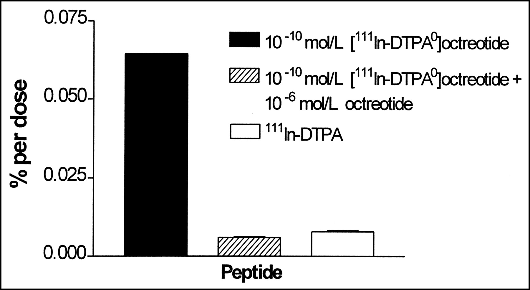
Figure 1 shows the internalization of [111In-DTPA0]octreotide and 111In-DTPA after a 1-h incubation. Cells were also incubated with an excess of octreotide (10−6 mol/L) to determine nonspecific internalization. The percentage of the dose that is internalized is approximately 0.06; the noninternalized radioactivity represented <10% of the total cellular uptake. Figure 1 shows that 111In-DTPA is much less internalized into the cells compared with that of [111In-DTPA0]octreotide.

Internalization in CA20948 cells after 1-h incubation at 37°C with [111In-DTPA0]octreotide and 111In-DTPA. Bars represent mean ± SEM.
To investigate the PRRT effects of the internalized radioactivity, and not of the radioactivity in the medium, a short incubation time in the medium and a low number of cells per well were necessary, the latter to rule out the option of crossfire between cells. In our PRRT studies with [111In-DTPA0]octreotide in vitro, we incubated 200 cells per well for 1 h. In this way the distance between the cells is 1.2 mm (the surface of a well in a 6-well plate is 9.62 cm2; 962 mm2/200 = 4.81 mm2 per cell; πr2 = 4.81; r2 = 1.5 mm; r = 1.2 mm); this is longer than the maximum pathlength of conversion electrons, which is 200–500 μm, which rules out the option of crossfire between the cells when distributed equally in the well.
The cells were incubated for 1 h with [111In-DTPA0]octreotide and after 3 d the medium was refreshed. We determined how much radioactivity is internalized in the cells directly after a 1-h incubation and also after 72 h in growth medium to be able to mimic the PRRT experiments with [111In-DTPA0]octreotide with a similar amount of 111In-DTPA in the medium.
To determine whether internalization of [111In-DTPA0]octreotide is dependent on the cell number in the wells we performed experiments using an increasing cell concentration (range, 200–10,000 cells per well). Figure 2A shows the amount of internalized counts per minute after 72 h with an increasing amount of [111In-DTPA0]octreotide (specific activity is kept constant). Figure 2A also demonstrates that when more cells are used, at constant radioactivity, more radioactivity is internalized. It also shows that when more radioactivity is used, at constant cell concentration, more radioactivity is internalized into the cells. In Figure 2B the amount of [111In-DTPA0]octreotide that is internalized into the cells is expressed as percentage of the dose. This shows that more or less the same percentage of the dose is internalized into the cells, which is approximately 0.003% of the dose for 200 cells per well. Directly after the 1-h incubation this percentage is higher—namely, 0.03% of the dose.
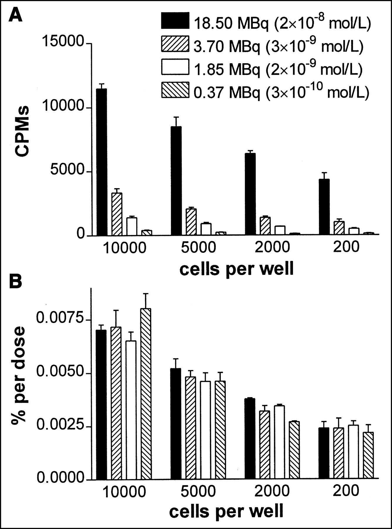
Amount of internalized [111In-DTPA0]octreotide after 72 h in growth medium with 200–10,000 cells per well. Cells were incubated for 1 h at 37°C with increasing concentrations of [111In-DTPA0]octreotide. Amount of internalized [111In-DTPA0]octreotide was determined after 72 h. (A) Amount of internalized counts per minute. (B) Internalized amount presented as percentage of given dose. Bars represent mean ± SEM.
PRRT In Vitro with [111In-DTPA0]Octreotide
Figure 3 shows the percentage of tumor cell survival of the CA20948 cell line after treatment with 3.7 MBq/10−8 mol/L (Fig. 3A), 3.7 MBq/10−7 mol/L (Fig. 3B), and 37 MBq/10−7 mol/L (Fig. 3C) [111In-DTPA0]octreotide for 1, 3, and 5 h. Survival values are shown as percentage survival compared with that of the control cells (no peptide added). Figures 3A and 3B share the same amount of radioactivity, but the latter has a 10 times higher peptide amount, so a lower specific activity. Figures 3B and 3C share the same amount of peptide, but the latter has a 10 times higher radioactivity, so a higher specific activity. Figures 3A and 3C share the same specific activity. When the tumor cell survival of Figures 3A and 3B is compared, Figure 3B shows a higher tumor cell survival than that of Figure 3A, despite the same amount of radioactivity that is used. Figure 3 also shows a time- and dose-dependent inhibition of the colony growth: When cells were incubated for 5 h with the highest concentration used, survival is virtually zero. Because the 1-h incubation time already shows a clear inhibition of the clonogenic cell survival we continued with a 1-h incubation time.
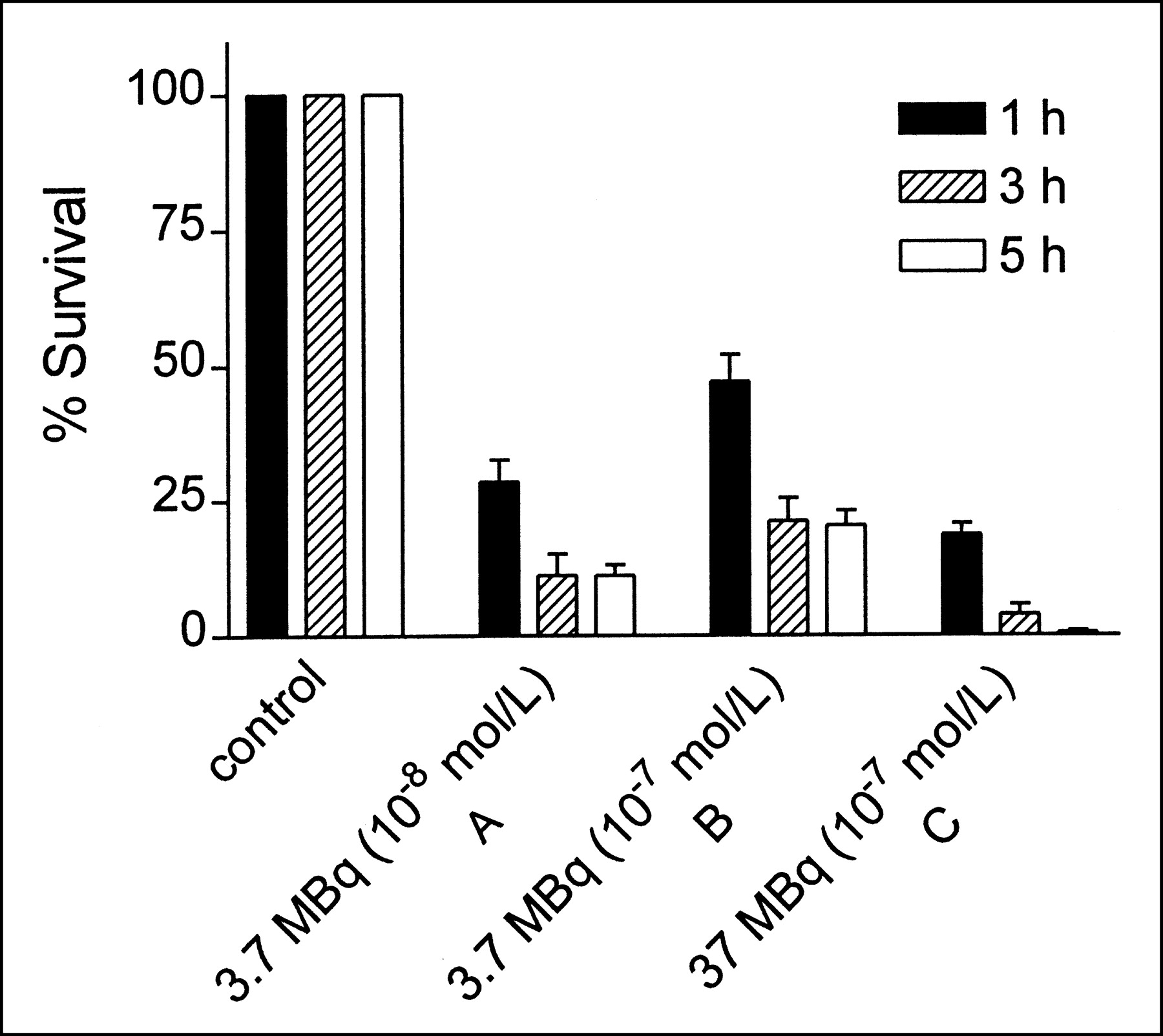
Effects of incubation time, concentration, and specific activity of [111In-DTPA0]octreotide on cell survival of CA20948 cells after PRRT. Cells were incubated for 1, 3, and 5 h with 0.37 or 3.7 MBq [111In-DTPA0]octreotide at 37°C using 2 different specific activities (specific activity of B is different from that of A and C). Bars represent mean ± SEM.
The same experiment was performed with a higher specific activity of [111In-DTPA0]octreotide, 370 MBq/0.5 μg. As is shown in Figure 4, CA20948 cells were incubated for 1 h with 18.5, 3.7, 1.85, and 0.37 MBq [111In-DTPA0]octreotide. The control cells received incubation medium without [111In-DTPA0]octreotide for 1 h. Figure 4 shows less tumor cell survival when a higher concentration of [111In-DTPA0]octreotide is used, indicating a clear dose-dependent relation.
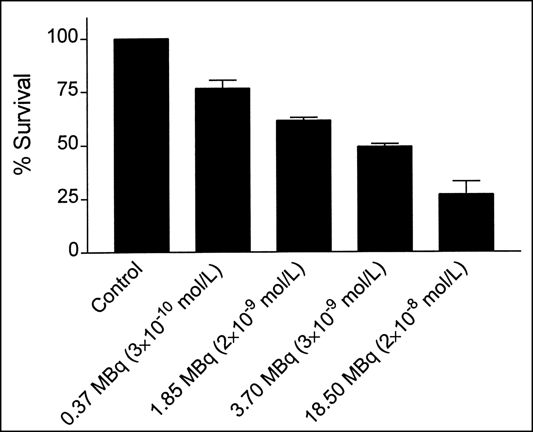
Inhibitory effect of [111In-DTPA0]octreotide on clonogenic cell survival of rat pancreatic tumor cell line CA20948. Cells were incubated for 1 h at 37°C with increasing amount of [111In-DTPA0]octreotide. Bars represent mean ± SEM.
Effects of 111In-DTPA and Low Amounts of Octreotide.
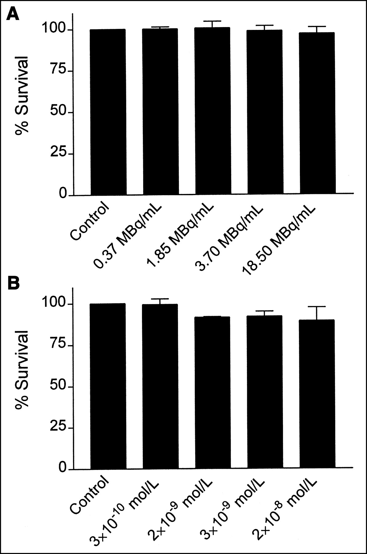
To investigate whether Auger or conversion electrons emitted by [111In-DTPA0]octreotide were responsible for the inhibitory effect, CA20948 cells were exposed to increasing concentrations of 111In-DTPA for 1 h, the same concentrations as used for [111In-DTPA0]octreotide (Fig. 4). Because 111In-DTPA is internalized (Fig. 1) to a much lesser extent than [111In-DTPA0]octreotide, the amount of radioactivity from [111In-DTPA0]octreotide that is normally internalized into the cells is added to the medium as 111In-DTPA after the 1-h incubation and also after 3 d, when the medium is normally refreshed (Fig. 2). Figure 5A clearly shows that there is no difference in tumor cell survival compared with the control, when CA20948 cells are incubated with an increasing amount of 111In-DTPA. Thus, 111In-DTPA does not have an effect on the tumor cell survival.

Effect of 111In-DTPA (A) and octreotide (B) on clonogenic cell survival of CA20948 cells. Cells were incubated for 1 h in increasing amount of 111In-DTPA (same amount was given as was used with [111In-DTPA]octreotide [Fig. 4]). Amount of radioactivity that is normally attached to cells was added to medium after 1-h incubation. 111In-DTPA was also added when medium was refreshed after 3 d. Bars represent mean ± SEM.
Because it is known that octreotide alone can have an inhibitory effect on the cell survival we also incubated the CA20948 cells with octreotide, again the same concentration that is used with [111In-DTPA0]octreotide (Fig. 4). Figure 3B shows that when CA20948 cells are incubated with low amounts of octreotide (10−10 mol/L to 10−8 mol/L) for 1 h there might be a slight inhibition of the tumor cell survival (≈5%).
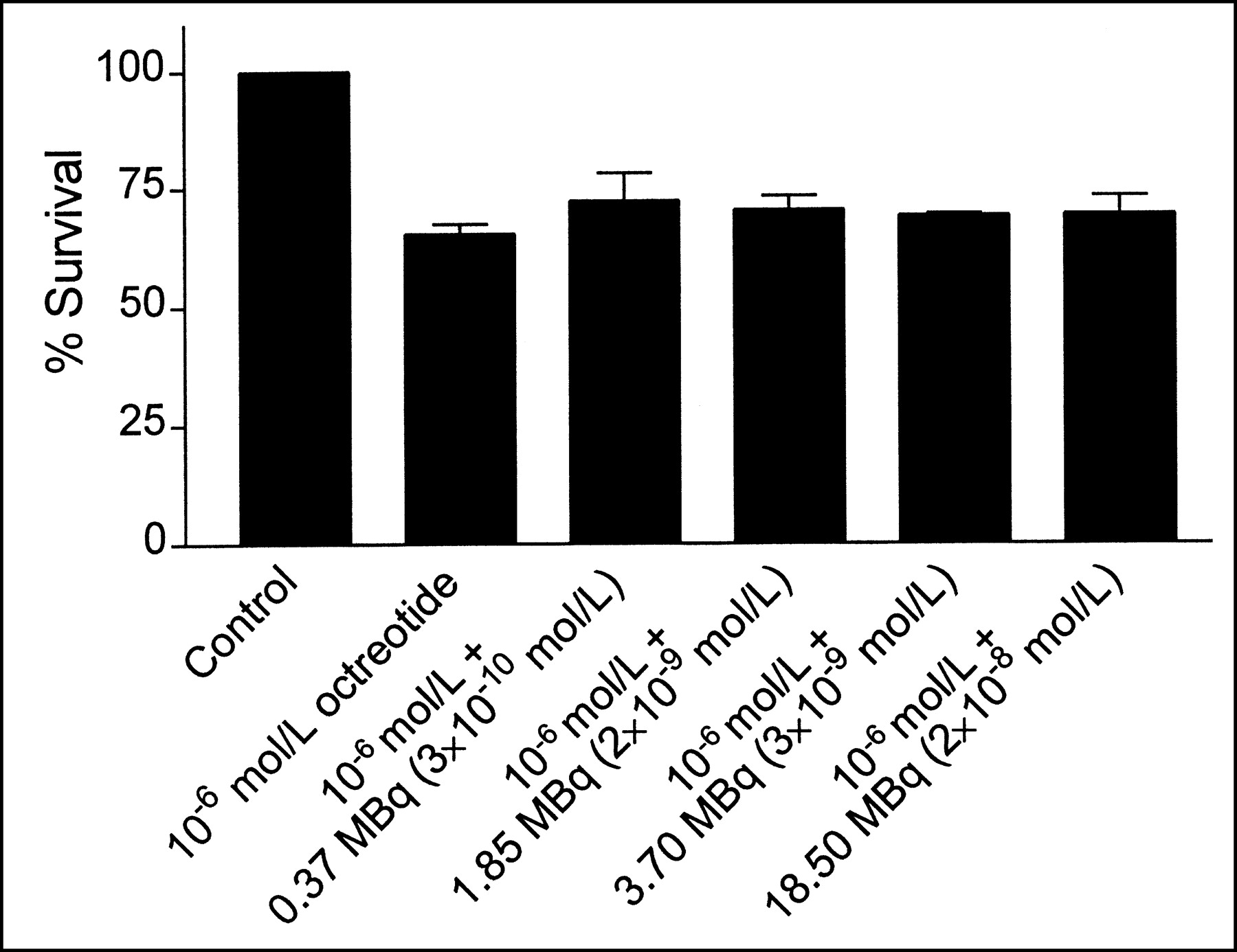
PRRT with [111In-DTPA0] Plus Excess Amount of Unlabeled Octreotide.
Figure 6 shows the effect of [111In-DTPA0]octreotide incubated together with an excess (10−6 mol/L) of unlabeled octreotide. The cells were incubated with the same amount of [111In-DTPA0]octreotide as used previously. In this way the receptors are occupied with octreotide and [111In-DTPA0]octreotide is not internalized into the cells. Figure 6 shows that a high amount of octreotide alone gives an inhibition of ≈30% on the tumor cell survival. When cells were incubated with 10−6 mol/L octreotide together with [111In-DTPA0]octreotide there is no further reduction of the tumor cell survival compared with only 10−6 mol/L octreotide.

Inhibitory effect of octreotide on tumor cell survival of CA20948 cell line. Cells were incubated for 1 h at 37°C with 10−6 mol/L octreotide (second bar from left) and cells were incubated with 10−6 mol/L octreotide plus increasing amount of [111In-DTPA]octreotide (third to fifth bars from left). Bars represent mean ± SEM.
DISCUSSION
Peptide receptor scintigraphy with the radioactive somatostatin analog [111In-DTPA0]octreotide is widely used to visualize sst-positive tumors in vivo. Besides γ-radiation, 111In emits also both therapeutic Auger electrons and internal conversion electrons; therefore, a new application of [111In-DTPA0]octreotide is PRRT.
In preclinical studies on rats, different experiments with [111In-DTPA0]octreotide were performed by determining the response of a solid octreotide receptor-positive tumor (CA20948) inoculated in the flank (13). For [111In-DTPA0]octreotide a dose–response was found, leading in rats bearing small tumors (<1 cm2) to 50% cure after the highest dose (3 injections of 370 MBq, given with an interval of 1 wk), whereas in rats bearing large tumors (>10 cm2) only a partial response could be achieved. In larger tumors, more clonogenic, presumably hypoxic, cells will be present, thereby limiting radiocurability. So, 111In is more suitable for smaller rather than large tumors. This is in agreement with the antiproliferative effects of [111In-DTPA0]octreotide found in a rat liver tumor metastases model (14). Administration of 370 MBq [111In-DTPA0]octreotide on day 1 or day 8 after intraportal CA20948 tumor cell inoculation induced a significant decrease in the number of hepatic metastases at day 21. These findings show that, after radionuclide therapy, reduction of tumor volume can be obtained because of the radiotherapeutic effect of 111In-octreotide. These findings hold promise for the application of radionuclide therapy with 111In-octreotide in an adjuvant, micrometastatic setting and are consistent with the findings in this study. We showed that [111In-DTPA0]octreotide is able to control tumor growth in our single-cell in vitro system of CA20948 cells. Apart from uptake, tumor response is also dependent on radiosensitivity. To determine the radiosensitivity of these tumor cells, we performed external beam radiation experiments. It appeared that CA20948 cells are relatively radioresistant, making our results more interesting.
In this in vitro system we also demonstrated that the Auger electrons emitted by 111In are responsible for the tumor response, not the conversion electrons. Because we use only 200 cells per well in our in vitro system the distance between the cells is 1.2 mm, which rules out the option of crossfire between the cells because the cells are approximately equally distributed in the well.
No tumor cell survival was found after a 5-h incubation with 37 MBq (coupled to 0.15 μg peptide) [111In-DTPA0]octreotide, whereas we found almost 50% survival with 3.7 MBq (coupled to 0.15 μg peptide) [111In-DTPA0]octreotide with a 1-h incubation. When a higher specific activity was used there was less tumor cell survival (Fig. 3). With a higher specific activity more radioactivity is internalized into the cells, leading to a lower tumor cell survival (24). Thus, the differences in tumor cell survival are dependent on the radiation dose, incubation time, and specific activity that are used. Because we found a clear effect of [111In-DTPA0]octreotide on the tumor cell survival after a 1-h incubation, we continued with this short incubation time to investigate the effects of the internalized radioactivity, not of the radioactivity in the medium.
Because 111In emits both Auger and conversion electrons we discriminated between the effects of both electrons by comparing internalized ([111In-DTPA0]octreotide) versus noninternalized (111In-DTPA) 111In. The effects of the conversion electrons were investigated using 111In-DTPA in the medium; with a tissue penetration of 200–500 μm they could reach the nucleus without being internalized. The results showed a 100% survival in the number of colonies after incubation for 1 h with 18.5 MBq with 111In-DTPA (Fig. 5A) compared with a 30% survival with 18.5 MBq [111In-DTPA0]octreotide (Fig. 4). McLean and Wilkinson (25) determined the radiation dose to cells in vitro from intracellular 111In. In these experiments, the radiation dose to the cell from the intranuclear decay of 111In was determined from cell survival studies to be 3.5 mGy per decay, using 60Co as a reference radiation. The average dose to the cells from extracellular 111In was calculated to be much lower—that is, 5.8 pGy per decay. So from 111In that remains extracellular, Auger electrons, with their short pathlengths, would have little biologic effect. For intracellular 111In, however, the release of Auger electrons into the nucleus or the DNA would have the potential to cause extensive biologic damage.
CA20948 cells were incubated with an excess of octreotide (10−6 mol/L) together with an increasing amount of [111In-DTPA0]octreotide in 1 experiment. The excess of octreotide was used to inhibit the internalization of [111In-DTPA0]octreotide. The results show that the excess of octreotide inhibited cell survival by 30%; addition of radiolabeled peptide ([111In-DTPA0]octreotide [10−10 mol/L] + 10−6 mol/L octreotide) did not further decrease survival, showing that PRRT effects were sst receptor mediated.
A phase I study using this radioligand on patients with neuroendocrine tumor has started in our center. Fifty patients with somatostatin receptor-positive tumors were treated with multiple doses of [111In-DTPA0]octreotide (26). Forty patients were evaluable after cumulative doses of at least 20 GBq up to 160 GBq. Therapeutic effects were seen in 21 patients: partial remission in 1 patient, minor remissions in 6 patients, and stabilization of previously progressive tumors in 14 patients. All patients in this study had advanced tumor, and many were end-stage patients with a large tumor burden.
Anthony et al. (27) reported on a phase II trial on patients with gastroenteropancreatic tumors in which 2 doses (6.66 GBq each) of [111In-DTPA0]octreotide were given. It improved symptoms in 62% of the patients, decreased hormonal markers with 81%, decreased Hounsfield units on CT scans in 27% of patients, with 8% partial radiographic responses and increased expected survival in gastroenteropancreatic cancer patients with somatostatin receptor-expressing tumors, showing the promise of this compound in patients with neuroendocrine tumors.
PRRT with β-emitting radionuclides, such as 90Y and 177Lu, can be even more effective because higher tumor radiation doses can be achieved and the longer range of the β-particles, typically 1–10 mm, may cause irradiation of neighboring receptor-negative tumor cells by crossfire. We are currently performing experiments with 90Y- and 177Lu- labeled somatostatin analogs in this in vitro system. In micrometastases or even single tumor cells the absorbed fraction of the radiation energy will be very low, especially for 90Y. Recently, Bernhardt et al. (28) have calculated with a mathematic model the tumor-to-normal tissue mean absorbed dose ratios (TNDs) for different tumor sizes. The results show that radionuclides suitable for therapy should emit few photons, emit many electrons with moderate energy, and have a rather long half-live (>2 d). In these calculations 161Tb and 177Lu obtained the highest TNDs. We had promising results with biodistribution studies in rats after administration of [161Tb-DTPA]octreotide (29). Both of these radionuclides will be of interest for somatostatin receptor–based therapy in the future.
However, in very small lesions PRRT with Auger electron–emitting radiopharmaceuticals may be the best choice. Recently, it was shown that after internalization 111In is translocated to the perinuclear area and into the nucleus (30); it was also shown that prolonged exposure to radiolabeled somatostatin analogs significantly increases their cellular internalization, nuclear translocation, and DNA binding (31). Behr et al. (32) demonstrated a therapeutic advantage of the Auger electron–emitting 111In over the β-emitter 90Y. It was shown that internalizing antibodies labeled with Auger electron–emitting radionuclides, such as 125I and 111In, have encouraging therapeutic results. They cause biologic effects similar to those of typical high linear-energy-transfer (LET) radiation, such as α-emitters. Auger electron emitters decaying in the neighborhood of the DNA produce similar amounts of reactive chemical radical species as do α-emitters, which are regarded as the classical form of high LET radiation (32). So it is conceivable to use [111In-DTPA0]octreotide, or other low-energy emitters such as 161Tb, 58mCo, 103mRh, 119Sb, and 189mOs, in cocktails combined with β-emitting radionuclides as a neoadjuvant therapy.
CONCLUSION
[111In-DTPA0]octreotide is able to control tumor growth in this in vitro system; the effects were dependent on radiation dose, incubation time, and specific activity used. An excess of unlabeled octreotide decreased survival to 60% of the control; addition of radiolabeled peptide did not further decrease survival, showing that PRRT was receptor mediated. 111In-DTPA, which is not internalized into sst2-positive tumor cells like [111In-DTPA0]octreotide, did not influence survival, showing that the therapeutic effect of 111In is dependent on internalization, enabling the Auger electrons, with their very short particle range, to reach the nucleus.
Footnotes
Received Mar. 18, 2002; revision accepted Jul. 26, 2002.
For correspondence or reprints contact: Astrid Capello, MD, Department of Nuclear Medicine, Erasmus Medical Center, 3015 GD Rotterdam, The Netherlands.
E-mail: capello{at}nuge.azr.nl
REFERENCES
In this issue


Jump to section
Related Articles
Cited By...
- Dosimetry in Peptide Radionuclide Receptor Therapy: A Review
- Anticancer Activity of Targeted Proapoptotic Peptides
- Trifunctional Somatostatin-Based Derivatives Designed for Targeted Radiotherapy Using Auger Electron Emitters
- Auger Electrons: Lethal, Low Energy, and Coming Soon to a Tumor Cell Nucleus Near You
- Increased Cell Death After Therapy with an Arg-Gly-Asp-Linked Somatostatin Analog