


Abstract
Receptor-targeted scintigraphy and radionuclide therapy with radiolabeled somatostatin analogs are successfully applied for somatostatin receptor-positive tumors. The synergistic effects of an apoptosis-inducing factor, for example, the Arg-Gly-Asp (RGD) motif, can increase the radiotherapeutic efficacy of these peptides. Hence, the tumoricidal effects of the hybrid peptide RGD-diethylaminetriaminepentaacetic acid (DTPA)-Tyr3-octreotate (cyclic[c](Arg-Gly-Asp-d-Tyr-Asp)-Lys(DTPA)-d-Phe-c(Cys-Tyr-d-Trp-Lys-Thr-Cys)-Thr), hereafter referred to as RGD-DTPA-octreotate, were evaluated in comparison with those of RGD (c(Arg-Gly-Asp-d-Tyr-Asp)) and Tyr3-octreotate (d-Phe-c(Cys-Tyr-d-Trp-Lys-Thr-Cys)-Thr). Methods: The therapeutic effects of RGD-111In-DTPA-octreotate, 111In-DTPA-RGD, and 111In-DTPA-Tyr3-octreotate were investigated with various cell lines by use of a colony-forming assay, and caspase-3 activity was also determined. Results: Tumoricidal effects were found with 111In-DTPA-RGD, 111In-DTPA-Tyr3-octreotate, and RGD-111In-DTPA-octreotate, in order from least effective to most effective. Also, the largest increase in caspase-3 levels was found with RGD-111In-DTPA-octreotate. Conclusion: RGD-111In-DTPA-octreotate has more pronounced tumoricidal effects than 111In-DTPA-RGD and 111In-DTPA-Tyr3-octreotate, because of increased apoptosis, as indicated by increased caspase-3 activity.
The 111In-labeled somatostatin analog octreotide has proven to be particularly useful in the diagnosis of neuroendocrine tumors that overexpress somatostatin receptors (1). Somatostatin receptor–positive neuroendocrine tumors are in general unresponsive to standard therapy regimens (2). Therefore, a variety of radiolabeled somatostatin derivatives have been prepared for radionuclide therapy purposes and are in various stages of preclinical and clinical investigations; these include 90Y-labeled 1,4,7,10-tetraazacyclododecane-N,N′,N″,N‴-tetraacetic acid (DOTA)-Tyr3-octreotide and 177Lu-labeled DOTA-Tyr3-octreotate.
Although the results of these studies are very promising, we hypothesize that the synergistic effects of an apoptosis-inducing factor, such as the Arg-Gly-Asp (RGD) motif, can increase the radiotherapeutic efficacy of these peptides. The RGD sequence is well known as a major integrin-binding site, such as the αvβ3-integrin receptor. In addition, Buckley et al. (3) demonstrated that RGD peptides are able to directly activate caspase-3 and induce apoptosis. Other work has shown that molecules specific for the GPIIb/IIIa integrins can also stimulate caspase-3 activity (4). Since caspase-3 is one of the key executioner proteases (5) in the apoptosis pathway, it seems likely that this enzyme will be an important site of action for targeted therapeutics that are designed to selectively induce cell death.
To combine these characteristics in one compound, a new peptide, RGD-diethylaminetriaminepentaacetic acid (DTPA)-octreotate (cyclic[c](Arg-Gly-Asp-d-Tyr-Asp)-Lys(DTPA)-d-Phe-c(Cys-Tyr-d-Trp-Lys-Thr-Cys)-Thr) (Fig. 1), was synthesized. This radiopharmaceutical consists of a somatostatin receptor–targeting peptide, Tyr3-octreotate, the chelator DTPA to enable radiolabeling with, for example, 111In, and an RGD peptide moiety. Recently, Bernard et al. reported the synthesis and characterization of this hybrid peptide in vitro and in vivo (6). RGD-DTPA-octreotate enables rapid and high-specific-activity labeling with 111In. The hybrid peptide retains an affinity for 2 receptors; octreotate binds with a high affinity to the somatostatin receptor subtype 2 (sst2 receptor), and RGD binds to the αvβ3-integrin receptor.
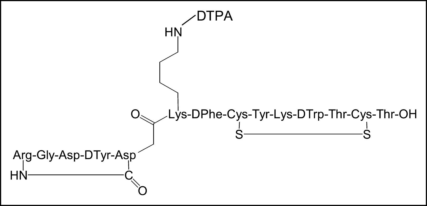
Structure of RGD-DTPA-octreotate.
In this study, we investigated the therapeutic effects of the 111In-labeled hybrid peptide in comparison with those of 111In-RGD (c(Arg-Gly-Asp-d-Tyr-Asp)) and Tyr3-octreotate (d-Phe-c(Cys-Tyr-d-Trp-Lys-Thr-Cys)-Thr) by using an in vitro model (7). We also determined caspase-3 activity after incubation with the 3 peptides for several cell lines.
MATERIALS AND METHODS
Peptides
RGD (c(Arg-Gly-Asp-d-Phe-Val)) was obtained from Bachem. RGD-DTPA-octreotate (Fig. 1), obtained from A. Srinivasan (Mallinckrodt), was synthesized as described previously (6). DTPA-RGD (c(Arg-Gly-Asp-d-Tyr-Lys)-ε-DTPA) and DTPA-Tyr3-octreotate (DTPA-d-Phe-c(Cys-Tyr-d-Trp-Lys-Thr-Cys)-Thr) were synthesized as described previously (8,9).
Radiochemical Analysis
111InCl3 was obtained from Mallinckrodt Medical BV. RGD-DTPA-octreotate, DTPA-Tyr3-octreotate, and DTPA-RGD were labeled as described previously (10) with 111InCl3 at a specific activity of 130 MBq per microgram of peptide. Peptides with more than 99% labeling efficiency and more than 90% radiochemical purity were used.
Cell Culture
CA20948 cells were grown in Dulbecco’s modified Eagle’s medium (Gibco BRL). The medium was supplemented with 10% heat-inactivated fetal bovine serum, glutamine at 2 mmol/L, sodium pyruvate at 1 mmol/L, amphotericin B (Fungizone; Gibco, Invitrogen) at 0.1 mg/L, and penicillin–streptomycin at 50 IU/mL. Chinese hamster ovary (CHO) cells transfected with the sst2 receptor (provided by J.E. Bugaj) were grown in RPMI 1640 medium (Life Technologies) supplemented with 5% heat-inactivated fetal bovine serum, glutamine at 2 mmol/L, sodium pyruvate at 1 mmol/L, amphotericin B at 0.1 mg/L, penicillin–streptomycin at 50 IU/mL, and 1:1,000 (v/v) gentamicin (Gibco BRL).
Peptide Receptor Radionuclide Therapy In Vitro
One day before the start of the experiment, cells were transferred to 6-well plates at a density of 200 or 400 cells per well. Cells were washed with phosphate-buffered saline at 37°C and incubated for 1 h in internalization medium—RPMI 1640 medium without fetal bovine serum but with 1% bovine serum albumin and 20 mmol of N-(2-hydroxyethyl)piperazine-N′-(2-ethanesulfonic acid)—containing increasing concentrations of RGD-111In-DTPA-octreotate, 111In-DTPA-RGD, or 111In-DTPA-Tyr3-octreotate. In additional experiments performed with CA20948 cells, RGD-111In-DTPA-octreotate and 111In-DTPA-Tyr3-octreotate were coincubated with an excess (10−6 mol/L) of unlabeled RGD. Control cells received only internalization medium for 1 h. Thereafter, cells were thoroughly washed with phosphate-buffered saline and allowed to form colonies over 12 d in medium. The medium was refreshed once after 3 d.
After 12 d, the cells were fixed with methanol:glacial acetic acid (3:1) and stained with hematoxylin. Colonies that contained more than 50 cells were visually scored as survivors. Experiments were performed in triplicate for each cell density.
Caspase-3 Assay
Cell extracts were prepared and caspase-3 activity was determined according to the package insert for the CASPASE-3 cellular activity kit of the BIOMOL QuantiZyme assay system (SanverTECH).
Cells were treated with 18.5 MBq of RGD-111In-DTPA-octreotate, 111In-DTPA-octreotate, or 111In-DTPA-RGD for 3 h. Caspase-3 activity was determined either directly after the incubation period or after 24 h of recovery. For preparation of the cell extracts, cells were harvested by trypsin treatment and then lysed with the lysis buffer provided in the kit. The assay is based on the cleavage of the substrate Asp-Glu-Val-Asp (DEVD) bound to the chromophore p-nitroanilide, which is measured by reading the absorbance of the samples at a wavelength of 405 nm (microplate reader; Bio-Rad).
Statistical Analysis
Statistical analysis was performed for the clonogenic survival data. Multiple-comparison analysis was performed for each concentration group by use of ANOVA and the Bonferroni correction.
RESULTS
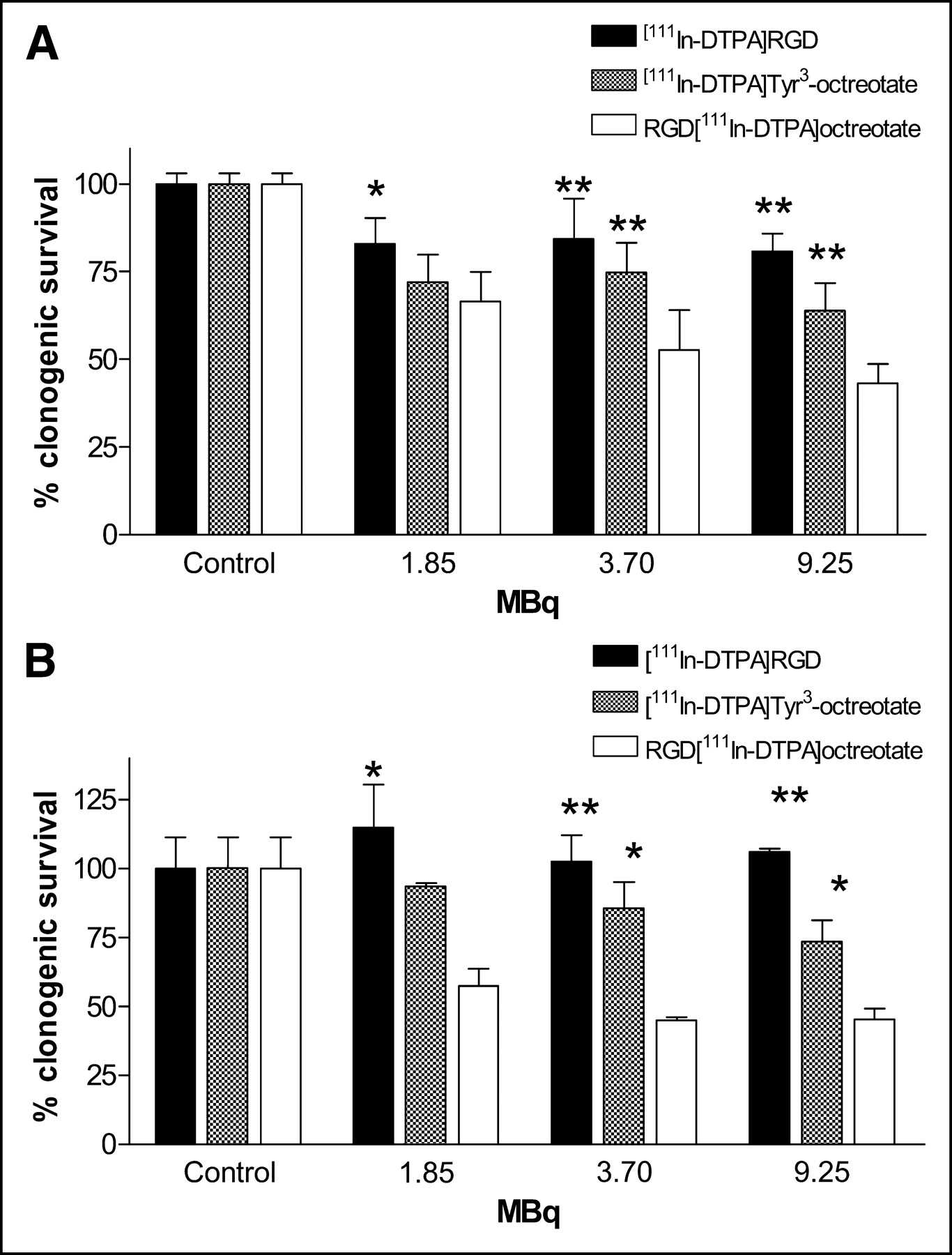
Figure 2 shows the clonogenic survival after peptide receptor radionuclide therapy studies in vitro. CA20948 cells (Fig. 2A) and CHO cells transfected with the sst2 receptor (Fig. 2B) were incubated for 1 h with the 3 different peptides—the hybrid peptide RGD-111In-DTPA-octreotate and the monopeptides 111In-DTPA-octreotate and 111In-DTPA-RGD. Survival values are shown as percentages of survival of treated cells compared with control cells. Experiments were performed with 200 or 400 cells per well, but because both cell densities showed the same results, only the results obtained with 200 cells per well are shown. Figure 2 clearly shows for both cell lines that increasing concentrations of 111In-DTPA-RGD had no effect on tumor cell survival, that 111In-DTPA-Tyr3-octreotate had a greater tumoricidal effect, but that the most pronounced tumoricidal effect was achieved with the hybrid peptide RGD-111In-DTPA-octreotate. As is also shown in Figure 2, a radioactivity dose dependence was seen for RGD-111In-DTPA-octreotate; the higher the administered dose, the lower the tumor cell survival. For the 2 highest concentrations (3.70 and 9.25 MBq), the effects of the hybrid peptide were significantly different from those of the 2 monopeptides (P < 0.01 and P < 0.05 for CA20948 cells and sst2 receptor-positive CHO cells, respectively).

Clonogenic tumoricidal effect of 111In-labeled compounds RGD, octreotate, and RGD-DTPA-octreotate in rat pancreatic cell line CA20948 (A) and sst2 receptor-positive CHO cells (B). Cells were incubated for 1 h with increasing concentrations of 3 peptides. Experiments were performed 2–4 times in triplicate; bars represent mean ± SEM. Single and double asterisks indicate P values of <0.05 and <0.01 for comparisons with RGD-111In-DTPA-octreotate within a concentration group.
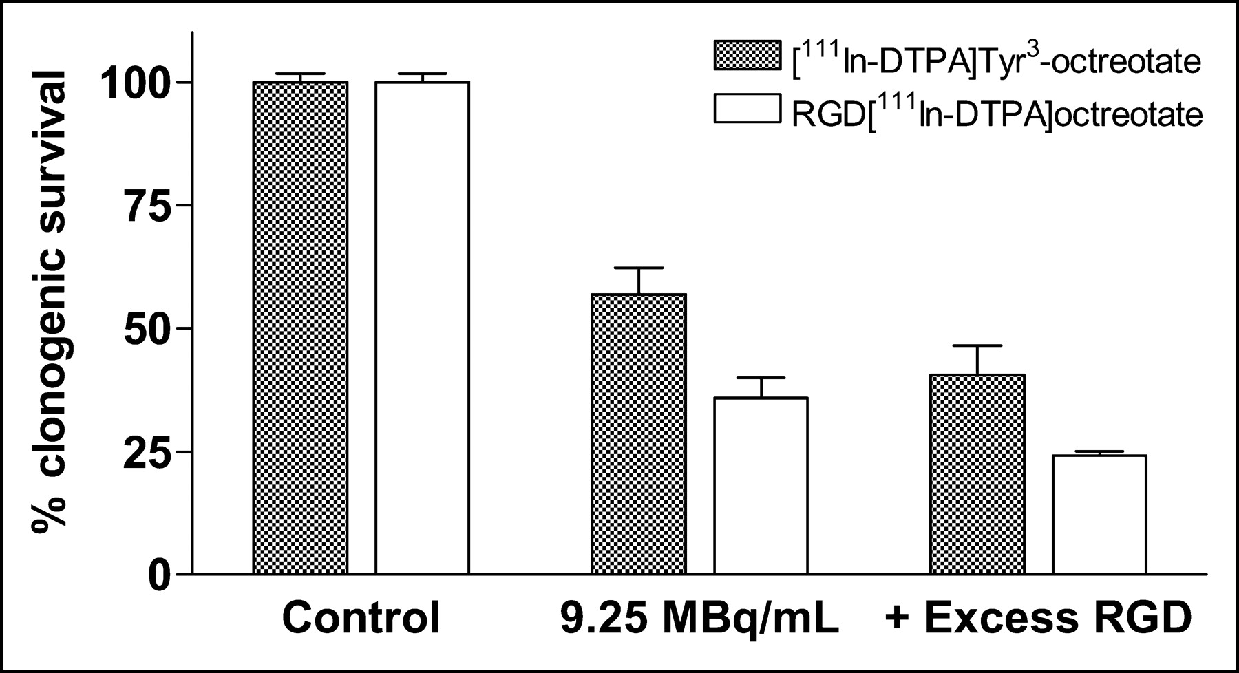
When the cells were coincubated with an excess of unlabeled RGD (10−6 mol/L), tumor cell survival decreased further (Fig. 3). This effect was seen when the cells were incubated with 111In-DTPA-Tyr3-octreotate but also when the cells were incubated with RGD-111In-DTPA-octreotate. The effects of the hybrid peptide were significantly different from those of 111In-DTPA-Tyr3-octreotate (P < 0.05), and the effects of the hybrid peptide with an excess of unlabeled RGD were significantly different from those of 111In-DTPA-Tyr3-octreotate with an excess of unlabeled RGD (P < 0.05).

Clonogenic tumor cell survival after 1 h of incubation of cell line CA20948 with no addition (control), with 111In-DTPA-Tyr3-octreotate or RGD-111In-DTPA-octreotate at 9.25 MBq/mL, or with either of those peptides with or without excess unlabeled RGD (10−6 mol/L). Experiments were performed 2 times in triplicate; bars represent mean ± SEM. Results for all peptides were significantly different from those for controls.
To investigate the mechanism of action of the hybrid peptide, we investigated possible caspase-3 activation. Figure 4 shows the caspase-3 activity after treatment with 111In-RGD, 111In-octreotate, and 111In-RGD-octreotate. Figure 4A shows the caspase-3 activity measured directly after the incubation period, and Figure 4B shows the caspase-3 activity measured after 24 h. The highest caspase-3 activity was seen for the hybrid peptide RGD-111In-DTPA-octreotate, and the lowest was seen for 111In-DTPA-RGD. The caspase-3 activity measured after 24 h was higher (Fig. 4B) than that measured directly after incubation (Fig. 4A). When the cells were incubated with the caspase-3 inhibitor N-acetyl (Ac)-DEVD-CHO (aldehyde), the caspase-3 levels remained at control levels.
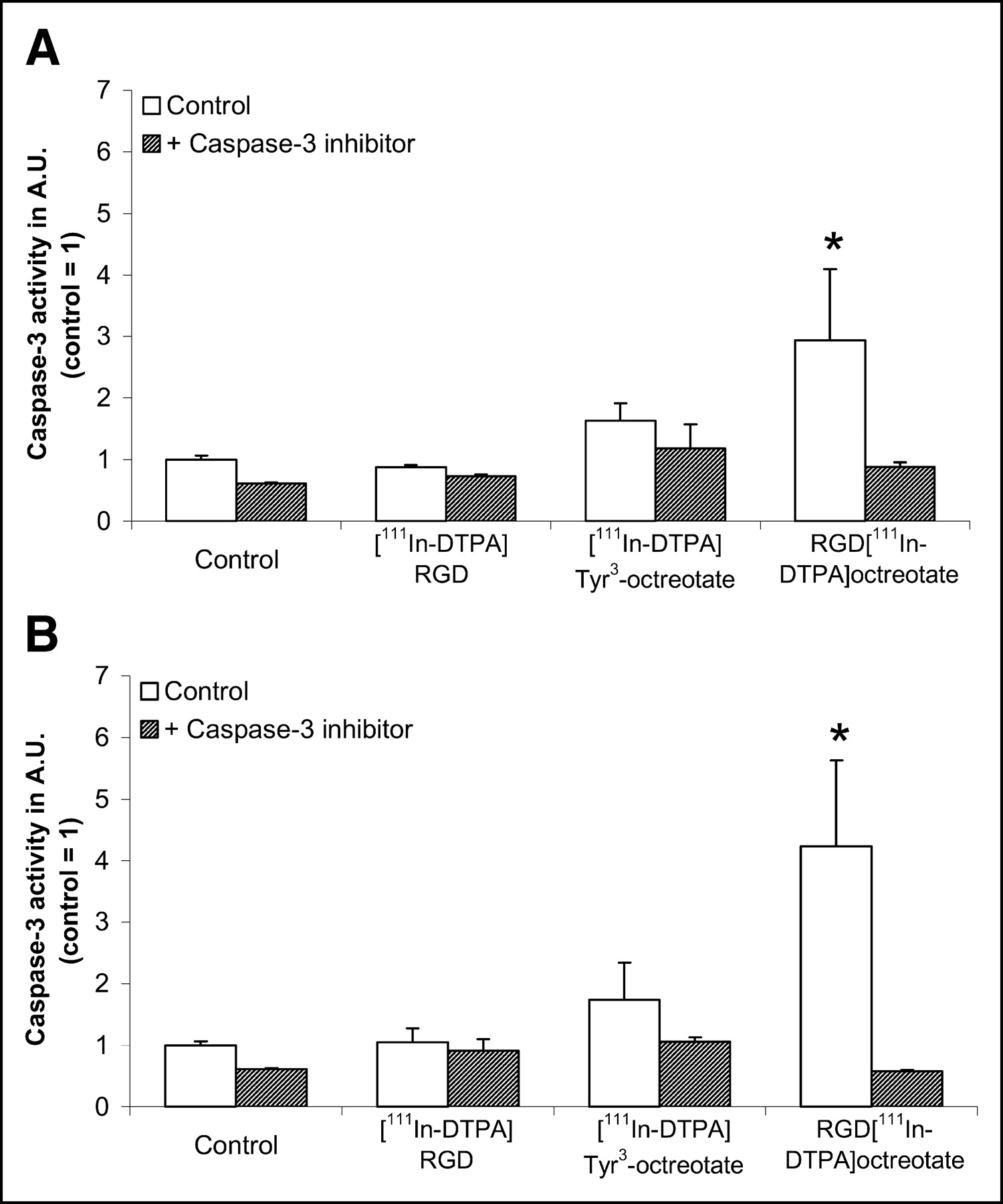
Caspase-3 activity after 3 h of incubation with 111In-RGD, Tyr3-octreotate, or RGD-DTPA-octreotate in cell line CA20948. Caspase-3 activity was measured directly after incubation (A) and after 24 h (B) and was expressed as a fraction of activity in control cells (A.U., arbitrary units). Treatment with a known inhibitor of caspase-3 (Ac-DEVD-CHO) was used to determine assay specificity. Experiments were performed in triplicate, and each sample was measured in triplicate; bars represent mean ± SEM. An asterisk indicates a P value of <0.05 for comparisons with controls.
DISCUSSION
We examined the therapeutic potential of a 111In-labeled hybrid peptide, an RGD peptide coupled to the somatostatin analog Tyr3-octreotate (RGD-111In-DTPA-octreotate). In a previous study (6), it was shown that this hybrid peptide exhibited high uptake and rapid, receptor-specific, time- and temperature-dependent internalization in sst2 receptor-positive CA20948 rat pancreatic tumor cells. Uptake and internalization of RGD-111In-DTPA-Tyr3-octreotate could be decreased almost completely with an excess of octreotide (10−6 mol/L) but not with an excess of RGD (10−6 mol/L). Therefore, internalization occurred mainly via the sst2 receptor (6). These findings might be explained by the somewhat higher affinity of Tyr3-octreotate for its receptor than of RGD for the αvβ3-integrin receptor. Another explanation, however, could be the difference in the rates of internalization of the receptors, on the basis of the differences in the biologic roles of the integrin receptors and the somatostatin receptors. Furthermore, a difference in receptor densities could contribute to the findings. Biodistribution studies with rats revealed high tumor uptake of RGD-111In-DTPA-octreotate in tumor-bearing rats. The level of retention of radioactivity for RGD-111In-DTPA-octreotate was at least as high as that for Tyr3-octreotate.
From these in vitro and in vivo studies, it can be concluded that octreotate can serve as a carrier for RGD internalization (6). In this study, it was shown that for both cell lines, the greatest tumoricidal effect was reached when the cells were incubated with the hybrid peptide RGD-111In-DTPA-octreotate. Also, a clear dose response was observed with the hybrid peptide, whereas 111In-RGD alone had almost no effect on clonogenic tumor cell survival and 111In-DTPA-Tyr3-octreotate had a less pronounced tumoricidal effect.
An excess of unlabeled RGD (10−6 mol/L) could further decrease tumor cell survival when the cells were incubated with 111In-DTPA-Tyr3-octreotate or RGD-111In-DTPA-octreotate. However, the tumoricidal effect of RGD-111In-DTPA-octreotate alone was still greater than that of 111In-DTPA-octreotate with an excess of unlabeled RGD. The further decrease in tumor cell survival seen with unlabeled RGD could be explained by the fact that RGD itself is also internalized (9). Because a much higher concentration of the unlabeled peptide than of the other radiotracers was used, RGD itself could have activated caspase-3 and thereby induced apoptosis, leading to more tumor killing.
According to Buckley et al. (3), RGD peptides are able to activate caspase-3 inside cells. Because caspase-3 is an important executioner caspase in the apoptosis pathway, this enzyme will be an important site of action for targeted therapeutics to selectively induce cell death. We therefore investigated whether RGD-111In-DTPA-octreotate indeed activated caspase-3.
Buckley et al. (3) showed a clear increase in caspase-3 levels after incubation periods of 2–24 h. We therefore incubated cells in the caspase-3 assay for 3 h instead of the 1 h used in the colony-forming assay. Because a much larger cell number (>106 cells) was used for the caspase-3 assay than for the clonogenic assay (200–400 cells), we doubled the amount of the peptide and the radioactivity administered. The same trend was found with 9.25 MBq per well. In this study, the effects on caspase-3 activity were studied with the CA20948 cell line. 111In-DTPA-RGD and 111In-DTPA-Tyr3-octreotate induced no significant increase in caspase-3 levels, in contrast to the hybrid peptide RGD-111In-DTPA-octreotate (P < 0.05). Samples treated with a caspase-3 inhibitor showed no increase in caspase-3 levels, a result that indicates the specificity of the assay. These findings are consistent with the results obtained in the colony-forming assay, as described above. The effects were reached with concentrations of RGD peptide lower than those used by Buckley et al. (3); they demonstrated that caspase-3 induction could be achieved with peptide concentrations between 0.1 and 1 mmol/L. Our results at lower peptide concentrations can be explained by the fact that cyclic derivatives of RGD peptides inhibit cell adhesion to vitronectin more than 100-fold more effectively than the linear variants (11). In this study, we used cyclic peptides; Buckley et al. used linear peptides (3). Furthermore, internalization studies showed that approximately 0.9% of 111In-DTPA-RGD (9) and 15.7% of RGD-DTPA-octreotate (6) were internalized after 60 min in the CA20948 cell line. Biodistribution studies in vivo showed that the uptake of 111In-DTPA-RGD after 60 min in CA20948 tumors in rats was 0.18% (9). The uptake of RGD-111In-DTPA-octreotate was 1.9%, comparable to the uptake of 111In-Tyr3-octreotate (6).
From this study, we conclude that 111In-RGD-DTPA-octreotate promotes apoptosis via an increase in caspase-3 levels. The 111In-labeled hybrid peptide therefore can significantly enhance the therapeutic efficacy of somatostatin-based agents.
Also very interesting is the possible use of the unlabeled hybrid peptide RGD-DTPA-octreotate for adjuvant therapy. Preliminary data showed that the unlabeled compound RGD-DTPA-octreotate also induced an increase in caspase-3 levels. The largest increase (5.3 times that in the control) in caspase-3 levels with unlabeled RGD-DTPA-octreotate (10−6 mol/L) was found after 24 h of incubation. RGD and Tyr3-octreotate induced no increase in caspase-3 levels (A. Capello, unpublished data, 2004).
RGD compounds have been shown to inhibit angiogenesis because of upregulation of αvβ3-integrin. Preclinical studies found that several RGD peptidomimetic agents and a monoclonal antibody to αvβ3-integrin can inhibit tumor growth by blocking tumor angiogenesis (12). Neovascular endothelium expresses both sst2 and αvβ3-integrin receptors; these data make RGD-octreotate a candidate for the treatment of neovascular disease.
Another promising application is the coupling of RGD peptides to other peptides, for instance, bombesin, neurotensin, cholecystokinin, and gastrin, to increase the therapeutic effects of the peptides. Bombesin is a neuropeptide with a high affinity for the gastrin-releasing peptide receptor; this receptor is expressed on a variety of tumors, including prostate cancer and breast cancer (13,14). Neurotensin receptors, on the other hand, are overexpressed in exocrine pancreatic cancer and Ewing’s sarcoma (15). Cholecystokinin B receptors are frequently expressed on medullary thyroid carcinomas, small-cell lung cancers, astrocytomas, stromal ovarian tumors, and some gastroenteropancreatic tumors (16,17).
CONCLUSION
Coupling RGD peptides to somatostatin analogs can increase the therapeutic potential of these peptides.
Acknowledgments
We thank Dr. Ananthachari Srinivasan for expertise and for kindly providing the hybrid peptide.
Footnotes
Received Jan. 5, 2004; revision accepted Jun. 29, 2004.
For correspondence or reprints contact: Astrid Capello, Department of Nuclear Medicine, Erasmus Medical Center, Dr. Molewaterplein 40, 3015 GD Rotterdam, The Netherlands.
E-mail: a.capello{at}erasmusmc.nl


