


Abstract
The 14-amino-acid peptide bombesin (BN) has a high affinity for the gastrin-releasing peptide (GRP) receptor that is expressed by a variety of tumors. Recently, high densities of GRP receptors were identified by in vitro receptor autoradiography in human prostate and breast carcinomas using [125I-Tyr4]BN as radioligand. Radiometal-labeled diethylenetriaminepentaacetic acid (DTPA)-BN derivatives are potentially useful radioligands for receptor-targeted scintigraphy and radiotherapy of GRP receptor-expressing tumors. Methods: [DTPA-Pro1,Tyr4]BN (A), [DOTA-Pro1,Tyr4]BN (B), [DTPA-ε-Lys3,Tyr4]BN (C), and [DOTA-ε-Lys3,Tyr4]BN (D) (where DOTA is dodecanetetraacetic acid) were synthesized and studied for competition with binding of [125I-Tyr4]BN to the GRP receptor. The 111In-labeled BN analogs were studied in vitro for binding and internalization by GRP receptor-expressing CA20948 and AR42J pancreatic tumor cells as well as in vivo for tissue distribution in rats. Specific tissue binding was tested by coinjection of 0.1 mg [Tyr4]BN. Results: All BN analogs competitively inhibited the binding of [125I-Tyr4]BN to the GRP receptor with 50% inhibitory concentration values in the range of 2–9 nmol/L. All 111In-labeled analogs showed high and specific time- and temperature-dependent binding and internalization by CA20948 and AR42J cells. In in vivo studies, high and specific binding was found in GRP receptor-positive tissues such as pancreas (0.90, 1.2, 0.54, and 0.79 percentage injected dose per gram for A–D, respectively). In a rat model, the AR42J tumor could clearly be visualized by scintigraphy using [111In-DTPA-Pro1,Tyr4]BN as the radioligand. Although [111In-DOTA-Pro1,Tyr4]BN showed the highest uptake of radioactivity in GRP receptor-positive tissues as well as higher target-to-blood ratios, [111In-DTPA-Pro1,Tyr4]BN was easier to handle and is more practical to use. Therefore, we decided to start phase I studies with this DTPA-conjugated radioligand. Conclusion: [111In-DTPA-Pro1,Tyr4]BN is a promising radioligand for scintigraphy of GRP receptor-expressing tumors. We are currently performing a phase I study on patients with invasive prostate carcinoma.
Bombesin (BN) is a 14-amino-acid neuropeptide with high affinity for the gastrin-releasing peptide (GRP) receptor (Fig. 1). The GRP receptor is expressed by a variety of cancers, including prostate and breast cancer. In a recent report, the expression of the GRP receptor in human prostate was studied by autoradiography using [125I-Tyr4]BN as the radioligand (1). Normal prostate and most hyperplastic prostate were GRP receptor negative, whereas GRP receptors were found in high density in invasive prostate carcinomas and intraepithelial proliferative lesions. GRP receptors may be markers for early molecular events in prostate carcinogenesis and, thus, useful in differentiating prostate hyperplasia from prostate neoplasia. In another autoradiographic study with human breast tumor tissue, a heterogeneous GRP receptor distribution was found. However, the lymph node metastases from 7 patients with GRP receptor-expressing carcinomas were all GRP receptor positive (2). The presence of the GRP receptor may therefore be of biologic significance and form a molecular basis for diagnosis and treatment of relevant tumors by, for example, GRP receptor-targeted scintigraphy, radionuclide therapy, and cytotoxic therapy (1–5).

Amino acid sequence of GRP and BN analogs. Common C-terminal 7 amino acids in GRP and BN analogs are indicated in boldface type.
Therefore, in analogy with the clinical usefulness of somatostatin receptor targeting (6–8), the application of GRP receptor-targeted (radiolabeled) BN analogs has been proposed. Because GRP may function as a paracrine/autocrine growth stimulator, also in neoplasms, it was initially thought that these analogs should preferably be GRP receptor antagonists. However, we recently showed that diethylenetriaminepentaacetic acid (DTPA)-conjugated GRP antagonists—for example, BN analogs with pseudopeptide bonds or N-terminal ethylamides—are not internalized by GRP receptor-expressing cells (4) in contrast to DTPA-conjugated GRP agonists. Therefore, DTPA-conjugated GRP receptor antagonists do not appear suitable for application in nuclear medicine. We also reported visualization of GRP receptor-positive tumors in a rat model using [111In-DTPA-Pro1,Tyr4]BN (4,5). 111In is a γ-emitter but also emits Auger electrons, and the antiproliferative effects of 111In-labeled peptides have been reported (9). However, a β−-particle emitter, such as 90Y, may be more effective for peptide receptor-targeted radiotherapy. Because 90Y-DTPA-conjugated peptides are not stable in vivo, resulting in hematopoietic toxicity in contrast to the 90Y- and 111In-labeled dodecanetetraacetic acid (DOTA) compounds, we have investigated the potential usefulness of DTPA- and DOTA-conjugated BN analogs in vitro and in rats.
MATERIALS AND METHODS
Synthesis of BN Analogs, Radiolabeling, and Purification
[Tyr4]BN was purchased from Sigma (St. Louis, MO). [DTPA-Pro1,Tyr4]BN, [DOTA-Pro1,Tyr4]BN, [DTPA-ε-Lys3,Tyr4]BN, and [DOTA-ε-Lys3,Tyr4]BN (Fig. 1) were synthesized by a solid-phase method as described (4,5). 111InCl3 (DRN 4901; 370 MBq/mL in HCl, pH 1.5–1.9) was obtained from Mallinckrodt (Petten, The Netherlands). The DTPA analogs were labeled with 111In up to 270 MBq/nmol essentially as described (10). The DOTA peptides were labeled for 25 min at 100°C up to 30 MBq/nmol as described (11). Quality control of the products was performed by instant thin-layer chromatography and Sep-Pak C18 reverse-phase chromatography as described (12). The radiolabeled BN analogs were analyzed by high-performance liquid chromatography (HPLC) on a 3.9 × 300 mm, 10-μm, 125-Å μBondapak C18 reverse-phase column (Waters, Etten-Leur, The Netherlands), using a linear gradient of 20%–40% acetonitrile in 0.1% trifluoroacetic acid (30 min) at a flow of 1.5 mL/min as described (10,12,13).
Simulation of Heating Procedure for Labeling of DOTA-BN
Labeling of DOTA-conjugated BN analogs with 90Y or 111In requires a heating procedure (14). To investigate the potential loss of biologic characteristics by this treatment, it was simulated by reaction of [111In-DTPA-Pro1,Tyr4]BN for 25 min at 100°C. The heated radioligand was analyzed by HPLC and tested for receptor binding. In addition, 2 groups of 3 male Wistar rats were injected with nonheated or heated radioligand, and tissue distribution was determined after 24 h.
BN Receptor Binding and Internalization Studies
Receptor binding was studied using membranes prepared from the AR42J rat pancreatic acinar cell line similarly as reported (15). Assays were performed using FC96 plates and the Multiscreen system (Millipore, Bedford, MA). Binding of [111In-DTPA-Pro1,Tyr4]BN to AR42J cell membranes (∼50 μg per well) was determined in the presence of increasing concentration of unlabeled competitors in buffer (50 mmol/L Tris-HCl, pH 7.4, 5 mmol/L MgCl2, 0.2 mg/mL bovine serum albumin) in a total volume of 200 μL per well. After incubation for 90 min at room temperature, membranes were filtered and washed with ice-cold buffer. The filters containing membrane-bound radioactivity were counted using a Cobra γ-counter (Packard, Meriden, CT). Fifty percent inhibitory concentration (IC50) values were calculated using a 4-parameter curve-fitting routine using the GraFit program (Erithacus Software, Horley, Sussex, U.K.).
Internalization of [111In-DTPA-Pro1,Tyr4]BN, [111In-DOTA-Pro1,Tyr4]BN, [111In-DTPA-ε-Lys3,Tyr4]BN, and [111In-DOTA-ε-Lys3,Tyr4]BN by GRP receptor-positive CA20948 and AR42J rat pancreas tumor cells and BN receptor-negative ARO human thyroid cells was studied as described (11,16). Briefly, after adjustment of the cells for 1 h at 5°C or 37°C to the internalization medium, they were incubated for 1 h with ∼0.1 nmol/L radioligand. To determine nonspecific binding and internalization, incubations were also performed in the presence of 1 μmol/L [Tyr4]BN. Cell surface-bound radioligand was removed by washing of the cells with acidic buffer (0.02 mol/L sodium acetate in saline, pH 5.0). Internalized radioligand was determined as the cell-associated radioactivity that was not removed by this procedure. The internalized and noninternalized radioactivities were determined in a well-type LKB-1282-Compu-gamma system (13) and expressed as the percentage of the dose per mg of cellular protein. The latter was determined using a commercially available kit (Bio-Rad, Veenendaal, The Netherlands). The experiments were performed 2–4 times in triplicate.
Tissue Distribution and Data Acquisition
The biodistribution of the labeled BN analogs was studied in male rats (210–260 g). Rats were anesthetized with ether, and the radioligands (0.1 μg peptide) were injected in 0.3–0.5 mL saline into the dorsal vein of the penis, as described (5). The rats were killed 24 h after injection. Blood was collected, and the pituitary, esophagus, adrenals, antrum, fundus, pancreas, jejunum, colon, kidneys, spleen, liver, tumor, femur muscle, and lungs were isolated. A GRP receptor-blocking dose of 0.1 mg [Tyr4]BN was coinjected with the radioligand to determine nonspecific uptake of radioactivity (4,5). Specific binding is defined as total binding minus nonspecific binding. Radioactivity was determined in the injection fluid, tissues, and blood using a well-type LKB-1282-Compu-gamma system (13). The ratios of the percentage injected dose (%ID) in tissue versus blood or soft tissue (thigh) were calculated for each individual rat. Tissue distribution was also studied with a gamma camera in AR42J tumor-bearing rats 24 h after the administration of the radioligand (12,13). Animals were kept, treated, and cared for in accordance with the guidelines approved by the European Community on November 24, 1986.
Data are presented as means ± SD. One-way ANOVA was used for statistical analysis. Means were compared using the Bonferroni t test or the Newman–Keuls method (11). P < 0.05 was considered significant.
RESULTS
Labeling and Biologic Activity of DTPA- and DOTA-BN Analogs
We found that the incorporation of 111In into the BN analogs dropped to <95% if the ratio of radioactivity versus peptide exceeded 270 MBq/nmol for the DTPA peptides or 30 MBq/nmol for the DOTA peptides. Simulation of the heating procedure resulted in no significant differences between heated and nonheated [111In-DTPA-Pro1,Tyr4]BN regarding the HPLC pattern, receptor binding, and tissue distribution (data not shown). This finding strongly suggests that the heating procedure required for labeling of the DOTA-BN analogs does not affect the biologic activity of these peptides.
Receptor Binding and Internalization
Receptor-binding studies were performed using AR42J rat pancreatic tumor cell membranes as the source of GRP receptor, [111In-DTPA-Pro1,Tyr4]BN as the radioligand, and unlabeled BN analogs as competitors. We found IC50 values of 1 nmol/L for [Tyr4]BN and values of 3–9 nmol/L for all 4 BN derivatives, without mutual significant differences. Figure 2A shows the binding and internalization of the 111In-labeled BN analogs by the GRP receptor-positive CA20948 rat pancreatic tumor cells. The radiolabeled agonists bound specifically to the GRP receptor and were internalized in a temperature-dependent manner. Figure 2B shows similar results in parallel experiments using the GRP receptor-positive AR42J rat pancreatic tumor cell line. We found neither specific binding nor temperature-dependent internalization of the labeled BN derivatives in the GRP receptor-negative ARO human thyroid tumor cells (data not shown).

Specific binding and internalization (dotted) of 111In-labeled BN analogs by GRP receptor-positive CA20948 (A) and AR42J (B) rat pancreatic tumor cells. 5° and 37° = 5°C and 37°C, respectively. Data are expressed as % dose per mg of protein.
Tissue Distribution in Rats
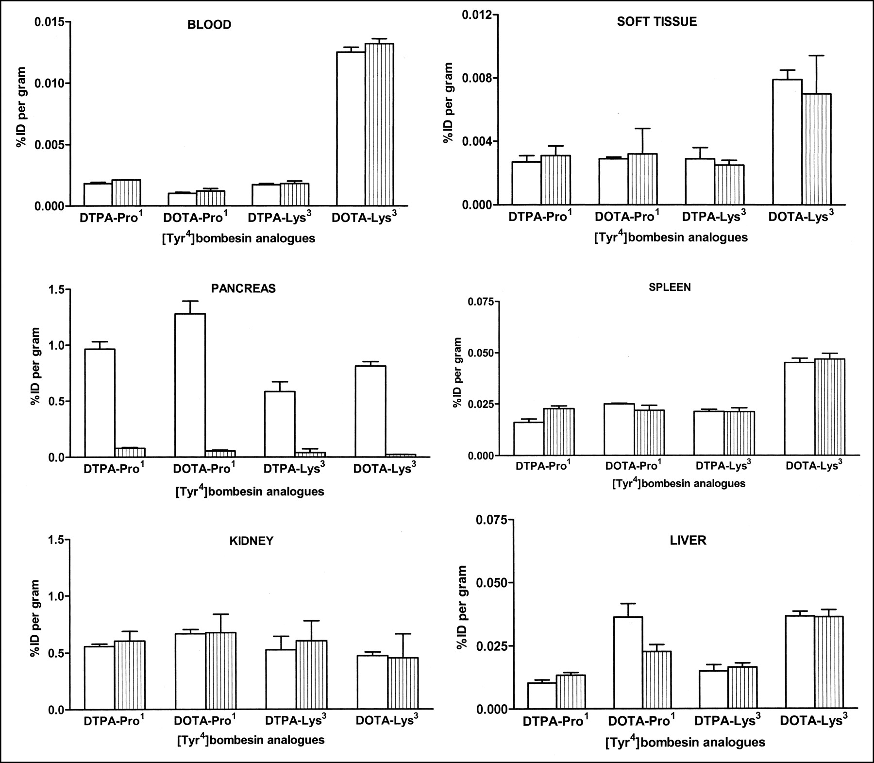
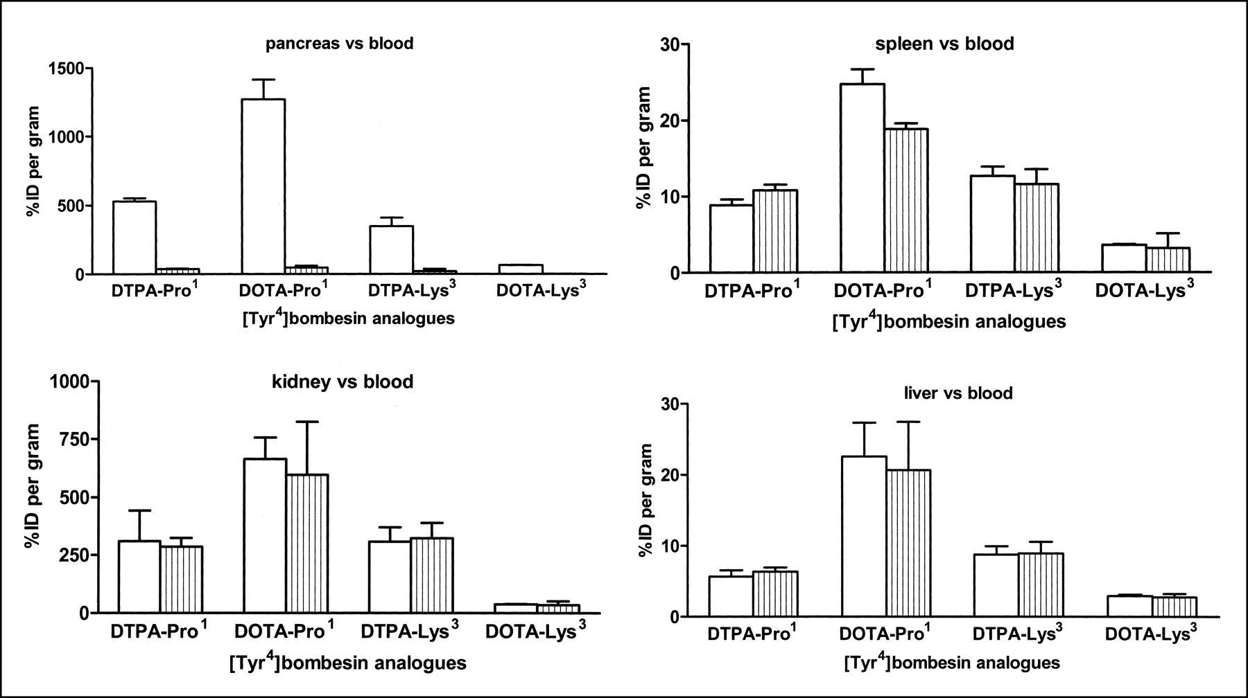
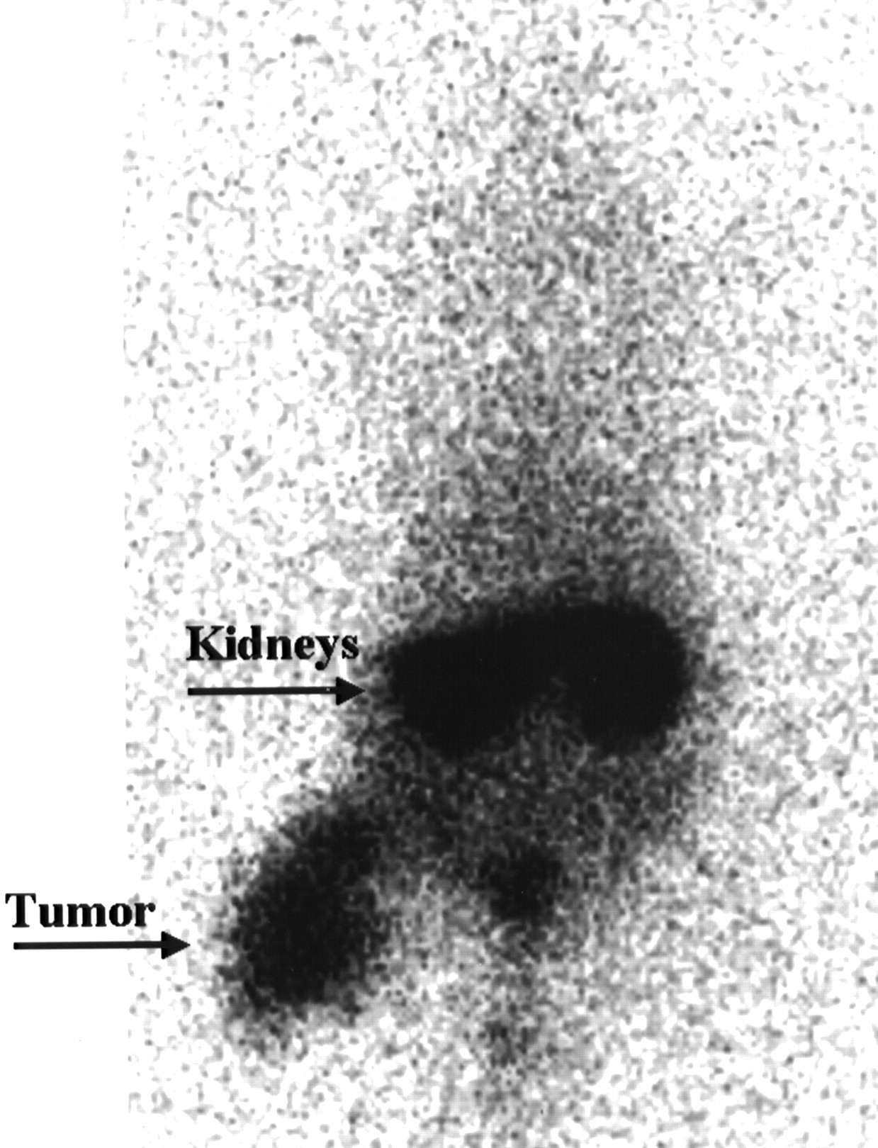
Figure 3A shows the remaining radioactivity in blood and soft tissue 24 h after injection of the 4 111In-labeled BN analogs. Radioactivity in blood after administration of [11IIn-DOTA-ε-Lys3,Tyr4]BN was ∼5 times higher than that after administration of the other radioligands. This difference was not found in soft tissue (thigh). In spleen, an organ with a high blood content, a pattern similar to that in blood was found (Fig. 3B). In the highly GRP receptor-positive pancreas, retention of radioactivity was highest for [111In-DOTA-Pro1,Tyr4]BN (1.2 %ID/g), followed by [111In-DTPA-Pro1,Tyr4]BN (0.9 %ID/g), [111In-DOTA-ε-Lys3,Tyr4]BN (0.79 %ID/g), and [111In-DTPA-ε-Lys3,Tyr4]BN (0.54 %ID/g) (Fig. 3B). Other GRP receptor-positive tissues with significant specific binding of the different radioligands—that is, antrum, fundus, jejunum, colon, cecum, adrenals, and pituitary—also showed the highest retention of radioactivity after administration of [111In-DOTA-Pro1,Tyr4]BN (data not shown). In the kidneys, no significant differences were found between the 4 analogs, whereas in liver both DTPA-BN analogs showed the lowest retention of radioactivity (Fig. 3C). No differences were found between the analogs in the other BN receptor-negative organs, showing insignificant specific binding—that is, heart, esophagus, femur, femur muscle, and lungs (data not shown). The ratios of tissue versus blood are presented in Figure 4A–4C. For pancreas, these ratios amounted to 529 for [111In-DTPA-Pro1,Tyr4]BN, 1,269 for [111In-DOTA-Pro1,Tyr4]BN, 346 for [111In-DTPA-ε-Lys3, Tyr4]BN, and 65 for [111In-DOTA-ε-Lys3,Tyr4]BN. In kidney and liver, the ratio versus blood was highest for [111In-DOTA-Pro1,Tyr4]BN and lowest for [111In-DOTA-ε-Lys3, Tyr4]BN (Fig. 4B). This pattern was also found in all GRP receptor-positive tissues studied, whereas no differences between the analogs were found in GRP receptor-negative tissues (data not shown). In a rat model, the AR42J tumor could clearly be visualized by scintigraphy using [111In-DTPA0,Pro1,Tyr4]BN as the radioligand (Fig. 5).

Tissue distribution of 111In-labeled [DTPA-Pro1,Tyr4]BN, [DOTA-Pro1,Tyr4]BN, [DTPA-ε-Lys3,Tyr4]BN, and [DOTA-ε-Lys3,Tyr4]BN in rats 24 h after administration of radioligand. Nonspecific uptake of radioactivity (striped) was determined by coinjection of GRP receptor-blocking dose of 0.1 mg [Tyr4]BN. Difference between total uptake and nonspecific uptake represents specific binding of radioligand to GRP receptor. Data are expressed as %ID per gram of tissue and presented as means ± SD (n = 3).

Tissue-to-blood ratio of 111In-labeled [DTPA-Pro1,Tyr4]BN, [DOTA-Pro1,Tyr4]BN, [DTPA-ε-Lys3,Tyr4]BN, and [DOTA-ε-Lys3,Tyr4]BN in rats 24 h after administration of radioligand. Nonspecific uptake of radioactivity (striped) was determined by coinjection of GRP receptor-blocking dose of 0.1 mg [Tyr4]BN. Data are expressed as ratio of %ID per gram of tissue versus that in blood and presented as means ± SD (n = 3).

Distribution of radioactivity in AR42J tumor-bearing rat 24 h after administration of 15 MBq (0.1 μg) [111In-DOTA-Pro1,Tyr4]BN. Note uptake in tumor and physiologic uptake in kidneys.
DISCUSSION
The 14-amino-acid neuropeptide BN has a high affinity for the GRP receptor, which is expressed by a variety of cancers. This has been demonstrated most convincingly for breast and prostate carcinomas by Reubi’s group in 2 recent autoradiographic studies using [125I-Tyr4]BN as the radioligand (1). These workers hypothesized that GRP receptors may be markers for early molecular events in carcinogenesis. Therefore, in analogy with studies using OctreoScan (TYCO, Health Care, Petten, The Netherlands) and other radiolabeled somatostatin analogs (6,7,12,13,17), expression of the GRP receptor on tumors may be the molecular basis for diagnosis and treatment of such tumors by GRP receptor-targeted radionuclide therapy and cytotoxic therapy (1,3). In a recent report, 10 patients (4 men with invasive prostate carcinoma, 6 women with breast cancer) were studied by scintigraphy using a 99mTc-labeled BN analog, 99mTc-RP527 (18). The results suggested that 99mTc-RP527 is able to localize GRP receptor-positive lesions. Therefore, we decided to study the potential usefulness of new DTPA- and DOTA-conjugated BN analogs in vitro and in rats. In a previous study, we found that DTPA-conjugated GRP receptor agonists, but not antagonists, were internalized by GRP receptor-expressing cells. We now demonstrate that DOTA-conjugated BN agonists are also internalized after binding to the GRP receptor. Internalization of radiolabeled BN analogs by tumor cells may be essential for application in nuclear medicine such as scintigraphy and radiotherapy of GRP receptor-expressing lesions. Tyrosine-containing peptides are also frequently labeled with radioiodine—for instance, with 125I for in vitro receptor studies or with 123I or 131I for in vivo applications such as receptor-targeted scintigraphy and radiotherapy. However, these radioiodinated ligands often have the drawback of being rapidly degraded in vivo, with release and subsequent deiodination of radioiodotyrosine, which greatly hampers their diagnostic and therapeutic use. This has also been reported for [125I-Tyr4]BN (19,20).
The retention of radioactivity in the kidney after administration of the labeled DTPA- and DOTA-conjugated BN analogs is most probably due to glomerular filtration and subsequent tubular reabsorption of the radioligands or their metabolites, as was also found for other DTPA-conjugated peptides (6,17,21). However, the different 111In-labeled BN analogs show relatively low kidney uptake of radioactivity—that is, 0.6 %ID/g compared with 1.8 %ID/g for [111In-DTPA0]octreotide (13), 1.1 %ID/g for [111In-DTPA0]substance P (21), and 5.3 %ID/g for [111In-DTPA0]RC-160 (13).
Radioactivity in blood and spleen 24 h after administration of [111In-DOTA-ε-Lys3,Tyr4]BN is higher than that for the other radioligands, which lowers the GRP receptor-positive target-to-blood ratio for this analog. In addition, uptake of radioactivity in the liver after the administration of [111In-DOTA-ε-Lys3,Tyr4]BN is also the highest of all 4 radioligands investigated. However, specific binding in BN receptor-positive tissues is still detectable. The other DOTA-analog, [DOTA-Pro1,Tyr4]BN, shows a very low concentration of radioactivity in blood and spleen.
When 111In/peptide ratios exceed 270 MBq/nmol for DTPA peptides and 30 MBq/nmol for DOTA analogs, incorporation of radioactivity drops to <95%, as was also found by others (22). In preparation for a phase I clinical trial with 111In-labeled BN analogs, we calculated putative peptide doses based on the required radioactive dose of 200 MBq, a molecular weight of 2 kDa for the BN analogs, and the above-mentioned maximal specific activities of the labeled DTPA-BN and DOTA-BN analogs, amounting to 1.5 and 13.3 μg peptide, respectively. The intravenous administration of BN to patients at a rate of 2.5 ng·kg−1·min−1 for 30 min was well tolerated (23). This amount allows control of the pharmacologic side effects of BN receptor agonists, such as stimulation of gastrointestinal hormone release, exocrine pancreatic secretion, smooth muscle contraction, and so forth. At the above infusion rate, 200 MBq [111In-DTPA-Pro1,Tyr4]BN should be administered to a patient over a period of 8 min versus 75 min for the DOTA counterpart. Although the latter, [111In-DOTA-Pro1,Tyr4]BN, shows a higher uptake of radioactivity in GRP receptor-positive tissues as well as higher target-to-blood ratios, [111In-DTPA-Pro1,Tyr4]BN is easier to handle and is more practical to use. Therefore, we decided to start phase I studies with this DTPA-conjugated radioligand. In preparation for human application, we recently reported on the preclinical evaluation of this radiolabeled BN analog. Male Lewis rats were injected with 0.02 or 0.1 μg [111In-DTPA-Pro1,Tyr4]BN as a bolus in 3 s or infused over a period of 4 min (4,5). The results showed no significant differences between these modes of administration regarding uptake in GRP receptor-positive or -negative tissues, either in absolute terms or as target-to-blood ratios. This finding suggests that the radioligand may also be administered by infusion without a loss in sensitivity of tumor detection, allowing a better control of the pharmacologic side effects of BN receptor agonists. In previous toxicology studies (4,5) and in this study on rats we found no signs of toxicity or discomfort during and after the intravenous administration of as much as 0.1 mg [Tyr4]BN (5). We found the uptake of radioactivity to be low in the lungs and high in the pancreas of rats. How this finding will affect visualization of GRP receptor-positive small cell lung cancer or pancreatic and other gastrointestinal cancers awaits studies in patients.
CONCLUSION
[111In-DTPA-Pro1,Tyr4]BN is a promising radioligand for scintigraphy of GRP receptor-expressing tumors. We are currently performing phase I studies in patients with invasive prostate carcinoma.
Acknowledgments
The authors acknowledge Marcel E. van der Pluijm, Arthur van Gameren, and Elizabeth G. Webb for expert technical assistance.
Footnotes
Received Feb. 19, 2002; revision accepted Jul. 5, 2002.
For correspondence or reprints contact: Wouter A.P. Breeman, PhD, Department of Nuclear Medicine, Erasmus University Medical Center Rotterdam, Dr. Molewaterplein 40, Rotterdam 3015 GD, The Netherlands.
E-mail: breeman{at}nuge.azr.nl
REFERENCES
In this issue


Jump to section
Related Articles
Cited By...
- Preparation and In Vitro and In Vivo Characterization of the Tumor-specific Antigen-derived Peptide as a Potential Candidate for Targeting Human Epidermal Growth Factor Receptor 2-positive Breast Carcinomas
- Targeted Radiotherapy of Prostate Cancer with a Gastrin-Releasing Peptide Receptor Antagonist Is Effective as Monotherapy and in Combination with Rapamycin
- In Vitro and In Vivo Evaluation of 64Cu-Labeled SarAr-Bombesin Analogs in Gastrin-Releasing Peptide Receptor-Expressing Prostate Cancer
- Design, Synthesis, Radiolabeling and In Vitro and In Vivo Characterization of Tumor-antigen- and Antibody-derived Peptides for the Detection of Breast Cancer
- International Union of Pharmacology. LXVIII. Mammalian Bombesin Receptors: Nomenclature, Distribution, Pharmacology, Signaling, and Functions in Normal and Disease States
- 177Lu-AMBA: Synthesis and Characterization of a Selective 177Lu-Labeled GRP-R Agonist for Systemic Radiotherapy of Prostate Cancer
- 111In-Benzyl-DTPA-ZHER2:342, an Affibody-Based Conjugate for In Vivo Imaging of HER2 Expression in Malignant Tumors
- Synergistic effects of light-emitting probes and peptides for targeting and monitoring integrin expression
- GRP Receptor-Targeted PET of a Rat Pancreas Carcinoma Xenograft in Nude Mice with a 68Ga-Labeled Bombesin(6-14) Analog
- 68Ga-Labeled Peptides in Tumor Imaging
- Increased Cell Death After Therapy with an Arg-Gly-Asp-Linked Somatostatin Analog
- Synthesis and Evaluation of Bombesin Derivatives on the Basis of Pan-Bombesin Peptides Labeled with Indium-111, Lutetium-177, and Yttrium-90 for Targeting Bombesin Receptor-Expressing Tumors
- microPET and Autoradiographic Imaging of GRP Receptor Expression with 64Cu-DOTA-[Lys3]Bombesin in Human Prostate Adenocarcinoma Xenografts