


Abstract
We evaluated whether spironolactone would improve cardiac sympathetic nerve activity and symptoms in patients with congestive heart failure (CHF). Methods: Thirty patients with CHF (left ventricular ejection fraction [LVEF] < 40%; mean, 30% ± 9%) were treated with an angiotensin-converting enzyme inhibitor, a loop diuretic, and, in most cases, digoxin. Fifteen patients (group A) were assigned to additionally receive spironolactone (12.5–50 mg/day), and the remaining 15 patients (group B) continued their current regimen. Patients were studied before and 6 mo after treatment. The delayed heart-to-mediastinum count ratio (H/M ratio), delayed total defect score (TDS), and washout rate (WR) were determined from 123I-meta-iodobenzylguanidine (MIBG) images. LVEF was determined by echocardiography, and New York Heart Association (NYHA) functional class was estimated. Results: Before treatment, LVEF, TDS, H/M ratio, WR, and NYHA functional class were similar in both groups. With treatment, LVEF did not significantly improve in either group. However, after treatment in group A, TDS decreased from 37 ± 9 to 25 ± 13 (P = 0.0001), H/M ratio increased from 1.62 ± 0.20 to 1.83 ± 0.27 (P < 0.0001), and WR decreased from 51 ± 9 to 40 ± 15 (P < 0.001). In group B, these parameters did not significantly change. NYHA functional class improved in both groups (in group A, from 3.3 ± 0.5 to 1.7 ± 0.5 [P < 0.0001]; in group B, from 3.3 ± 0.5 to 2.4 ± 0.6 [P = 0.01]); this was a significantly greater improvement in group A than in group B (P < 0.01). Conclusion: Spironolactone improves cardiac sympathetic nerve activity and symptoms in patients with CHF.
Since the Randomized Aldactone Evaluation Study (RALES) (1) showed the effectiveness of spironolactone, this drug has often been used as a treatment for patients with severe congestive heart failure (CHF). Aldosterone is important in the pathophysiology of CHF (2–5). Aldosterone promotes retention of sodium, loss of magnesium and potassium, myocardial and vascular fibrosis, baroreceptor dysfunction, vascular damage and arterial compliance, sympathetic activation, and parasympathetic inhibition (5–9). RALES reported that the addition of spironolactone (an aldosterone-receptor blocker) reduced the risk of death from cardiac causes, hospitalization for cardiac causes, and the combined endpoint of death from cardiac causes among patients who had severe left ventricular systolic dysfunction and who were receiving standard therapy including an angiotensin-converting enzyme inhibitor. Spironolactone also improved the symptoms of heart failure, as measured by changes in the New York Heart Association (NYHA) functional class.
Myocardial imaging with 123I-meta-iodobenzylguanidine (MIBG), an analog of norepinephrine, is a useful tool for detecting abnormalities of the myocardial adrenergic nervous system in patients with CHF (10–16). However, there are no reports on cardiac 123I-MIBG scintigraphy evaluating the effects of chronic spironolactone therapy in patients with CHF. This study was performed to determine whether spironolactone can improve cardiac sympathetic nerve activity and symptoms in patients with severe CHF.
MATERIALS AND METHODS
Study Population
Thirty patients, 17 men and 13 women (mean age, 69 ± 13 y; age range, 42–88 y), with CHF were included in the study. A detailed history and physical examination were obtained. Chest radiography, standard electrocardiography, echocardiography, and 201Tl and 123I-MIBG scintigraphy were performed on all patients. Patients were in NYHA functional class III or IV at the time of enrollment and had echocardiographic left ventricular ejection fraction (LVEF) < 40% (mean, 30% ± 9%). The causes of CHF were old myocardial infarction (n = 16), idiopathic dilated cardiomyopathy (n = 8), and valvular disease (n = 6). All patients were being treated with an angiotensin-converting enzyme inhibitor and a loop diuretic. Treatment with digitalis and vasodilators was allowed, but potassium-sparing diuretics were not permitted (Table 1).
Demographics and Clinical Characteristics
Patients were excluded from the study if they had primary operable valvular heart disease, congenital heart disease, unstable angina, recent acute myocardial infarction, primary hepatic failure, or active cancer.
Study Protocol
Fifteen patients (group A) were randomized to additionally receive spironolactone (12.5–50 mg/day), and the remaining 15 patients (group B) continued their current drug regimen. We performed a series of examinations before and 6 mo after treatment. In this study, no patient received a β-blocker.
123I-MIBG Imaging
The 123I-MIBG was obtained commercially (Daiichi Radioisotope Laboratories, Tokyo, Japan). Patients were injected intravenously with 123I-MIBG (111 MBq) while upright. Anterior planar imaging and SPECT were performed beginning at 15 min and were repeated 4 h later. SPECT was performed with a dedicated single-head imaging system (Millennium MPR; General Electric Medical Systems, Waukesha, WI). The energy, uniformity, and linearity were constantly corrected. Images were acquired for 40 s each at 32 steps over a 180° orbit and were recorded at a digital resolution of 128 × 128 from the anterior planar 123I-MIBG image.
From anterior planar delayed 123I-MIBG images, the heart-to-mediastinum count ratio (H/M ratio) was determined (Fig. 1). Washout rate (WR) was calculated by the following: {([H] − [M])early − ([H] − [M])delayed}/([H] − [M])early × 100 (%), where [H] = mean count per pixel in the left ventricle and [M] = mean count per pixel in the upper mediastinum. In our laboratory, the normal value of the delayed H/M ratio is from 2.00 to 2.80, and the normal value of WR is from 22% to 32%.

Cardiac 123I-MIBG uptake was quantified as H/M ratio 4 h after injection, using regions of interest positioned over heart (H) and upper mediastinum (M).
The myocardial delayed SPECT images for each patient were divided into 20 segments (Fig. 2). The short-axis images at the basal, middle, and apical ventricular levels were divided into 6 segments. The apical segment of the vertical long-axis image was divided into 2 segments. Regional tracer uptake was assessed semiquantitatively using a 4-point scoring system (0 = normal uptake; 1 = mildly reduced uptake; 2 = moderately reduced uptake; 3 = severely reduced uptake). The total defect score (TDS) was calculated as the sum of the scores for all 20 segments.
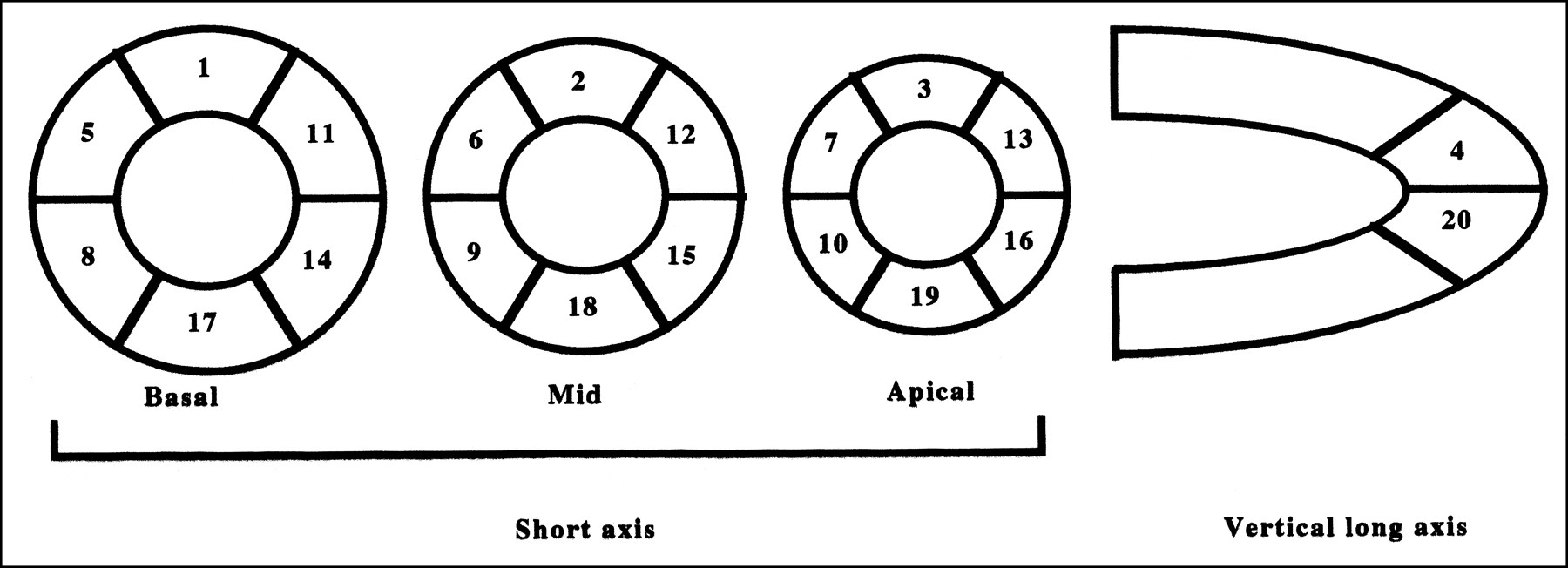
Segmentation scheme used to quantitate regional 123I-MIBG uptake.
Interobserver variability was determined in a masked manner by 2 independent observers. The interobserver correlation was represented by r = 0.90 (P < 0.001).
Echocardiography
Echocardiographic measurement was performed using standard methods. LVEF was calculated using the modified Simpson method (17).
Statistical Analysis
Statistical analysis was performed using StatView (Abacus Concepts, Berkeley, CA) for Macintosh (Apple Computer, Inc., Cupertino, CA). Unpaired t and χ2 tests were used to compare the 2 groups. All values are reported as mean ± SD. P < 0.05 was considered statistically significant.
RESULTS
The hemodynamic characteristics of the 2 groups did not significantly differ. Before treatment, LVEF, TDS, H/M ratio, WR, and NYHA functional class were similar in both groups. LVEF is reported in Table 2. The baseline value did not significantly differ from the value after 6 mo of treatment in either group.
Changes in LVEF and NYHA Functional Class in Patients with Heart Failure
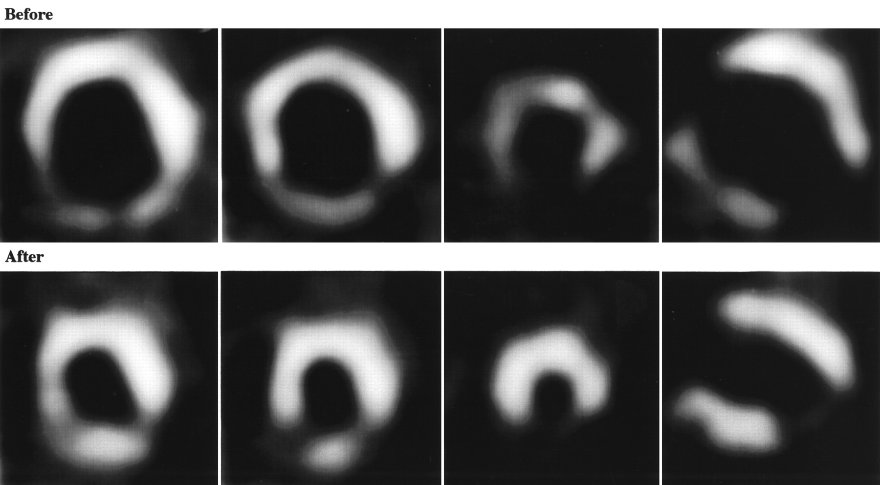
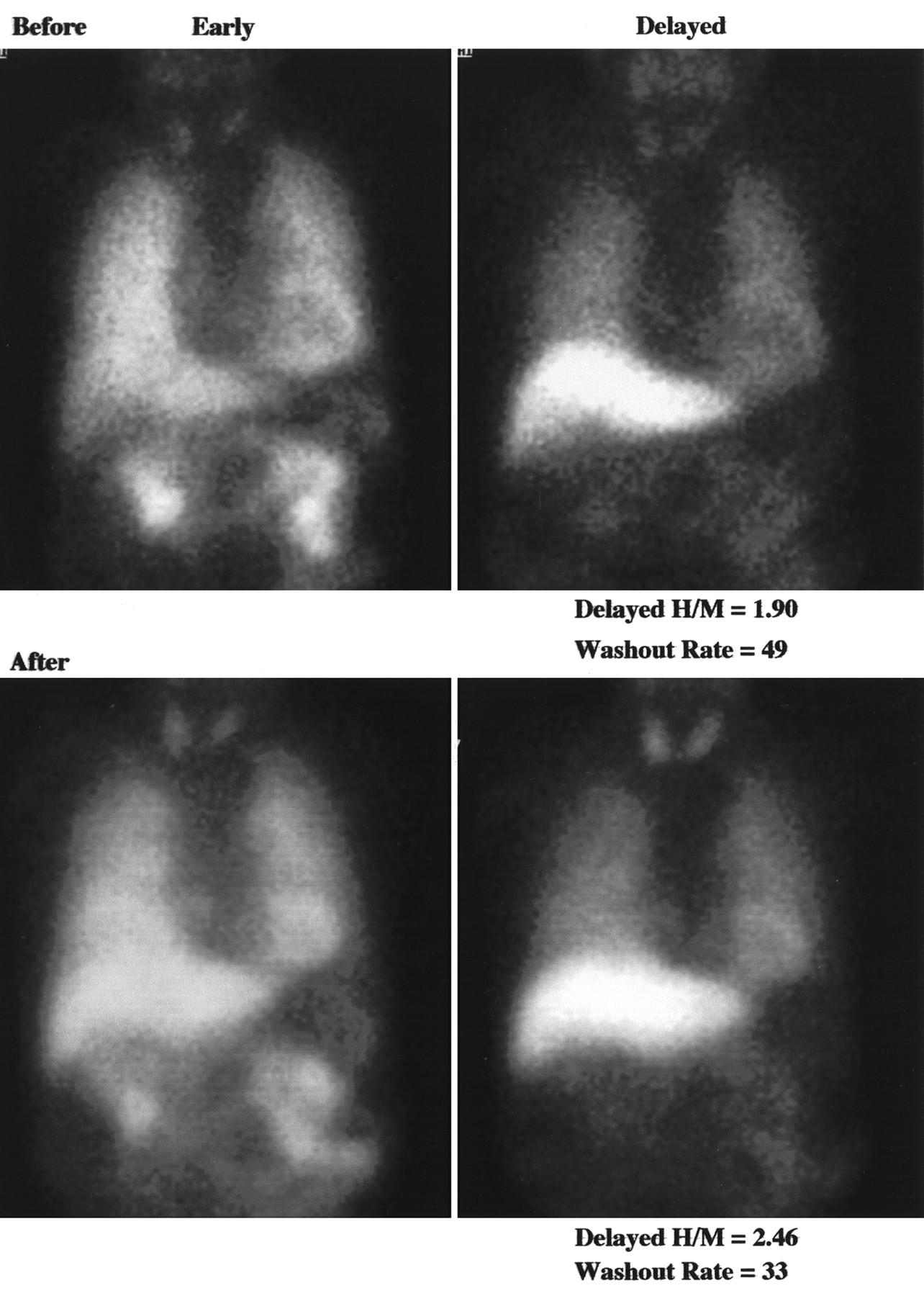
TDS, H/M ratio, and WR are reported in Table 3. In group A, the TDS decreased significantly after 6 mo (25 ± 13) from the baseline value (37 ± 9) (P = 0.0001). In the segmental analysis of TDS, though it tended to improve the uptake of the inferior wall, the improvement was not statistically significant. The H/M ratio increased significantly after 6 mo (1.83 ± 0.27) from the baseline (1.62 ± 0.20) (P < 0.0001). The WR decreased significantly after 6 mo (40 ± 15) compared with baseline (51 ± 9) (P < 0.001). In contrast, in group B, there were no significant differences between baseline and values after 6 mo of treatment. Furthermore, after 6 mo of treatment, the WR of group A was significantly lower than that of group B (P < 0.05). Representative 123I-MIBG images before and after spironolactone treatment are shown in Figures 3 and 4.

Representative SPECT 123I-MIBG images before and after spironolactone treatment in dilated cardiomyopathy.

Representative anterior planar 123I-MIBG images before and after spironolactone treatment in dilated cardiomyopathy. In this example, delayed H/M ratio increased from 1.90 to 2.46, and WR decreased from 49 to 33.
Changes in TDS, H/M Ratio, and WR for 123I-MIBG Imaging in Patients with Heart Failure
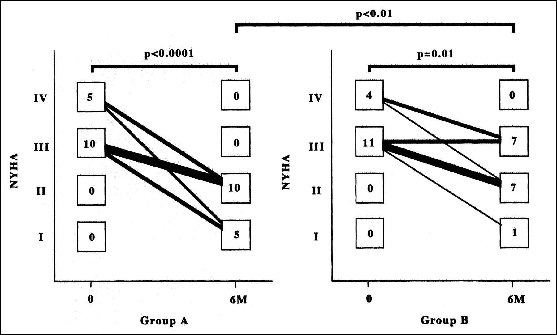
The NYHA functional class of the patients is shown in Table 2 and Figure 5. Patients in both groups showed improvement after 6 mo of treatment compared with baseline (in group A, from 3.3 ± 0.5 to 1.7 ± 0.5 [P < 0.0001]; in group B, from 3.3 ± 0.5 to 2.4 ± 0.6 [P = 0.01]). After treatment, the NYHA functional class of patients in group A was better than that in group B (P < 0.01).

Changes in NYHA functional class during treatment in the 2 groups. 6M = after 6 mo of therapy.
DISCUSSION
Aldosterone causes myocardial and vascular fibrosis (18,19), direct vascular damage (9), and baroreceptor dysfunction (7) and prevents myocardial uptake of norepinephrine (5,20). RALES (1) found that spironolactone reduced the risk of death in patients with CHF from cardiac causes. Spironolactone may prevent myocardial fibrosis by blocking aldosterone; myocardial fibrosis may predispose patients to variation in ventricular-conduction times and, hence, to reentry ventricular arrhythmias (20–23). Spironolactone may also prevent sudden death by increasing myocardial uptake of norepinephrine (1). Thus, we examined whether cardiac MIBG imaging was useful for evaluating increasing myocardial uptake of norepinephrine.
Cardiac MIBG, an analog of norepinephrine, is a useful tool for detecting abnormalities of the myocardial adrenergic nervous system in patients with CHF (10–16). Several reports have suggested that β-blocker therapy can improve cardiac sympathetic nerve activity by cardiac MIBG scintigraphy in patients with CHF (24–27). In this study, the TDS, H/M ratio, and WR of cardiac MIBG scintigraphy improved in the spironolactone treatment group as opposed to the control group. However, there was no significant difference in the recovery of cardiac function before and after treatment in either group. β-blocker treatment improves both cardiac MIBG scintigraphy and cardiac function by echocardiography (24,27). Thus, spironolactone may lack some of the beneficial actions of β-blockers: increased myocardial energy for synthetic and reparative processes and improved diastolic relaxation, filling, and compliance (28–30). Instead, spironolactone may be effective by increasing myocardial uptake of norepinephrine.
Tsutamoto et al. (31) reported that spironolactone could improve cardiac function in patients with CHF. However, that study included patients with mild to moderate nonischemic CHF. In contrast, our study included many patients with CHF due to old myocardial infarction; therefore, spironolactone might not recognize the improvement in cardiac function. Correlation between cardiac sympathetic nerve activity and cardiac function in patients with idiopathic dilated cardiomyopathy has been reported (32). However, that study also excluded patients with old myocardial infarction and thus seemed to be different from our study. Because of the results of our study and the other 2 studies, we consider that spironolactone treatment could not improve cardiac function in patients with CHF due to old myocardial infarction because the infarcted areas had already undergone irreversible necrosis after the treatment.
In this study, delayed MIBG images were used to obtain TDS and H/M ratio. There are 2 types of norepinephrine or MIBG uptake. Uptake-1 (neuronal uptake), which takes place even if the concentration of norepinephrine or MIBG is low, depends on sodium and adenosine triphosphate and is suppressed by tricyclic antidepressants. Uptake-2 (extraneuronal uptake), which takes place only when the concentration is high, represents a diffusion system and is unaffected by tricyclic agents (33–35). Early images result from both uptake-1 and uptake-2 (36,37), whereas delayed images involve less of uptake-2 and therefore show the status of cardiac sympathetic nerve activity more accurately. For these reasons, we used delayed MIBG imaging in this study. Several reports suggest that WR is the most clinically useful for severity and improvement of CHF (38,39). Increased norepinephrine turnover at cardiac sympathetic nerve endings may decrease the uptake in delayed images such that the increase in turnover, that is, the increase in the WR, reflects CHF severity. In this study, all 3 parameters (delayed TDS, delayed H/M ratio, and WR) improved with spironolactone. We infer that spironolactone increases myocardial uptake of norepinephrine as a mechanism for CHF improvement.
CONCLUSION
Spironolactone improves cardiac sympathetic nerve activity and symptoms in patients with CHF.
Footnotes
Received Jan. 9, 2002; revision accepted Jun. 4, 2002.
For correspondence or reprints contact: Shu Kasama, MD, Second Department of Internal Medicine, Gunma University School of Medicine, 3-39-15, Shouwa-machi, Maebashi, Gunma 371-0034, Japan.
E-mail: s-kasama{at}bay.wind.ne.jp
REFERENCES
In this issue


Jump to section
Related Articles
Cited By...
- Comparative effects of long-acting and short-acting loop diuretics on cardiac sympathetic nerve activity in patients with chronic heart failure
- 123I-MIBG SPECT for Evaluation of Patients with Heart Failure
- Introduction to Cardiac Neuronal Imaging: A Clinical Perspective
- A Pooled Analysis of Multicenter Cohort Studies of 123I-mIBG Imaging of Sympathetic Innervation for Assessment of Long-Term Prognosis in Heart Failure
- Effects of spironolactone on cardiac sympathetic nerve activity and left ventricular remodelling after reperfusion therapy in patients with first ST-segment elevation myocardial infarction
- Myocardial Iodine-123 Meta-Iodobenzylguanidine Imaging and Cardiac Events in Heart Failure: Results of the Prospective ADMIRE-HF (AdreView Myocardial Imaging for Risk Evaluation in Heart Failure) Study
- 123I-mIBG Scintigraphy to Predict Inducibility of Ventricular Arrhythmias on Cardiac Electrophysiology Testing: A Prospective Multicenter Pilot Study
- Prognostic Value of Serial Cardiac 123I-MIBG Imaging in Patients with Stabilized Chronic Heart Failure and Reduced Left Ventricular Ejection Fraction
- Additive Effects of Spironolactone and Candesartan on Cardiac Sympathetic Nerve Activity and Left Ventricular Remodeling in Patients with Congestive Heart Failure
- Long-Term Nicorandil Therapy Improves Cardiac Sympathetic Nerve Activity After Reperfusion Therapy in Patients with First Acute Myocardial Infarction
- Effects of Intravenous Atrial Natriuretic Peptide on Cardiac Sympathetic Nerve Activity and Left Ventricular Remodeling in Patients With First Anterior Acute Myocardial Infarction
- Effects of torasemide on cardiac sympathetic nerve activity and left ventricular remodelling in patients with congestive heart failure
- Comparative effects of valsartan and enalapril on cardiac sympathetic nerve activity and plasma brain natriuretic peptide in patients with congestive heart failure
- Effects of candesartan on cardiac sympathetic nerve activity in patients with congestive heart failure and preserved left ventricular ejection fraction
- Dobutamine Stress 99mTc-Tetrofosmin Quantitative Gated SPECT Predicts Improvement of Cardiac Function After Carvedilol Treatment in Patients with Dilated Cardiomyopathy
- Effects of Intravenous Atrial Natriuretic Peptide on Cardiac Sympathetic Nerve Activity in Patients with Decompensated Congestive Heart Failure
- Sympatholysis and cardiac sympathetic nerve function in the treatment of congestive heart failure
- Addition of Valsartan to an Angiotensin-Converting Enzyme Inhibitor Improves Cardiac Sympathetic Nerve Activity and Left Ventricular Function in Patients with Congestive Heart Failure