
Main menu

  • Home
  • Content
    • Current
    • Ahead of print
    • Past Issues
    • JNM Supplement
    • SNMMI Annual Meeting Abstracts
    • Continuing Education
    • JNM Podcasts
  • Subscriptions
    • Subscribers
    • Institutional and Non-member
    • Rates
    • Journal Claims
    • Corporate & Special Sales
  • Authors
    • Submit to JNM
    • Information for Authors
    • Assignment of Copyright
    • AQARA requirements
  • Info
    • Reviewers
    • Permissions
    • Advertisers
  • About
    • About Us
    • Editorial Board
    • Contact Information
  • More
    • Alerts
    • Feedback
    • Help
    • SNMMI Journals
  • SNMMI
    • JNM
    • JNMT
    • SNMMI Journals
    • SNMMI

User menu

  • Subscribe
  • My alerts
  • Log in
  • Log out
  • My Cart

Search

  • Advanced search
Journal of Nuclear Medicine
  • SNMMI
    • JNM
    • JNMT
    • SNMMI Journals
    • SNMMI
  • Subscribe
  • My alerts
  • Log in
  • Log out
  • My Cart
Journal of Nuclear Medicine

Advanced Search

  • Home
  • Content
    • Current
    • Ahead of print
    • Past Issues
    • JNM Supplement
    • SNMMI Annual Meeting Abstracts
    • Continuing Education
    • JNM Podcasts
  • Subscriptions
    • Subscribers
    • Institutional and Non-member
    • Rates
    • Journal Claims
    • Corporate & Special Sales
  • Authors
    • Submit to JNM
    • Information for Authors
    • Assignment of Copyright
    • AQARA requirements
  • Info
    • Reviewers
    • Permissions
    • Advertisers
  • About
    • About Us
    • Editorial Board
    • Contact Information
  • More
    • Alerts
    • Feedback
    • Help
    • SNMMI Journals
  • View or Listen to JNM Podcast
  • Visit JNM on Facebook
  • Join JNM on LinkedIn
  • Follow JNM on Twitter
  • Subscribe to our RSS feeds
Meeting ReportPhysics, Instrumentation & Data Sciences

A 4D Statistical Atlas of the Human Heart from Gated PET Myocardial Perfusion Imaging

Fangxu Xing, Jonghye Woo, Nicolas Guehl, Matthieu Pelletier-Galarneau, Georges El Fakhri and Nathaniel Alpert
Journal of Nuclear Medicine May 2019, 60 (supplement 1) 257;
Fangxu Xing
1Gordon Center for Medical Imaging Massachusetts General Hospital Boston MA United States
  • Find this author on Google Scholar
  • Find this author on PubMed
  • Search for this author on this site
Jonghye Woo
1Gordon Center for Medical Imaging Massachusetts General Hospital Boston MA United States
  • Find this author on Google Scholar
  • Find this author on PubMed
  • Search for this author on this site
Nicolas Guehl
1Gordon Center for Medical Imaging Massachusetts General Hospital Boston MA United States
  • Find this author on Google Scholar
  • Find this author on PubMed
  • Search for this author on this site
Matthieu Pelletier-Galarneau
1Gordon Center for Medical Imaging Massachusetts General Hospital Boston MA United States
  • Find this author on Google Scholar
  • Find this author on PubMed
  • Search for this author on this site
Georges El Fakhri
1Gordon Center for Medical Imaging Massachusetts General Hospital Boston MA United States
  • Find this author on Google Scholar
  • Find this author on PubMed
  • Search for this author on this site
Nathaniel Alpert
1Gordon Center for Medical Imaging Massachusetts General Hospital Boston MA United States
  • Find this author on Google Scholar
  • Find this author on PubMed
  • Search for this author on this site
  • Article
  • Figures & Data
  • Info & Metrics
Loading

Abstract

257

Objectives: PET myocardial perfusion imaging (MPI) has emerged as an excellent imaging modality to diagnose coronary artery disease and provide prognostic information in a wide range of pathologies. Although PET MPI allows absolute quantification of myocardial blood flow and quantitative assessment of left ventricular ejection fraction, regional wall motion analysis is primarily performed qualitatively. The ability to automate quantitative assessment of regional wall motion could provide additional diagnostic and prognostic information. We are developing methodology for a normative, intersubject, and spatiotemporal four-dimensional (4D) atlas of the left ventricle to support statistical comparison and quantitative evaluation of the characteristics of human cardiac function. The goal of this work is to report a 4D atlas of the human heart using gated PET MPI in which normal intersubject variability is summarized in the voxelwise mean and variance of the functional endpoints across various subjects. Statistics of the myocardial motion pattern in a healthy population can be succinctly summarized by the principle components (PCs) of an orthogonal decomposition of the 4D atlas.

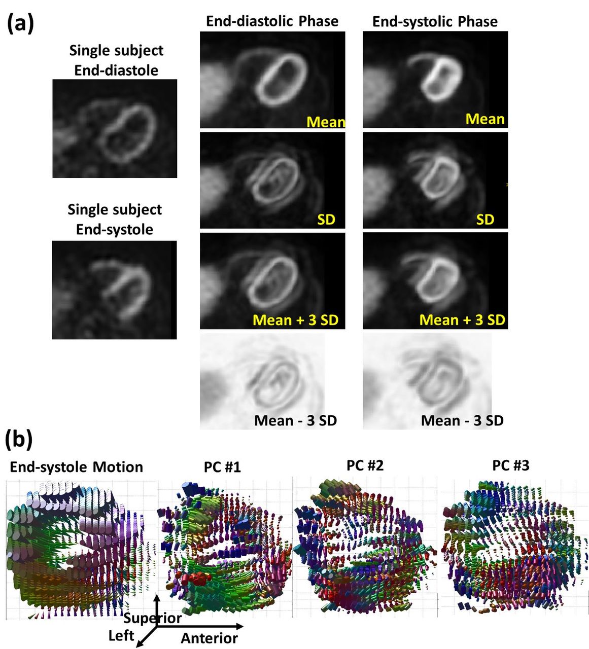
Methods: Twelve healthy human subjects were imaged on a Siemens Biograph PET/CT system using 13N-NH3. The scan included a standard stress acquisition with the single-scan, two-injection method that was previously proposed and validated[1-3]. The acquisition is performed at peak stress with an adenosine infusion (140 mcg/kg/min) and 13N-NH3 injection (600 MBq). Gated PET image volumes were dynamically reconstructed with 16 gates per cardiac cycle. To define a basic structural framework for atlas construction, we first created a common physical space using a diffeomorphic groupwise registration method. The gated PET volumes of all subjects were transformed into this common space using the deformations fields computed simultaneously during the registration process. At each of the 16 time bins, the deformed PET volumes were fused together, yielding a gated 4D PET atlas. The mean and variance of the fused volumes were computed to summarize the statistical properties of the atlas. In addition, we incorporated a principal component analysis (PCA) framework to characterize myocardial motion. Consecutive diffeomorphic registrations from the end-diastole (ED) as reference phase to the end-systole (ES) phase are compared. The variability of the myocardial motion within a population are reflected in the weights and directions of the PCs.

Results: The proposed 4D PET atlas was successfully constructed with high quality mean intensity image and a standard deviation image describing intersubject variability (Figure 1(a)). Specifically, statistics of the mean and standard deviation images at ED and ES are listed in Table 1. Subject-specific myocardial motion was computed at ES phase. The motion field and PC directions were visualized (Figure 1(b)) and the weight percentages of the PCs are shown in Table 2. In general, PC #1 follows the myocardium circumferentially and PC #3 follows it vertically.

Conclusions: The 4D gated cardiac PET atlas provides myocardial images of a standard size and orientation combining anatomic and physiological data. This is the first atlas combining anatomic and functional images in a standard space. This provides a tool to compare the differences and variability of a variety of parameters in a population and allows us to analyze the variability of the myocardial anatomies and cardiac wall motion. This method can potentially be extended to clinical applications involving automated image interpretation. Acknowledgments: This work is supported by NIH P41EB022544 and R01HL137230. We thank Marina T. Macdonald-Soccorso for her effort on PET MPI reconstruction.

Figure
  • Download figure
  • Open in new tab
  • Download powerpoint
View this table:
  • View inline
  • View popup

Table 1. Statistics of the 4D Atlas at ED and ES Phase

View this table:
  • View inline
  • View popup

Table 2. Principal Component Weights of Myocardial Motion

Previous
Back to top

In this issue

Journal of Nuclear Medicine
Vol. 60, Issue supplement 1
May 1, 2019
  • Table of Contents
  • Index by author
Article Alerts
Sign In to Email Alerts with your Email Address
Email Article

Thank you for your interest in spreading the word on Journal of Nuclear Medicine.

NOTE: We only request your email address so that the person you are recommending the page to knows that you wanted them to see it, and that it is not junk mail. We do not capture any email address.

Enter multiple addresses on separate lines or separate them with commas.
A 4D Statistical Atlas of the Human Heart from Gated PET Myocardial Perfusion Imaging
(Your Name) has sent you a message from Journal of Nuclear Medicine
(Your Name) thought you would like to see the Journal of Nuclear Medicine web site.
Citation Tools
A 4D Statistical Atlas of the Human Heart from Gated PET Myocardial Perfusion Imaging
Fangxu Xing, Jonghye Woo, Nicolas Guehl, Matthieu Pelletier-Galarneau, Georges El Fakhri, Nathaniel Alpert
Journal of Nuclear Medicine May 2019, 60 (supplement 1) 257;

Citation Manager Formats

  • BibTeX
  • Bookends
  • EasyBib
  • EndNote (tagged)
  • EndNote 8 (xml)
  • Medlars
  • Mendeley
  • Papers
  • RefWorks Tagged
  • Ref Manager
  • RIS
  • Zotero
Share
A 4D Statistical Atlas of the Human Heart from Gated PET Myocardial Perfusion Imaging
Fangxu Xing, Jonghye Woo, Nicolas Guehl, Matthieu Pelletier-Galarneau, Georges El Fakhri, Nathaniel Alpert
Journal of Nuclear Medicine May 2019, 60 (supplement 1) 257;
Twitter logo Facebook logo LinkedIn logo Mendeley logo
  • Tweet Widget
  • Facebook Like
  • Google Plus One
Bookmark this article

Jump to section

  • Article
  • Figures & Data
  • Info & Metrics

Related Articles

  • No related articles found.
  • Google Scholar

Cited By...

  • No citing articles found.
  • Google Scholar

More in this TOC Section

Physics, Instrumentation & Data Sciences

  • 3D Structural Convolutional Sparse Coding for PET Image Reconstruction
  • Exploration of Multi-objective Optimization with Genetic Algorithms for PET Image Reconstruction
  • AI-based methods for nuclear-medicine imaging: Need for objective task-specific evaluation
Show more Physics, Instrumentation & Data Sciences

Cardiac and Cancer Imaging (Data Management, PIDS)

  • SPECT/CT studies for dosimetry after PRRT: impact of reduced number of studies on organ dose calculation and patient management
  • On the Accuracy of Voxel-Based Kidney Dosimetry
Show more Cardiac and Cancer Imaging (Data Management, PIDS)

Similar Articles

SNMMI

© 2025 SNMMI

Powered by HighWire