
Main menu

  • Home
  • Content
    • Current
    • Ahead of print
    • Past Issues
    • JNM Supplement
    • SNMMI Annual Meeting Abstracts
    • Continuing Education
    • JNM Podcasts
  • Subscriptions
    • Subscribers
    • Institutional and Non-member
    • Rates
    • Journal Claims
    • Corporate & Special Sales
  • Authors
    • Submit to JNM
    • Information for Authors
    • Assignment of Copyright
    • AQARA requirements
  • Info
    • Reviewers
    • Permissions
    • Advertisers
  • About
    • About Us
    • Editorial Board
    • Contact Information
  • More
    • Alerts
    • Feedback
    • Help
    • SNMMI Journals
  • SNMMI
    • JNM
    • JNMT
    • SNMMI Journals
    • SNMMI

User menu

  • Subscribe
  • My alerts
  • Log in
  • Log out
  • My Cart

Search

  • Advanced search
Journal of Nuclear Medicine
  • SNMMI
    • JNM
    • JNMT
    • SNMMI Journals
    • SNMMI
  • Subscribe
  • My alerts
  • Log in
  • Log out
  • My Cart
Journal of Nuclear Medicine

Advanced Search

  • Home
  • Content
    • Current
    • Ahead of print
    • Past Issues
    • JNM Supplement
    • SNMMI Annual Meeting Abstracts
    • Continuing Education
    • JNM Podcasts
  • Subscriptions
    • Subscribers
    • Institutional and Non-member
    • Rates
    • Journal Claims
    • Corporate & Special Sales
  • Authors
    • Submit to JNM
    • Information for Authors
    • Assignment of Copyright
    • AQARA requirements
  • Info
    • Reviewers
    • Permissions
    • Advertisers
  • About
    • About Us
    • Editorial Board
    • Contact Information
  • More
    • Alerts
    • Feedback
    • Help
    • SNMMI Journals
  • View or Listen to JNM Podcast
  • Visit JNM on Facebook
  • Join JNM on LinkedIn
  • Follow JNM on Twitter
  • Subscribe to our RSS feeds
Meeting ReportOncology: Clinical Therapy and Diagnosis

Integration of dynamic parameters in the analysis of 18F-FDopa PET imaging improves the prediction of the WHO-2016 classification of gliomas

Merwan Ginet, Timothée Zaragori, Pierre-Yves Marie, Laetitia Imbert and Antoine Verger
Journal of Nuclear Medicine May 2019, 60 (supplement 1) 361;
Merwan Ginet
1CHRU Nancy Vandoeuvre les Nancy France
  • Find this author on Google Scholar
  • Find this author on PubMed
  • Search for this author on this site
Timothée Zaragori
1CHRU Nancy Vandoeuvre les Nancy France
  • Find this author on Google Scholar
  • Find this author on PubMed
  • Search for this author on this site
Pierre-Yves Marie
1CHRU Nancy Vandoeuvre les Nancy France
  • Find this author on Google Scholar
  • Find this author on PubMed
  • Search for this author on this site
Laetitia Imbert
1CHRU Nancy Vandoeuvre les Nancy France
  • Find this author on Google Scholar
  • Find this author on PubMed
  • Search for this author on this site
Antoine Verger
1CHRU Nancy Vandoeuvre les Nancy France
  • Find this author on Google Scholar
  • Find this author on PubMed
  • Search for this author on this site
  • Article
  • Figures & Data
  • Info & Metrics
Loading

Abstract

361

Introduction: 18F-FDopa PET imaging of gliomas is routinely interpreted with standard uptake value (SUV) derived indices. This study aimed to determine the added-value of dynamic 18F-FDopa PET parameters for predicting the characterization of newly-diagnosed gliomas achieved with the World Health Organization (WHO) 2016 classification.

Methods: We retrospectively included 58 patients having undergone a 18F-FDopa PET for the initial diagnosis of a glioma staging according to the WHO-2016 classification (20 IDH-mutant and 11 IDH-wildtype astrocytomas, 16 IDH-mutant and 1p/19q co-deleted oligodendrogliomas, 10 IDH-wildtype and 1 IDH-mutant glioblastomas). Dynamic parameters, involving time-to-peak (TTP) values and curve slopes, were tested for the prediction of glioma types in addition to current static parameters -i.e. tumor-to-normal brain or tumor-to-striatum standardized uptake values (SUV) ratios and metabolic tumor volume (MTV).

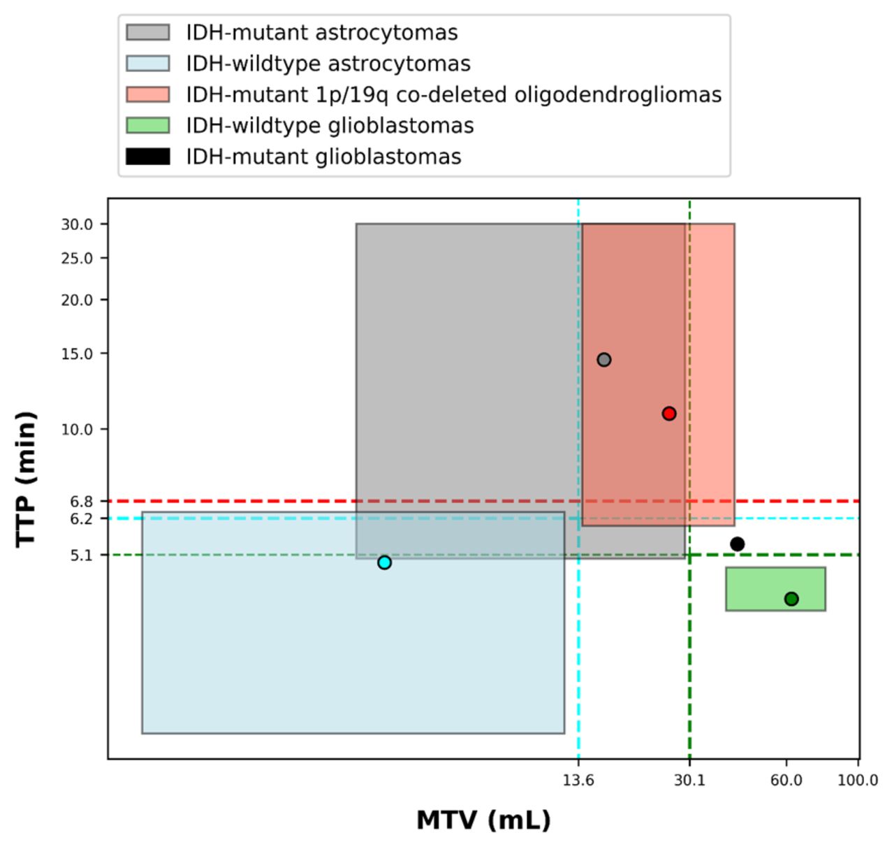
Results: The combined analysis of MTV and TTP provided the best prediction for IDH-wildtype glioblastomas (areas under curve (AUC) of 0.938 for the dual criterion of MTV ≥ 30.1 mL and TTP ≤ 5.1 min), as well as for IDH-wildtype astrocytomas (AUC of 0.907 for the dual criterion of MTV ≤ 13.6 mL and TTP ≤ 6.2 min), whereas TTP was the sole significant predictor of oligodendrogliomas (AUC of 0.679 for TTP ≥ 6.8min). Distribution of gliomas groups according to both MTV and TTP is available in Figure 1. Representative examples of each glioma subgroup are provided in Figure 2.

Conclusions: The prediction of the WHO 2016 classification of newly-diagnosed gliomas by 18F-FDopa PET is significantly enhanced when combining the analyses of dynamic with static PET parameters and especially, time-to-peak with the metabolic tumor volume. Figure 1. Distribution of median values (points) and interquartile ranges (colored rectangles) according to both metabolic tumor volume (MTV) and time-to-peak (TTP) expressed with natural logarithm, for IDH-wildtype (blue) and IDH-mutant (grey) astrocytomas, IDH-mutant and 1p/19q co-deleted oligodendrogliomas (red) and IDH-wildtype-glioblastomas (green). The single case of IDH-mutant glioblastomas is represented with a black point and the threshold diagnostic values provided by the ROC curves analyses are represented by dashed lines.Figure 2. Representative examples for each gliomas group of the metabolic tumor volume delineation on an axial slice of 18F-FDopa PET (left row) with the dynamic TBRmean curve (middle row) providing the time-to-peak delay-time (blue line) and the 10-to-30 min slope (dark blue line) and with for illustration, the same slice location recorded on contrast-enhanced T1-weighted MRI (right row).

Figure
  • Download figure
  • Open in new tab
  • Download powerpoint
Figure
  • Download figure
  • Open in new tab
  • Download powerpoint
Previous
Back to top

In this issue

Journal of Nuclear Medicine
Vol. 60, Issue supplement 1
May 1, 2019
  • Table of Contents
  • Index by author
Article Alerts
Sign In to Email Alerts with your Email Address
Email Article

Thank you for your interest in spreading the word on Journal of Nuclear Medicine.

NOTE: We only request your email address so that the person you are recommending the page to knows that you wanted them to see it, and that it is not junk mail. We do not capture any email address.

Enter multiple addresses on separate lines or separate them with commas.
Integration of dynamic parameters in the analysis of 18F-FDopa PET imaging improves the prediction of the WHO-2016 classification of gliomas
(Your Name) has sent you a message from Journal of Nuclear Medicine
(Your Name) thought you would like to see the Journal of Nuclear Medicine web site.
Citation Tools
Integration of dynamic parameters in the analysis of 18F-FDopa PET imaging improves the prediction of the WHO-2016 classification of gliomas
Merwan Ginet, Timothée Zaragori, Pierre-Yves Marie, Laetitia Imbert, Antoine Verger
Journal of Nuclear Medicine May 2019, 60 (supplement 1) 361;

Citation Manager Formats

  • BibTeX
  • Bookends
  • EasyBib
  • EndNote (tagged)
  • EndNote 8 (xml)
  • Medlars
  • Mendeley
  • Papers
  • RefWorks Tagged
  • Ref Manager
  • RIS
  • Zotero
Share
Integration of dynamic parameters in the analysis of 18F-FDopa PET imaging improves the prediction of the WHO-2016 classification of gliomas
Merwan Ginet, Timothée Zaragori, Pierre-Yves Marie, Laetitia Imbert, Antoine Verger
Journal of Nuclear Medicine May 2019, 60 (supplement 1) 361;
Twitter logo Facebook logo LinkedIn logo Mendeley logo
  • Tweet Widget
  • Facebook Like
  • Google Plus One
Bookmark this article

Jump to section

  • Article
  • Figures & Data
  • Info & Metrics

Related Articles

  • No related articles found.
  • Google Scholar

Cited By...

  • No citing articles found.
  • Google Scholar

More in this TOC Section

Oncology: Clinical Therapy and Diagnosis

  • Added value of single photon emission computed tomography (SPECT) / Computed Tomography (CT) with radioiodine whole body scan in follow up of patients with differentiated thyroid cancer
  • Simultaneous PET/MR reveals significant correlation between the intravoxel incoherent motion parameters and standardized uptake values in nasopharyngeal carcinoma patients
  • Accuracy of 18F-fluoroethyltyrosine PET for detecting high grade glioma recurrence: size matters
Show more Oncology: Clinical Therapy and Diagnosis

Head and Neck Cancer

  • Differential diagnosis of radiotherapy injury and recurrent glioma by diffusion kurtosis imaging and 11C-Methionine metabolic imaging with PET/MR
  • Feasibility of Rapid Integrated Radiation Therapy Planning with follow up FDG PET / CT to improve overall treatment assessment in Head and Neck Cancer
  • The Association of Metabolic and Genetic Heterogeneity in Head and Neck Squamous Cell Carcinoma with Prognostic Implications: Integration of FDG PET and Genomic Analysis
Show more Head and Neck Cancer

Similar Articles

SNMMI

© 2025 SNMMI

Powered by HighWire