
Main menu

  • Home
  • Content
    • Current
    • Ahead of print
    • Past Issues
    • JNM Supplement
    • SNMMI Annual Meeting Abstracts
    • Continuing Education
    • JNM Podcasts
  • Subscriptions
    • Subscribers
    • Institutional and Non-member
    • Rates
    • Journal Claims
    • Corporate & Special Sales
  • Authors
    • Submit to JNM
    • Information for Authors
    • Assignment of Copyright
    • AQARA requirements
  • Info
    • Reviewers
    • Permissions
    • Advertisers
  • About
    • About Us
    • Editorial Board
    • Contact Information
  • More
    • Alerts
    • Feedback
    • Help
    • SNMMI Journals
  • SNMMI
    • JNM
    • JNMT
    • SNMMI Journals
    • SNMMI

User menu

  • Subscribe
  • My alerts
  • Log in
  • My Cart

Search

  • Advanced search
Journal of Nuclear Medicine
  • SNMMI
    • JNM
    • JNMT
    • SNMMI Journals
    • SNMMI
  • Subscribe
  • My alerts
  • Log in
  • My Cart
Journal of Nuclear Medicine

Advanced Search

  • Home
  • Content
    • Current
    • Ahead of print
    • Past Issues
    • JNM Supplement
    • SNMMI Annual Meeting Abstracts
    • Continuing Education
    • JNM Podcasts
  • Subscriptions
    • Subscribers
    • Institutional and Non-member
    • Rates
    • Journal Claims
    • Corporate & Special Sales
  • Authors
    • Submit to JNM
    • Information for Authors
    • Assignment of Copyright
    • AQARA requirements
  • Info
    • Reviewers
    • Permissions
    • Advertisers
  • About
    • About Us
    • Editorial Board
    • Contact Information
  • More
    • Alerts
    • Feedback
    • Help
    • SNMMI Journals
  • View or Listen to JNM Podcast
  • Visit JNM on Facebook
  • Join JNM on LinkedIn
  • Follow JNM on Twitter
  • Subscribe to our RSS feeds
Research ArticleHot Topics

Whole-Body PET Imaging: A Catalyst for Whole-Person Research?

Lalith Kumar Shiyam Sundar, Marcus Hacker and Thomas Beyer
Journal of Nuclear Medicine February 2023, 64 (2) 197-199; DOI: https://doi.org/10.2967/jnumed.122.264555
Lalith Kumar Shiyam Sundar
1Quantitative Imaging and Medical Physics Team, Medical University of Vienna, Vienna, Austria; and
  • Find this author on Google Scholar
  • Find this author on PubMed
  • Search for this author on this site
Marcus Hacker
2Division of Nuclear Medicine, Department of Biomedical Imaging and Image-Guided Therapy, Medical University of Vienna, Vienna, Austria
  • Find this author on Google Scholar
  • Find this author on PubMed
  • Search for this author on this site
Thomas Beyer
1Quantitative Imaging and Medical Physics Team, Medical University of Vienna, Vienna, Austria; and
  • Find this author on Google Scholar
  • Find this author on PubMed
  • Search for this author on this site
  • Article
  • Figures & Data
  • Info & Metrics
  • PDF
Loading

Visual Abstract

Figure
  • Download figure
  • Open in new tab
  • Download powerpoint

You don’t understand anything until you learn it more than one way.

—Marvin Minsky (1927–2016)

Living organisms maintain homeostasis through dynamic multiorgan systemic interactions (1). Considerable energy is needed to fuel these interactions to promptly orchestrate multiple organs to respond to perturbations (allostatic load) (2). For example, inflammation in response to infection or tissue damage is a critical survival mechanism to return to the original homeostatic state. In the case of ill-compensated systemic feedback loops (allostatic overload), persistent disruptions in baseline homeostasis may occur, which gives rise to chronic diseases such as arthritis, cancer, cardiovascular disease, or diabetes (3). These pathologies can, in theory, be characterized by deviations in parameters that describe a normative multiorgan network and that extend beyond their usual range.

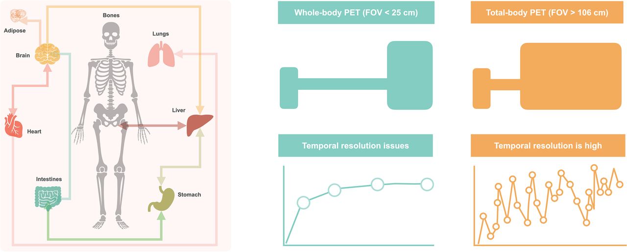
Molecular imaging modalities such as PET can provide valuable insights into the underlying homeostasis of living subjects using target-specific radiotracer imaging (4). After the commercial inception of PET, most of its clinical investigations focused on imaging with a single-organ field of view (FOV) (cardiology and neurology). With the introduction of a whole-body (WB) acquisition mode—that is, the successive translation of the subject through the axial FOV of a PET system with slightly overlapping bed positions (5)—the identification of hypermetabolic tumor lesions in oncology patients became the primary application of PET. Such a reductionist “lumpology” approach (6), however, caused a wealth of molecular information available from PET to be overlooked and the concept of human physiology imaging to be discarded.

The recent extension of the WB PET concept to extended axial-imaging ranges with larger-FOV systems, colloquially referred to as total-body (TB) PET, has sparked interest in the PET community to conduct multiorgan systemic investigations. TB PET systems cover axial scan ranges of 1 m (7,8) to 2 m (9), allowing synchronous measurement of signals from multiple organs. In addition, the richness of the multiorgan data derived from WB PET notwithstanding (10,11), TB PET is particularly unique as it satisfies 2 critical criteria for such causal investigations: the simultaneous acquisition of signals from multiple investigated distant organs and a high temporal resolution across the FOV (12). The combination of increased sensitivity and subsecond temporal sampling (13) provided by TB PET could potentially aid in probing real-time multiorgan interactions (Fig. 1).

FIGURE 1.
  • Download figure
  • Open in new tab
  • Download powerpoint
FIGURE 1.

Thanks to markedly increased performance, TB PET allows assessment of multiple organs synchronously, giving way to noninvasive exploration of systemic, interorgan interactions.

MULTIORGAN ANALYSIS WITH STANDARD WB PET

Traditional WB PET with an axial FOV of about 20 cm can already be used for multiorgan analysis. For example, simple intergroup comparisons of organ-based SUV can provide crucial information on the underlying pathology. A recent study demonstrated that in a patient cohort with resected breast cancer, a high metabolic tumor volume and increased spleen glucose metabolism at baseline were associated with poor 5-y recurrence-free survival (14). That study hinted toward a possible interaction between the tumor and the host immune system through upregulation of hematopoiesis. Diseases formerly conceived as focal, such as myocardial infarction, have distributed effects throughout the body that are mediated through disease-specific networks (15). And finally, mental and societal stress triggers have been linked to various diseases associated with chronic inflammation that can already be assessed by WB PET (16).

INTERORGAN NETWORKS THROUGH PET

Current multiorgan network investigations using WB or TB PET are mostly fishing expeditions aiming to pinpoint stable correlations between organs (10,11). In general, correlation analyses explore gross systemic effects between 2 groups without causal explanation. When correlation analyses are performed, the chosen sample should represent the investigated population (e.g., healthy or pathologic) (17). Other factors, such as variability, linearity, and variance of the samples, must also be considered. Since most multiorgan correlation network studies seek to pinpoint monotonic relationships between investigated organs, Spearman correlation should be chosen over Pearson correlation, as the former is nonparametric and insensitive to the linearity and homogeneity of the variance of observed data.

The ultimate goal of interorgan analysis is to identify causal relationships between organs that can facilitate the development of impactful interventions in medicine. Here, structure learning of Bayesian networks (18) in combination with graph models as visual representations of causal links in complex processes can be an attractive approach (19), which, however, still mandates the integration of a clinical expert to denounce spurious causal links.

Both causal and correlation networks should be considered hypothesis-generating tools rather than tools that provide solid endpoints. Such hypotheses must be proven or disproven in rigorous validation studies (Fig. 2) in which investigators should be conscious of the confounders affecting the accuracy of SUV or kinetic parameters as part of a multiorgan analysis (20).

FIGURE 2.
  • Download figure
  • Open in new tab
  • Download powerpoint
FIGURE 2.

Categoric pathway to adopting WB or TB PET for exploring human connectome: several advanced and automated tools are required to extract robust data for hypothesis building and validation in translational setting. ECG = electrocardiography; EEG = electroencephalography; fMRI = functional MRI.

THE PROMISE OF TB PET

Despite the increasing installed base of TB PET systems, the number of studies that explore TB PET beyond dose reduction and higher throughput for the sake of assessing the human connectome studies is limited. Preliminary studies have demonstrated the potential of using the temporal domain, namely raw time–activity curves, to derive metabolic associations between different bone compartments (21) or to construct normative networks for healthy male and female controls (22). Although neither study explained causality, dynamic TB PET has the potential to create personalized causal networks from a single subject. Such a paradigm requires, however, the subject to be challenged by a task, pharmacologic intervention, or external stressor (e.g., pain or cold). By challenging (perturbing) the system, simultaneous or delayed changes in signals from different organs can be measured and used to establish causality.

For decades, such studies have been performed with functional MRI to derive effectivity connectivity by conducting baseline and task paradigms in a single imaging session (23). Recent innovative brain studies in functional PET have shown the possibility of using 18F-FDG PET to study dynamic changes in glucose metabolism within a single session with the aid of constant-infusion protocols (24). However, conducting such challenge-based studies is nontrivial in a TB PET setting, particularly in view of unknown response times and downstream interactions. Therefore, test studies on well-understood paradigms (25) should be performed before exploratory connectome investigations are conducted using TB PET.

ROAD MAP TO THE FUTURE: CONNECT TO THE CONNECTOME

To date, the PET imaging community is fragmented by vendor, geography, and skill set. There needs to be more meaningful sharing of code, data, and expertise to address the novel challenges and opportunities that arise with this technology. To fully leverage the potential of WB and TB PET alike for health care, new analysis methods are required, and new skills in the workforce are needed (Fig. 2). Automated data analytics pipelines, including automatic WB semantic segmentation (26) as well as WB and TB PET motion correction and spatial normalization, are prerequisites to robust TB PET connectome studies.

The community needs to be open to the repurposing of existing solutions (e.g., Statistical Parametric Mapping, version 12 (27)) and to be prepared to fail in this high-risk–high-gain approach to using PET far from the comfortable notion of a high-sensitivity lesion tracker. Fostering rigorous experiments to prove the validity of correlations and causalities while also sharing negative results must be encouraged. Also, rich data from healthy and pathologic cohorts should be pooled to amass large sample sizes that help us to better understand the actual distribution of the data and, therefore, to arrive at logical conclusions.

CONCLUSION

The introduction of TB PET offers unique opportunities to investigate multiorgan interactions—the organ connectome for understanding human physiology and pathology. Novel study protocols and paradigms, and translational research pipelines, will be required to support causal interpretations of interorgan relationships. As a community, we should unite to prioritize progress over our vanities. The same was said in the early days of PET/CT and PET/MRI, and it still holds true. Novel and open-minded collaborative efforts beyond the nuclear medicine comfort zone are required to unlock the power of WB and TB PET imaging. Adopting this concept requires significant personal and infrastructural investments; the concept may fail, but if it does not, it will benefit our patients and medicine at large.

DISCLOSURE

Research support was received from NIH R01CA29422, EraPErMED PETictCAC, and ANR-FWF I-6174B. No other potential conflict of interest relevant to this article was reported.

ACKNOWLEDGMENTS

We sincerely thank Irene Buvat (Paris), Ramsey Badawi (U.C. Davis), Simon Cherry (U.C. Davis), Terry Jones (U.K.), and Stefan Grünert (Vienna) for their continuous input and motivation.

Footnotes

  • Published online Dec. 2, 2022.

  • © 2023 by the Society of Nuclear Medicine and Molecular Imaging.

REFERENCES

  1. 1.↵
    1. Cannon WB
    . The Wisdom of the Body. Revised and enlarged ed. W.W. Norton and Co.; 1963.
  2. 2.↵
    1. Goodman L
    . Regulation and control in physiological systems: 1960–1980. Ann Biomed Eng. 1980;8:281–290.
    OpenUrlPubMed
  3. 3.↵
    1. Billman GE
    . Homeostasis: the underappreciated and far too often ignored central organizing principle of physiology. Front Physiol. 2020;11:200.
    OpenUrl
  4. 4.↵
    1. Phelps ME,
    2. Hoffman EJ,
    3. Mullani NA,
    4. Ter-Pogossian MM
    . Application of annihilation coincidence detection to transaxial reconstruction tomography. J Nucl Med. 1975;16:210–224.
    OpenUrlAbstract/FREE Full Text
  5. 5.↵
    1. Dahlbom M,
    2. Hoffman EJ,
    3. Hoh CK,
    4. et al
    . Whole-body positron emission tomography: part I. Methods and performance characteristics. J Nucl Med. 1992;33:1191–1199.
    OpenUrlAbstract/FREE Full Text
  6. 6.↵
    1. Hofman MS,
    2. Hicks RJ
    . Moving beyond “lumpology”: PET/CT imaging of pheochromocytoma and paraganglioma. Clin Cancer Res. 2015;21:3815–3817.
    OpenUrlAbstract/FREE Full Text
  7. 7.↵
    1. Karp JS,
    2. Viswanath V,
    3. Geagan MJ,
    4. et al
    . PennPET Explorer: design and preliminary performance of a whole-body imager. J Nucl Med. 2020;61:136–143.
    OpenUrlAbstract/FREE Full Text
  8. 8.↵
    1. Prenosil GA,
    2. Sari H,
    3. Fürstner M,
    4. et al
    . Performance characteristics of the Biograph Vision Quadra PET/CT system with a long axial field of view using the NEMA NU 2-2018 standard. J Nucl Med. 2022;63:476–484.
    OpenUrlAbstract/FREE Full Text
  9. 9.↵
    1. Spencer BA,
    2. Berg E,
    3. Schmall JP,
    4. et al
    . Performance evaluation of the uEXPLORER total-body PET/CT scanner based on NEMA NU 2-2018 with additional tests to characterize PET scanners with a long axial field of view. J Nucl Med. 2021;62:861–870.
    OpenUrlAbstract/FREE Full Text
  10. 10.↵
    1. Sun T,
    2. Wang Z,
    3. Wu Y,
    4. et al
    . Identifying the individual metabolic abnormities from a systemic perspective using whole-body PET imaging. Eur J Nucl Med Mol Imaging. 2022;49:2994–3004.
    OpenUrl
  11. 11.↵
    1. Dias AH,
    2. Hansen AK,
    3. Munk OL,
    4. Gormsen LC
    . Normal values for 18F-FDG uptake in organs and tissues measured by dynamic whole body multiparametric FDG PET in 126 patients. EJNMMI Res. 2022;12:15.
    OpenUrl
  12. 12.↵
    1. Bashan A,
    2. Bartsch RP,
    3. Kantelhardt JW,
    4. et al
    . Network physiology reveals relations between network topology and physiological function. Nat Commun. 2012;3:702.
    OpenUrlCrossRefPubMed
  13. 13.↵
    1. Zhang X,
    2. Cherry SR,
    3. Xie Z,
    4. et al
    . Subsecond total-body imaging using ultrasensitive positron emission tomography. Proc Natl Acad Sci USA. 2020;117:2265–2267.
    OpenUrlAbstract/FREE Full Text
  14. 14.↵
    1. Seban R-D,
    2. Rouzier R,
    3. Latouche A,
    4. et al
    . Total metabolic tumor volume and spleen metabolism on baseline [18F]-FDG PET/CT as independent prognostic biomarkers of recurrence in resected breast cancer. Eur J Nucl Med Mol Imaging. 2021;48:3560–3570.
    OpenUrl
  15. 15.↵
    1. Thackeray JT
    . Imaging the molecular footprints of the heart–brain axis in cardiovascular disease. J Nucl Med. 2019;60:728–729.
    OpenUrlFREE Full Text
  16. 16.↵
    1. Tawakol A,
    2. Ishai A,
    3. Takx RA,
    4. et al
    . Relation between resting amygdalar activity and cardiovascular events: a longitudinal and cohort study. Lancet. 2017;389:834–845.
  17. 17.↵
    1. Goodwin LD,
    2. Leech NL
    . Understanding correlation: factors that affect the size of r. J Exp Educ. 2006;74:249–266.
    OpenUrlCrossRef
  18. 18.↵
    1. Scanagatta M,
    2. Salmerón A,
    3. Stella F
    . A survey on Bayesian network structure learning from data. Prog Artif Intelligence. 2019;8:425–439.
    OpenUrl
  19. 19.↵
    1. Zheng X,
    2. Aragam B,
    3. Ravikumar PK,
    4. Xing EP,
    5. et al.
    , eds. Advances in Neural Information Processing Systems. Vol 31. Curran Associates, Inc.; 2018:1–6.
    OpenUrl
  20. 20.↵
    1. Sarikaya I,
    2. Albatineh AN,
    3. Sarikaya A
    . Revisiting weight-normalized SUV and lean-body-mass-normalized SUV in PET studies. J Nucl Med Technol. 2020;48:163–167.
    OpenUrlAbstract/FREE Full Text
  21. 21.↵
    1. Suchacki KJ,
    2. Alcaide-Corral CJ,
    3. Nimale S,
    4. et al
    . A systems-level analysis of total-body PET data reveals complex skeletal metabolism networks in vivo. Front Med (Lausanne). 2021;8:740615.
    OpenUrl
  22. 22.↵
    1. Sundar LS,
    2. Badawi RD,
    3. Spencer BA,
    4. et al
    . Enhance-PET: exploring the human functional connectome using total-body [18F] FDG-PET [abstract]. Eur J Nucl Med Mol Imaging. 2021;48(suppl):S201.
    OpenUrl
  23. 23.↵
    1. Glover GH
    . Overview of functional magnetic resonance imaging. Neurosurg Clin N Am. 2011;22:133–139.
    OpenUrlCrossRefPubMed
  24. 24.↵
    1. Rischka L,
    2. Gryglewski G,
    3. Pfaff S,
    4. et al
    . Reduced task durations in functional PET imaging with [18F]FDG approaching that of functional MRI. Neuroimage. 2018;181:323–330.
    OpenUrl
  25. 25.↵
    1. Castellani JW,
    2. Young AJ
    . Human physiological responses to cold exposure: acute responses and acclimatization to prolonged exposure. Auton Neurosci. 2016;196:63–74.
    OpenUrl
  26. 26.↵
    1. Sundar LKS,
    2. Yu J,
    3. Muzik O,
    4. et al
    . Fully automated, semantic segmentation of whole-body 18F-FDG PET/CT images based on data-centric artificial intelligence. J Nucl Med . 2022;63:1941–1948.
    OpenUrlAbstract/FREE Full Text
  27. 27.↵
    1. Ashburner J
    . SPM: a history. Neuroimage. 2012;62:791–800.
    OpenUrlCrossRefPubMed
  • Received for publication October 6, 2022.
  • Revision received November 17, 2022.
PreviousNext
Back to top

In this issue

Journal of Nuclear Medicine: 64 (2)
Journal of Nuclear Medicine
Vol. 64, Issue 2
February 1, 2023
  • Table of Contents
  • Table of Contents (PDF)
  • About the Cover
  • Index by author
  • Complete Issue (PDF)
Print
Download PDF
Article Alerts
Sign In to Email Alerts with your Email Address
Email Article

Thank you for your interest in spreading the word on Journal of Nuclear Medicine.

NOTE: We only request your email address so that the person you are recommending the page to knows that you wanted them to see it, and that it is not junk mail. We do not capture any email address.

Enter multiple addresses on separate lines or separate them with commas.
Whole-Body PET Imaging: A Catalyst for Whole-Person Research?
(Your Name) has sent you a message from Journal of Nuclear Medicine
(Your Name) thought you would like to see the Journal of Nuclear Medicine web site.
Citation Tools
Whole-Body PET Imaging: A Catalyst for Whole-Person Research?
Lalith Kumar Shiyam Sundar, Marcus Hacker, Thomas Beyer
Journal of Nuclear Medicine Feb 2023, 64 (2) 197-199; DOI: 10.2967/jnumed.122.264555

Citation Manager Formats

  • BibTeX
  • Bookends
  • EasyBib
  • EndNote (tagged)
  • EndNote 8 (xml)
  • Medlars
  • Mendeley
  • Papers
  • RefWorks Tagged
  • Ref Manager
  • RIS
  • Zotero
Share
Whole-Body PET Imaging: A Catalyst for Whole-Person Research?
Lalith Kumar Shiyam Sundar, Marcus Hacker, Thomas Beyer
Journal of Nuclear Medicine Feb 2023, 64 (2) 197-199; DOI: 10.2967/jnumed.122.264555
Twitter logo Facebook logo LinkedIn logo Mendeley logo
  • Tweet Widget
  • Facebook Like
  • Google Plus One
Bookmark this article

Jump to section

  • Article
    • Visual Abstract
    • MULTIORGAN ANALYSIS WITH STANDARD WB PET
    • INTERORGAN NETWORKS THROUGH PET
    • THE PROMISE OF TB PET
    • ROAD MAP TO THE FUTURE: CONNECT TO THE CONNECTOME
    • CONCLUSION
    • DISCLOSURE
    • ACKNOWLEDGMENTS
    • Footnotes
    • REFERENCES
  • Figures & Data
  • Info & Metrics
  • PDF

Related Articles

  • No related articles found.
  • PubMed
  • Google Scholar

Cited By...

  • Clinical Implementation of Total-Body PET in China
  • SEGMENTATION OF DYNAMIC TOTAL-BODY [18F]-FDG PET IMAGES USING UNSUPERVISED CLUSTERING
  • Google Scholar

More in this TOC Section

  • RECIP 1.0: A Roadmap for Clinical Implementation
  • Diagnostic Radiopharmaceutical Trial Design: Is It Time to Change Nomenclature?
  • From Stabilization to Depletion: Molecular Imaging to Measure Therapeutic Response in ATTR-CA
Show more Hot Topics

Similar Articles

SNMMI

© 2025 SNMMI

Powered by HighWire